Lumbar Spine Degenerative Disease: Effect on Bone Mineral Density Measurements in the Lumbar Spine and Femoral Neck1
Seon-Kwan Juhng, M.D., Peter Koplyay, M.D.2, J. Jeffrey Carr, M.D.2 Leon Lenchik, M.D.2
Purpose: To determine the effect of degenerative disease of the lumbar spine on bone miner- al density in the lumbar spine and femoral neck.
Materials and Methods: We reviewed radiographs and dual energy x-ray absorptiometry scans of the lumbar spine and hip in 305 Caucasian women with suspected osteoporosis.
One hundred and eighty-six patients remained after excluding women less than 40 years of age (n = 18) and those with hip osteoarthritis, scoliosis, lumbar spine fractures, lumbar spinal instrumentation, hip arthroplasty, metabolic bone disease other than osteoporosis, or medications known to influence bone metabolism (n = 101). On the basis of lumbar spine radiographs, those with absent/mild degenerative disease were assigned to the control group and those with moderate/severe degenerative disease to the degenerative group. Spine radi- ographs were evaluated for degenerative disease by two radiologists working independently;
discrepant evaluations were resolved by consensus. Lumbar spine and femoral neck bone mineral density was compared between the two groups.
Results: Forty-five (24%) of 186 women were assigned to the degenerative group and 141 (76%) to the control group. In the degenerative group, mean bone mineral density measured 1.075 g/cm2in the spine and 0.788 g/cm2in the femoral neck, while for controls the corre- sponding figures were 0.989 g/cm2and 0.765 g/cm2. Adjusted for age, weight and height by means of analysis of variance, degenerative disease of the lumbar spine was a significant pre- dictor of increased bone mineral density in the spine (p = 0.0001) and femoral neck (p = 0.0287).
Conclusion: Our results indicate a positive relationship between degenerative disease of the lumbar spine and bone mineral density in the lumbar spine and femoral neck, and suggest that degenerative disease in that region, which leads to an intrinsic increase in bone mineral density in the femoral neck, may be a good negative predictor of osteoporotic hip fractures.
Index words : Spine, arthritis Spine, radiography Hip, arthritis
Bones, absorptiometry
1Department of Radiology, Wonkwang University School of Medicine, Iksan, Jeonbuk, Korea
2Department of Radiology, Division of Radiologic Sciences, Wake Forest University School of Medicine, Winston-Salem, NC, U.S.A.
This paper was supported in part by Soongsan fellowship in Wonkwang University and Research Fund (CURIMS-`97-B-121) of Chonnam University Research Institute of Medical Sciences.
Received January 2, 2001; Accepted March 21, 2001
Address reprint requests to : Seon-Kwan Juhng, M.D., Department of Radiology, Wonkwang University School of Medicine, 344-2 Sinyong Dong, Iksan, Jeonbuk 570-711, Korea.
Tel. 82-63-850-1514 Fax. 82-63-851-4749 E-mail: [email protected]
The use of dual energy x-ray absorptiometry (DXA) for the diagnosis of osteoporosis is now standard in most clinical settings. Measurement of bone mineral density (BMD) with DXA allows the diagnosis of osteoporosis, the estimation of risk of fracture, and the monitoring of response to therapy. BMD is usually measured at the lumbar spine and proximal femur because these sites are affected early in the course of osteoporosis and are the most common sites of osteoporotic fractures. A com- mon pitfall of DXA scan interpretation in elderly pa- tients is the elevation of spine BMD measurements ow- ing to the presence of spinal degenerative disease, a phe- nomenon demonstrated previous studies (1-4).
Furthermore, reports of several studies have reported that spinal degenerative disease results in elevated BMD measurements not only locally in the spine, but general- ly throughout the skeleton (5-7). If degenerative disease is indeed a generalized disorder affecting the entire skeleton, the ramifications of this will be important in the diagnosis of osteoporosis. For example, patients with spinal degenerative disease would be expected to have elevated BMD in the hip, and this, in turn might offer protection from osteoporotic fracture. Some studies, however, have reported no apparent association be- tween degenerative disease and BMD (8-13). The pur- pose of this study was to evaluate the effect of degenera- tive disease of the lumbar spine, as diagnosed with radi- ographs, on BMD in the lumbar spine and femoral neck, as measured with DXA.
Materials and Methods
We retrospectively evaluated radiographs and DXA scans of the lumbar spine and hip in 305 Caucasian women with suspected osteoporosis. The following sub- jects were excluded from the study: those younger than 40 years of age (n = 18), those with osteoarthritis of the hip (n = 27), scoliosis (n = 11), fractures of the lumbar spine (n = 36), lumbar spinal instrumentation (n = 5), arthroplasty of the hip (n = 2), metabolic bone disease other than osteoporosis (n=8), and those treated with medications, including corticosteroids, known to influ- ence bone metabolism (n=12). In the remaining 186 women, anteroposterior (AP) and lateral radiographs of the lumbar spine were independently evaluated for the presence of degenerative disease by two musculoskele- tal radiologists unaware of the results of DXA scans.
The radiographic criteria for degenerative disease in- cluded disc space narrowing, subchondral sclerosis of
the vertebral endplates, osteophytes, and facet joint os- teoarthritis (14). In each patient, the severity of degener- ative disease from the L1-L2 level to the L4-L5 level was categorized as absent/mild (Fig. 1A) or moderate/severe (Fig. 2A). Those with absent/mild degenerative disease were assigned to the control group and those with mod- erate/severe degenerative disease to the degenerative group. Discrepant interpretations were discussed by the two radiologists, who then reached a consensus. In each patient, BMD of the lumbar spine and femoral neck was measured using a Lunar DPX densitometer (Lunar Corporation, Madison, Wis., U.S.A.). Lumbar spine and femoral neck scans were obtained with the patient in the supine position. Lumbar spine measurement was analyzed from the L2 to the L4 vertebra, and BMD val- ues were expressed as area density (g/cm2).
BMD measurements of the lumbar spine and femoral neck in the degenerative group were compared with those in the control group; by means of analysis of vari- ance (ANOVA), measurements were adjusted for the pa- tient’s age, weight and height. For all data management and statistical computations, JMP statistics software (SAS Institute, Cary, N.C.) was used.
Results
With regard to the presence of degenerative disease, interobserver agreement was good (kappa = 0.81).
Forty-five (24%) of 186 women were included in the de- generative group, and 141 (76%) in the control group.
Table 1 compares the mean age, height and weight of women in the degenerative and control group.
In the former group (Fig. 2), the mean BMD measure- ment was 1.075 g/cm2 in the spine and 0.788 g/cm2 in the femoral neck (Table 2), while in the control group (Fig. 1), the corresponding measurements were 0.989 g/cm2and 0.765 g/cm2.
After adjusting for each patient’s age, weight and height by means of ANOVA, degenerative disease of the lumbar spine was found to be a significant predictor of
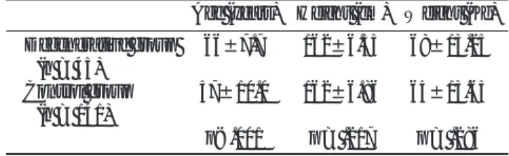
Table 1. Comparison of Age, Height, and Weight in Degenerative Group and Control Group
Age (years) Height (cm) Weight (Kg) Degenerative group 66±7.7 162±6.35 68±13.25
(n = 45)
Control group 57±10.0 162±6.86 65±13.65 (n = 141)
p<.001 p = .217 p = .286 Note. - Numbers are mean±SD.
increased spinal BMD (p = 0.0001). Furthermore, in pa- tients whose hip radiographs were normal, degenerative disease of the spine was a significant predictor of in- creased BMD in the femoral neck (p = 0.0287).
Discussion
Our results in 186 postmenopausal women indicate that those with radiographic findings of moderate to se- vere spinal degenerative disease had significantly higher BMD in the spine than did women whose radiographic findings were consistent with absent or mild degenera- tive disease. The results were significant even after the two groups were adjusted for age, weight and height (Tables). Our results support those of prior studies in which spinal degenerative disease, diagnosed with radi- ographs, was associated with elevated spinal BMD, measured with DXA (4, 6, 7). In a study of 130 normal postmenopausal women, Reid et al. (4) reported elevat- ed lumbar BMD in women with vertebral osteophytes.
A
B
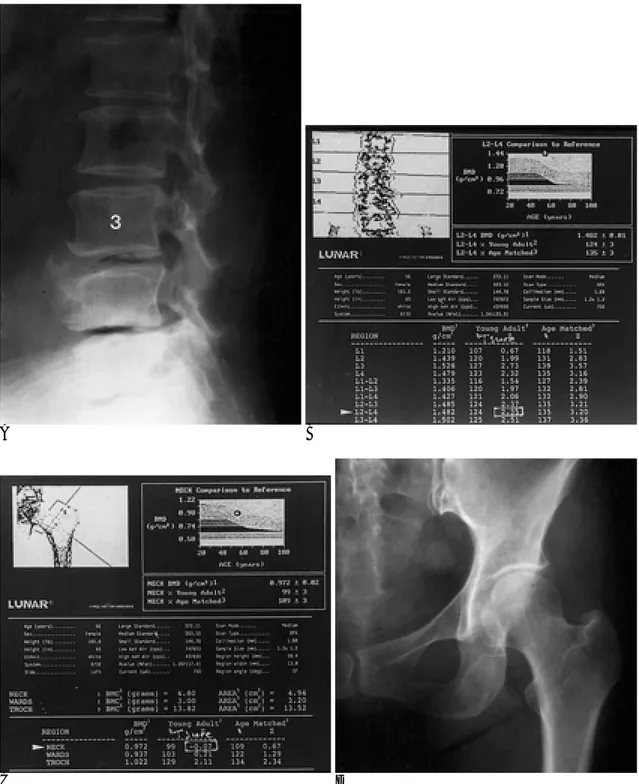
C Fig. 1. Control group.
A. Lateral spine radiograph in a 50-year-old woman shows preservation of disk spaces, minimal osteophytes, and minimal facet osteoarthritis.
Based on these radiographic findings, the patient was placed into control group.
B. Dual energy x-ray absorptiometry scan of lumbar spine. BMD as mea- sured from L2 to L4 is 106% of young-adult reference (T-score is 0.62.) (arrowhead).
C. Dual energy x-ray absorptiometry scan of left hip. BMD as measured in femoral neck is 77% of young-adult reference (T-score is -1.92.) (ar- rowhead).
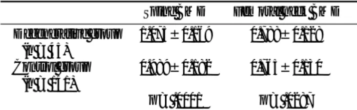
Table 2. Comparison of Spine BMD and Femoral Neck BMD in Degenerative Group and Control Group
Spine BMD Femoral neck BMD Degenerative group 1.075±0.169 0.788±0.128
(n = 45)
Control group 0.989±0.182 0.765±0.130 (n = 141)
p = .0001 p = .0287 Note. - BMD = bone mineral density. Numbers are mean±SD.
Similarly, in a study of 93 postmenopausal women with osteoporosis and at least one vertebral fracture, Masud et al. (6) reported elevated lumbar BMD in women who
also had vertebral osteophytes. In a study of 113 men and 187 women over 60 years of age, Jones et al. (7) re- ported that lumbar spine osteophytes, disk narrowing
A
D B
C
Fig. 2. Degenerative group.
A. Lateral spine radiograph in a 56-year-old woman shows extensive disk space narrowing, subchondral sclerosis, and osteophytes at L3-L4 and L4-L5 levels. Based on these radiographic findings, the patient was placed into degenerative disease group.
B. Dual energy x-ray absorptiometry scan of lumbar spine. BMD as measured from L2 to L4 is 124% of young-adult reference (T- score is 2.35.) (arrowhead).
C. Dual energy x-ray absorptiometry scan of left hip. BMD as measured in femoral neck is 99% of young-adult reference (T-score is -0.07.) (arrowhead).
D. Frontal radiograph of left hip shows no radiographic evidence for hip osteoarthritis.
and apophyseal disease were associated with elevated spinal BMD. In most studies, the increase in spinal BMD in patients with spinal degenerative disease was attributed to the presence of osteophytes, to narrowing of the intervertebral joint space, or to sclerosis of the facet joint (4, 6, 7, 15, 16). Because lumbar osteophytes, subchondral sclerosis and facet osteoarthritis result in a relative increase in cortical bone, elevated spinal BMD in patients with these findings was expected.
The fact that BMD measurements are elevated in the presence of local degenerative disease is further sup- ported by studies of anatomic regions other than the lumbar spine. In a population-based study of men and women aged 55 years or more, Burger et al. (17) report- ed that the grade of hip osteoarthritis, as determined ra- diographically, showed a significant correlation with femoral neck BMD, as measured with DXA.
Considerably more significant than the finding of ele- vated spinal BMD in the degenerative group was our finding that women with radiographic findings of mod- erate to severe spinal degenerative disease had elevated BMD in the femoral neck, despite normal radiographs of the hip (Fig. 2). Our findings support those of prior studies in which elevated BMD measurements were re- ported at sites shown by radiographs to be unaffected by degenerative disease (5, 7, 18). Hart et al. (18) reported that BMD was significantly higher in patients with de- generative disease of the lumbar spine than in controls, not only in the lumbar spine (+7.8%) but also in the femoral neck (+6.3%). Similarly, Jones et al. (7) reported increased BMD in the femoral neck in women with spinal degenerative disease. Peel et al. (5) recently re- ported that patients with spinal degenerative disease showed increased BMD in the lumbar spine, femoral neck, and total body, and suggested that the results may reflect a generalized increase in BMD or may be due to degenerative changes such as osteophytes in regions other than the spine. In our study, patients with radi- ographic evidence of hip degenerative disease were ex- cluded, and a generalized elevation of BMD is thus the more likely explanation.
Further supporting the argument that spinal degenera- tive disease results in a generalized increase in BMD is the fact that the presence of osteophytes in the region of the hip should not affect BMD in the femoral neck. In the spine, elevated BMD is often considered a conse- quence of facet sclerosis and osteophytes, but the femoral neck measurement is removed from osteo- phytes and subchondral sclerosis and BMD at that site
should not be elevated, even in patients with radi- ographically apparent degenerative disease of the hip (5). In our study, women with radiographic evidence of hip osteoarthritis were excluded, and increased BMD in the femoral neck is thus not due to degenerative disease of the hip. Our results and those of prior investigators suggest an intrinsic increase in femoral neck BMD in women with spinal degenerative disease and the pres- ence of spinal degenerative disease might thus be an in- dicator of decreased risk for osteoporotic hip fractures.
This is important in that BMD is considered the major determinant of osteoporotic fracture risk and osteo- porotic fracture mortality is associated primarily with hip fractures (19).
The theory that degenerative disease results in a gen- eralized increase in BMD is also supported by studies in which radiographic findings of degenerative disease were correlated with BMD measurements at other anatomic sites. Nevitt et al. (20) reported that elderly Caucasian women with moderate to severe osteoarthri- tis of the hip, as determined radiographically, had high- er BMD in the spine and appendicular skeleton.
Similarly, Gotfredsen et al. (21) reported that patients with osteoarthritis of the hip had higher bone mineral content, as measured in the total body. Hannan et al.
(22) reported that femoral BMD was higher in women with degenerative disease of the knee, and even degen- erative disease of the hand has been associated with ele- vated axial BMD (18, 23). Hart et al. (18) reported that degenerative disease of the distal interphalangeal joints was associated with increased lumbar spine BMD, and degenerative disease of the first carpometacarpal joint was associated with higher spine and femoral neck BMD, after adjustment for age, obesity, and vertebral osteophytes.
Some studies, however, have failed to demonstrate an association between degenerative disease and BMD (1, 8). Hochberg et al. (9), in a study of degenerative disease of the hand (Kellgren-Lawrence grade), found no signifi- cant association between metacarpal bone mass (per- cent cortical area of the second metacarpal) and distal radius BMD ( determined by single-photon absorptiom- etry). Other investigators (10-12) reported no increase in BMD of the distal radius or total body in patients with generalized osteoarthritis involving the hand. These conflicting reports may be attributable to differences in the selection of patients and controls, the selection of measurement sites, and the expression of results (13).
Our results are in line with those of prior studies indi-
cating an inverse relationship between osteoporosis and osteoarthritis, one that has important ramifications for the evaluation of patients in whom osteoporosis is sus- pected. Generalized degenerative disease in women usually manifests at the time of the menopause, before major bone loss occurs; its presence may therefore be a good negative predictor of osteoporosis (13). Because BMD is considered the major determinant of fracture risk (19), patients with elevated BMD owing to the pres- ence of degenerative disease may be protected from os- teoporotic fractures.
In conclusion, in our study of 186 women over 40 years of age, spinal BMD was significantly higher in those with radiologic evidence of degenerative disease of the lumbar spine. More importantly, femoral neck BMD was also significantly higher in patients with spinal degenerative disease and with no radiographic evidence of osteoarthritis of the hip. Our findings sug- gest that degenerative disease of the lumbar spine, which leads to an intrinsic increase in BMD in the femoral neck, may thus be a good negative predictor of osteoporotic hip fractures. Whether elevated BMD in patients with osteoporosis offers protection from osteo- porotic fracture remains to be determined.
Acknowledgment
We thank Donna S. Garrison and Terry Poovey for their editorial assistance.
References
1. Orwoll ES, Oviatt SK, Mann T. The imapct of osteophytic and vas- cular calcifications on vertebral mineral density measurements in men. J Clin Endocrinol Metab 1990;70:1202-1207
2. Dawson-Hughes B, Dallal GE. Effect of radiographic abnormali- ties on rate of bone loss from the spine. Calcif Tissue Int 1990;46:
280-281
3. Laitinen K, Valimaki M, Keto P. Bone mineral density measured by dual energy X-ray absorptiometry in healthy Finnish women.
Calcif Tissue Int 1991;48:224-231
4. Reid IR, Evans MC, Ames R, Wattie DJ. The influence of osteo- phytes and aortic calcification on spinal mineral density in post- menopausal women. J Clin Endocrinol Metab 1991; 72:1372-1374 5. Peel NFA, Barrington NA, Blumsohn A, Colwell A, Hannona R,
Eastell R. Bone mineral density and bone turnover in spinal os- teoarthrosis. Ann Rheum Dis 1995;54:867-871
6. Masud T, Langley S, Wiltshire P, Doyle DV, Spector TD. Effect of spinal osteophytosis on bone mineral density measurements in
vertebral osteoporosis. Br Med J 1993;307:172-173
7. Jones G, Nguyen T, Sambrook PN, Kelly PJ, Eisman JA. A longitu- dinal study of the effect of spinal degenerative disease on bone density in the elderly. J Rheumatol 1995;22:932-936
8. Franck H, Munz M, Scherrer M. Evaluation of dual-energy X-ray absorptiometery bone mineral measurement-comparison of a sin- gle-beam and fan-beam design: the effect of osteophytic calcifica- tion on spine bone mineral density. Calcif Tissue Int 1995;56:192- 195
9. Hochberg MC, Lethbridge-Cejku M, Scott WW, Jr., Plato CC, Tobin JD. Appendicular bone mass and osteoarthritis of the hands in women: data from the Baltimore longitudinal study of aging. J Rheumatol 1994;21:1532-1536
10. Cooper C, Poll V, McLaren M, Daunt SO’N, Cawley MID.
Alterations in appendicular skeletal mass in patients with rheuma- toid, psoriatic and osteoarthropathy. Ann Rheum Dis 1988;47:481- 484
11. Price T, Hesp R, Michell R. Bone density in generalised os- teoarthritis. J Rheumatol 1987;14:560-562
12. Reid DM, Kennedy NSJ, Smith MA, Tothill P, Nuki G. Bone mass in nodal primary generalized osteoarthritis. Ann Rheum Dis 1984;
43:240-242
13. Dequeker J, Boonen S, Aerssens J, Westhovens R. Inverse relation- ship osteoarthritis-osteoporosis: What is the evidence? What are the consequences? Br J Rheumatol 1996;35:813-820
14. Lane NE, Kremer LB. Radiographic indices for osteoarthritis.
Rheum Dis Clin North Am 1995;21:379-394
15. Yu W, Gluer C-C, Fuerst T, et al. Influence of degenerative joint disease on spinal bone mineral measurements in postmenopausal women. Calcif Tissue Int 1995;57:169-174
16. von der Recke P, Hansen MA, Overgaard K, Christiansen C. The impact of degenerative conditions in the spine on bone mineral density and fracture risk prediction. Osteoporosis Int 1996;6:43-49 17. Burger H, van Daele PLA, Odding E, et al. Association of radi-
ographically evident osteoarthritis and higher bone mineral densi- ty and increased bone loss with age. Arthritis Rheum 1996;39:81-86 18. Hart DJ, Mootoosamy I, Doyle DV, Spector TD. The relationship between osteoarthritis and osteoporosis in the general population:
The Chingford Study. Ann Rheum Dis 1994;53:158-162
19. Ross PD, Davis JW, Epstein RS, Wasnich RD. Pre-existing frac- tures and bone mass predict vertebral fracture incidence in women. Ann Intern Med 1991;114:919-923
20. Nevitt MC, Lane NE, Scott JC, et al. Radiographic osteoarthritis of the hip and bone mineral density. Arthritis Rheum 1995;38:907-916 21. Gotfredsen A, Riis BJ, Christiansen C, Rodbro P. Does a single lo- cal absorptiometric bone measurement indicate the overall skele- tal status? Implications for osteoporosis and osteoarthritis of the hip. Clin Rheumatol 1990;9:193-203
22. Hannan MT, Anderson JJ, Zhang Y, Levy D, Felson DT. Bone mineral density and knee osteoarthritis in elderly men and women: the Framingham study. Arthritis Rheum 1993;36:1671- 1680
23. Gevers G, Dequeker J, Geusens P, Nyssen-Behets C, Dhem A.
Physical and histomorphological characteristics of iliac crest bone differ according to the grade of osteoarthritis at the hand. Bone 1989;10:173-177
대한방사선의학회지 2001;44:531-537
요추의 퇴행성 질환이 요추와 대퇴골 경부의 골밀도에 미치는 영향1
1원광대학교 의과대학 방사선과학 교실
2Department of Radiology, Division of Radiologic Sciences, Wake Forest University School of Medicine, Winston-Salem, NC, U.S.A.
정선관・Peter Koplyay, M.D.2・J. Jeffrey Carr, M.D.2・Leon Lenchik, M.D.2
목적: 골다공증을 진단하려고 할때 일반적으로 요추와 근위부 대퇴골의 골밀도를 측정하지만 동반되는 퇴행성 질환에 의해 골밀도가 증가할 수 있다. 요추에 발생한 퇴행성 질환이 요추와 대퇴골 경부의 골밀도에 미치는 영향을 알아보고 자 한다.
대상과 방법: 골다공증이 의심된 여성 305명의 요추와 고관절의 단순방사선사진과 이중 X-선 골밀도 측정을 후향적으
로 분석하였다. 40세 미만인 18명과 고관절의 골관절염, 요추의 측만증이나 골절, 고관절성형술 및 요추에 기구사용술 을 받은 101명을 제외한 186명을 대상으로 하였다. 두명의 방사선과 전문의가 각각 요추의 단순 방사선 사진에서 퇴행 성 질환의 정도를 4단계(정상, 경도, 중등도, 고도)로 분류하였으며, 일치하지 않은 경우에는 협의하여 결정하였다. 정 상이거나 경도의 퇴행성 변화를 보인 경우를 대조군으로, 중등도 이상의 경우를 퇴행군으로 나눈 뒤, 두 군 간의 요추와 대퇴골 경부의 골밀도를 비교하였다.
결과: 45명(24%)이 퇴행군에, 141명(76%)은 대조군에 속하였다. 퇴행군에서 요추의 평균 골밀도는 1.075 g/cm2, 대 퇴골 경부에서는 0.788 g/cm2이었으며, 대조군에서는 각각 0.989 g/cm2, 0.765 g/cm2이었다. 두 군 사이의 나이, 체중, 신장 등의 변수를 보정하였을 때(ANOVA), 요추의 퇴행성 질환이 요추(p=0.0001)와 대퇴골 경부(p=0.0287)에서 골 밀도를 증가시킨 의의있는 원인이었다.
결론: 요추에 퇴행성 질환이 있을 때 요추의 골밀도뿐만 아니라 퇴행성 질환이 없는 대퇴골 경부에서도 골밀도가 증가 되어 요추의 퇴행성질환이 대퇴골에 발생하는 골다공증성 골절의 가능성을 낮추는 요인임을 시사한다.