흉곽에서 정맥기형은 우연히 발견되거나, 다른 기형과 동반 되어 나타난다. 흔히 볼 수 있는 흉곽의 정맥기형은 지속성 좌 상대정맥, 대동맥후방 혹은 대동맥하방 완두정맥(retroaortic or subaortic brachiocephalic vein), 홀허파엽(azygos lobe), 기정맥 혹은 반기정맥과 하대정맥의 연결(azygos and hemiazygous continuation of the IVC), 폐정맥류와 폐정 맥의 수적변이, 폐정맥환류이상(anomalous pulmonary vein return) 등이다(1-3). 이러한 정맥기형들은 종양과 유사 하게 보이거나 종격동의 종괴 혹은 림프절 종대와의 감별을 필 요로 한다(4, 5).
정맥기형은 많은 수에서 단순흉부촬영으로 볼 수 있으나, CT는 우수한 해부학적 영상을 제공하며, 관상면상 또는 시상 면상의 재구성 영상을 얻을 수 있어 정맥기형의 주행 및 주변 조직과의 관계를 명료하게 보여 준다. 이에 정상 정맥해부학에 대하여 알아보고 다양한 정맥기형에서 보이는 영상학적 소견 을 통해 방사선학적 진단에 도움을 주고자 한다.
상대정맥과 완두정맥(무명정맥)
해부 및 발생
완두정맥은 흉쇄관절(sternoclavicular joint) 후방에서 내 경정맥과 쇄골하정맥이 만나서 형성된다. 좌측 완두정맥은 대 동맥 전방으로 비스듬하게 아래로 주행하며, 우측 완두정맥은 복장뼈자루(manubrium) 뒤에서 가파르게 아래로 지나간다.
이들은 첫 번째 늑연골 높이에서 상종격동의 우측에서 합쳐져 서 상대정맥을 이룬다. 그 후 상대정맥은 약간 전내측으로 내
려가, 세 번째 늑연골 높이의 중종격동에서 우심방으로 들어간 다(6).
정상적으로 우측 전주정맥(right anterior cardinal vein) 과 총주정맥(common cardinal vein)이 상대정맥을 형성하고 좌측 전주정맥은 퇴화하는데(1), 좌측 전주정맥이 지속하면 양측 상대정맥이 생기고 우측 전주정맥이 퇴화하면 좌상대정 맥만이 형성된다.
양측 전주정맥사이에 2개 이상의 정맥총(venous plexus)이 있다가, 아래 부위의 정맥총이 퇴화하면서 정상적인 완두정맥 이 형성된다. 이러한 발생과정은 정상적인 흉곽 혈관 형성에서
─ 27 ─
흉곽 정맥 기형
1김 혜 린∙김 영 통2∙조 성 식2
고식적인 단순흉부촬영에서 보이는 정상 또는 기형 혈관구조들은 종괴로 잘못 해석될 수 있 다. 그러므로 그 해부학을 이해하고 방사선 소견을 숙지하는 것이 정확한 진단에 중요하며, 불 필요한 추가 검사를 줄이는 데 도움을 줄 것이다. 이에 이번 임상 화보를 통하여 흉곽 내의 폐 또는 체정맥에 생기는 다양한 기형들의 CT 및 단순흉부촬영 소견을 보여주어 방사선학적 진단 에 도움을 주고자 한다.
1순천향대학교 부천병원 영상의학과
2순천향대학교 천안병원 영상의학과
이 논문은 2008년 6월 11일 접수하여 2008년 9월 1일에 채택되었음.
Fig. 1. Retroaortic or subaortic brachiocephalic vein in a 32- year-old woman.
CT scan shows anomalous brachiocephalic vein draining into the superior vena cava(SVC), inferior and posterior to aortic arch.
대동맥궁이 짧아지고 대동맥과 폐동맥이 커지면서 압력이 증 가하여 대동맥궁 아래에 있던 기존의 정맥총이 퇴화하면서 대 동맥궁 상부의 완두정맥이 정상적으로 형성되는데, 경부 대동
맥궁, 우측 대동맥궁, 폐동맥 협착 혹은 폐쇄와 같이 대동맥궁 아래의 공간이 커질 수 있는 상황에서 대동맥후방 혹은 대동맥 하방 완두정맥이 형성될 수 있다(7).
A B
Fig. 3. Retroaortic brachiocephalic vein with right sided aortic arch in a 69-year-old woman.
A. Chest radiograph shows a central venous catheter within SVC via the retroaortic innominate vein below the right-sided aortic arch.
B. CT scan shows the retroaortic brachiocephalic vein and right-sided aortic arch.
A B
C D
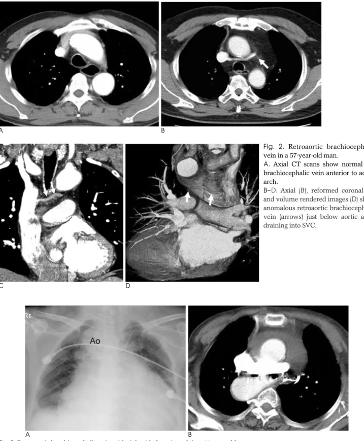
Fig. 2. Retroaortic brachiocephalic vein in a 57-year-old man.
A. Axial CT scans show normal left brachiocephalic vein anterior to aortic arch.
B--D. Axial (B), reformed coronal (C) and volume rendered images (D) show anomalous retroaortic brachiocephalic vein (arrows) just below aortic arch, draining into SVC.
대동맥후방 혹은 대동맥궁하방 완두정맥
모든 선천적 심장기형의 0.2-1%에서 보이며(7), CT와 MRI 같은 비침습적인 영상기법의 사용으로 발견이 증가하고 있다.
대동맥후방 완두정맥은 좌측 완두정맥의 기형 중 가장 많은 유형으로, 정상적으로 좌측 완두정맥이 대동맥궁의 전방으로 주행하여 우측 완두정맥과 만나서 합쳐지는 것과 달리, 상행대
─ 29 ─
A B
Fig. 4. Persistent left SVC in a 42-year-old man.
A. Chest radiograph shows an additional interface (arrowheads) lateral to the aortic arch.
B. CT scan shows left SVC (arrow) on the left side of the aortic arch.
Fig. 5. Persistent left SVC in a 2-year-old boy.
Lateral volume-rendered CT image shows left SVC (arrow- heads), which descends lateral to the aortic arch and anterior to the hilum and drains into the coronary sinus. There is no crossing left brachiocephalic vein between right (arrow) and left SVC.
Fig. 6. Azygos venous system in prevertebral space in a 56- year-old woman.
Volume-rendered CT image shows the azygos venous system in prevertebral space. The azygos vein (arrow) is formed by the union of the ascending lumbar veins with the right sub- costal veins at the level of the 12th thoracic vertebra, ascend- ing in the posterior mediastinum, and arching over the root of the right lung to join SVC. The hemiazygos vein (double ar- rowheads) joins the azygos vein at the level of T9 vertebra.
The accessory hemiazygos vein (arrowhead) begins at the me- dial end of the 4th or 5th intercostal space and joins the azygos vein at the level of T7 or T8 vertebrae. Sometimes the accesso- ry hemiazygos vein joins the hemiazygos or the superior inter- costal vein.
동맥의 후방, 아래에서 상대정맥으로 유입되기 때문에 대동맥 하방(subaortic) 완두정맥이라고도 한다(7) (Fig. 1, 2). 정상 적인 좌측 완두정맥이 없기도 하고 같이 있기도 한다(Fig.
2A). 대동맥후방 완두정맥은 우측 대동맥궁(Fig. 3), 경부 대 동맥궁 혹은 미성숙폐동맥과 동반되어 나타나기도 한다.
지속성 좌상대정맥
총인구의 약 0.5% 이하에서 발견되며, 선천적 심장기형을 가진 환자에서 약 4% 정도 증가한다(1). 좌상대정맥은 대동맥 궁의 외측으로 내려가다, 폐문에서 앞으로 향하여 후방실고랑 (posterior atrioventricular groove)을 통해 심낭막으로 들 어온다. 정상적으로 좌상대정맥은 대부분이 관상정맥동으로 들어가며, 관상정맥동이 커지게 된다(Fig. 4, 5). 대부분은 우 측 상대정맥와 같이 있으며, 이때 좌측 완두정맥이 작거나 없 다. CT나 MRI에서 커진 관상정맥동이 보이면 좌상대정맥의 가능성을 생각할 수 있다. 단순흉부촬영에서 대동맥 융기 (aortic knob) 왼편에 종격동의 국소적인 확장으로 보일 수 있으며(5) (Fig. 4A), 정맥 내 카테터를 삽입하면서 발견할 수도 있다. 일반적으로 좌상대정맥은 임상적으로 문제 되지 않
으나, 관상정맥동으로 들어가는 구멍(ostium)이 작으면 정맥 내 카테터 혹은 심방조율기 등을 삽입시키기 어려울 수 있다.
기정맥계
해부 및 발생
기정맥계는 기정맥, 반기정맥, 부반기정맥으로 나뉘며, 늑간 정맥과 종격동의 분지들을 받아 흉곽의 뒤쪽에 있는 한 쌍으로 된 척추주위의 정맥통로이다. 기정맥은 우상행요정맥(right ascending lumbar vein)과 늑골하정맥이 합쳐지면서 기인하 며, 대동맥 열공을 통해 흉곽 내로 들어온다. 그 후 흉추골의 전외측을 따라 올라가다가. 제5, 6 흉추골에서 우기관지 상방 에서 활 모양을 그리면서 상대정맥으로 들어가지만, 드물게 우 측 완두정맥, 우측 쇄골하정맥, 심장막내 상대정맥, 혹은 우심 방으로 들어간다(6).
기정맥과 유사하게, 반기정맥은 좌측 상행요정맥과 늑골하 정맥의 접합부에서 기인하며, 좌신정맥과 하대정맥으로부터 지류를 받기도 한다. 반기정맥은 흉추골의 전외측을 따라 올라 가다가 제8, 9 흉추골부위에서 하행흉부대동맥 후방에서 중앙
A
C
B
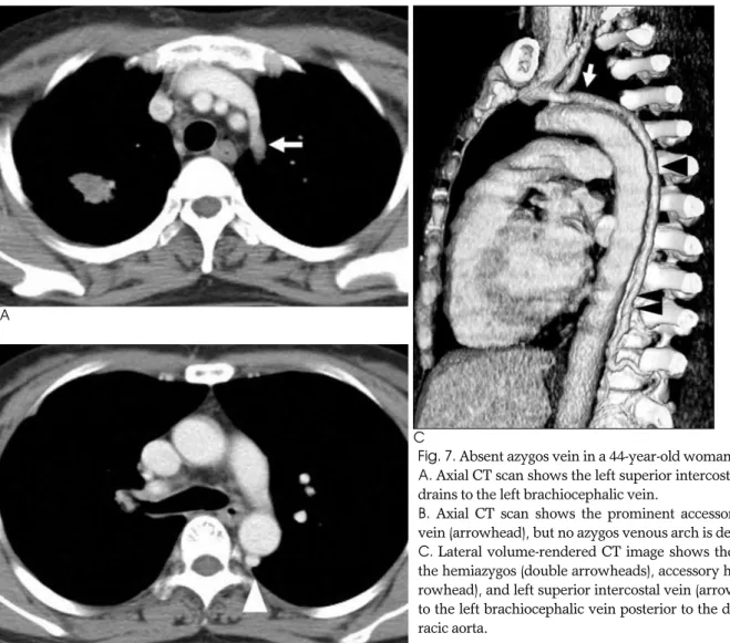
Fig. 7. Absent azygos vein in a 44-year-old woman.
A. Axial CT scan shows the left superior intercostal vein (arrow) drains to the left brachiocephalic vein.
B. Axial CT scan shows the prominent accessory hemiazygos vein (arrowhead), but no azygos venous arch is demonstrated.
C. Lateral volume-rendered CT image shows the continuity of the hemiazygos (double arrowheads), accessory hemiazygos (ar- rowhead), and left superior intercostal vein (arrow), draining in- to the left brachiocephalic vein posterior to the descending tho- racic aorta.
선을 교차하여 기정맥과 결합한다. 반기정맥이 부반기정맥으 로 상방으로 연장되어 기정맥과 통하기도 한다(Fig. 6).
기정맥계의 발생은 정확히 알려지지 않았다. 우측 상주정맥
(supracardinal vein)은 하대정맥의 신후분절로, 우측 상하 주정맥 문합은 신분절로, 우측 하주정맥(subcarinal vein)은 신전분절로, 우측 간정맥은 간분절로 발달한다. 상주정맥의 하 부가 하대정맥의 신후분절로 남아 존속되게 되면, 우측 상주정 맥은 기정맥으로, 좌측 상주정맥은 반기정맥으로 발생한다.
기정맥결여(Absent azygos vein)
선천적 기정맥결여는 드물며, 좌하대정맥이나 다비증과 연 관되어 나타나기도 한다. 기정맥이 없으면, 반기정맥이 혈액학 적으로 중요한 역할을 하여 반기정맥과 좌측 상늑간정맥으로 혈류가 증가한다(Fig. 7). 좌측 상늑간정맥 비대는 흉부사진 에서 확장된 aortic nipple로 보일 수 있다(1, 8).
홀허파엽
우측 후주정맥(posterior cardinal vein)이 우측 폐 첨부로 의 내측이동 실패로 생기며, 총인구의 약 0.4%-1.0%에서 보 인다(1). 홀허파엽은 영상검사에서 쉽게 확인할 수 있는데 (Fig. 8), 기정맥은 정상보다 외측에 있고, 정상 기정맥궁 위 치보다 상방으로 올라가 상대정맥이나 우측 완두정맥으로 들 어간다. 기정맥궁이 정상보다 외측에 있어 폐를 종격동으로 밀 어내고 CT에서 오목한 종격동 윤곽을 볼 수 있다. 비정상으로
─ 31 ─
A B
C
Fig. 9. Azygos lobe in a 65-year-old man.
A--C. Axial (A), coronal (B), and sagittal (C) CT images show the displaced azygos vein (arrowhead) draining to the right brachiocephalic vein, and azy- gos fissure (arrow) separating the azygos lobe (*) from the right upper lobe.
Fig. 8. Azygos lobe in a 14-year-old boy.
Chest radiograph shows the azygos fissure (arrow), curving down from the right apex toward the mediastinal shadow. The azygos vein is projected as a teardrop shadow at the inferior end of the azygos fissure.
위치한 혈관은 장측과 벽측 흉막을 움푹 들어가게 하여, 네 개 의 늑막층이 장간막과 유사한 홀허파간막(mesoazygos)을 만 들고, 홀허파엽과 남은 우폐 사이에 끼여 영상 검사에서 기정 맥열로 나타난다(Fig. 9). 반홀허파엽(hemiazygos lobe)은 빈도가 더 낮으며, 좌측 상늑간정맥의 위치이상에 의해 발생한 다.
기정맥, 반기정맥과 하대정맥의 연결
(Azygos and hemiazygous continuation of the IVC) 대부분이 다른 기형과 동반되어 나타나지만, 드물게 단독으 로 보일 수도 있다. 기정맥연결은 다비증 혹은 무비증, 역위증, 중복으로 또는 좌측 하대정맥 등과 동반될 수 있다.
우측 하주정맥과 간정맥간의 문합이 이루어지지 못하면 하 대정맥의 간분절이 발달하지 못하여 하대정맥의 단절이 생긴 다. 그러면 혈류는 우측 상주정맥으로 유입되고, 우측 상주정 맥의 존속으로 말미암아 기정맥과 하대정맥의 연결이 발생한
다(Fig. 10).
기정맥과 하대정맥이 연결되면 기정맥, 기정맥궁과 상대정 맥으로 혈류가 증가하여 혈관들이 확장된다. 이때, 간정맥은 간상부 하대정맥을 통해 우심방으로 들어간다.
반기정맥과 좌측 하대정맥이 연결되면 세 개의 경로를 통해 우심방으로 혈류가 유입된다. 첫째는 기정맥으로, 두 번째는 부반기정맥과 좌측 상대정맥을 통하여 관상정맥동으로, 셋째 는 부반기정맥, 좌측 상늑간정맥, 좌측 완두정맥을 통해 정상 우하대정맥으로 들어가는 것이다.
폐정맥
해부 및 발생
우측 상폐정맥은 우상엽과 우중엽에서, 좌측 상폐정맥은 좌 상엽과 설상엽(lingular segment)에서, 양쪽 하폐정맥은 각 각의 하엽에서 혈류를 받아 좌심방으로 들어간다. 폐문에서 상
A B
C D
Fig. 10. Hemiazygos and azygos continuation of the double inferior vena cava in a 15-year-old girl.
A. Axial CT scan at the level of lower thorax shows the dilated azygos and hemiazygous veins on both sides of the descending tho- racic aorta.
B. Axial CT scan at the level of upper abdomen can not demonstrate the evidence of the intrahepatic inferior vena cava.
C. Axial CT scan at the level of renal hilum shows the double inferior vena cava on both sides of the abdominal aorta.
D. Chest radiograph shows an enlargement of the azygos vein at the inferior margin of the right paratracheal stripe (arrow) and an abnormal mediastinal line (arrowheads) by the enlarged azygos vein.
─ 33 ─
A B
Fig. 11. Anomalous pulmonary vein of the right upper lobe.
A. Axial CT scan shows an anomalous pulmonary vein (arrow) posterior to the right bronchus intermedius, and the normal right superior pulmonary vein (arrowhead) anterior to pulmonary artery.
B. Posterior volume-rendered CT image shows an anomalous pulmonary vein (arrow) of the posterior segment of the right upper lobe.
A
C
B
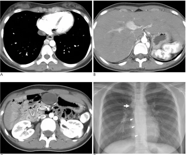
Fig. 12. Partial anomalous pulmonary venous return of the left upper lobe in a 51-year-old man.
A. Axial CT scan shows an anomalous vertical vein (arrow) lat- eral to the aortic arch. It is differentiated from the persistent left SVC.
B. Axial CT scan shows the absence of the left superior pul- monary vein in front of the left hilum.
C. Volume-rendered CT image shows a vertical anomalous vein (arrow) from the left upper lobe, draining to the left brachio- cephalic vein.
폐정맥은 폐동맥의 전방에 있고, 하폐정맥의 소공(ostia)은 상 폐정맥보다 좀 더 안쪽, 뒤쪽에 있다.
태생 8주 동안 폐싹(lung bud)은 앞창자(foregut)에서 발 생하여, 체정맥으로부터 혈류를 받는다. 동시에 좌심방에서 공 통폐정맥의 원기(primodium)가 돌출된다. 앞창자가 위장관 과 호흡기관으로 분리되면서 체정맥 혈류가 차단되고, 폐싹은 공통폐정맥과 융합된다. 공통폐정맥에서 양폐로 좌우폐정맥이 나가고, 각각 다시 나뉘어 네 개의 폐정맥이 발생하는데, 심방 이 자라면서 공통폐정맥과 첫 번째 분지 근위부까지 흡수하여 결국 좌심방구멍에서 네 개의 폐정맥이 나가게 된다.
폐정맥 수적 변이
폐정맥 분지 발생 과정에서 폐색이나 비정상 흡수 등을 통해 폐정맥의 수가 증가하거나 감소하게 된다. 가장 많은 수적 변 이는 우측 폐정맥이 세 개인 경우이며, 그 외 편측 또는 양측 단일 폐정맥 등이 있다(1). 우상엽의 후분절에서 유입되는 폐 정맥이 따로 우측 중간기관지의 후방으로 주행하여 좌심방으 로 들어가는 해부학적 변이를 볼 수 있다(Fig. 11). 폐정맥 소 공과 인접한 부정맥 병소에 고주파 절제술을 시행하거나, 수술
후 폐정맥협착 등의 합병증을 예측하기 위해 폐정맥 수적 변이 를 정확히 아는 것이 중요하다(9).
폐정맥류
폐정맥의 동맥류성 확장을 일컬으며, 대부분 폐정맥고혈압 에 의해 이차적으로 나타나며, 승모판협착과 승모판부전에서 잘 동반된다. 많은 경우에서 증상이 없으며, 선천적이든 후천 적이든 단순흉부촬영에서 경계가 잘 그려지는 폐 결절과 유사 하게 보이며(7), 동정맥 기형과 감별하는 것이 중요하다.
폐정맥환류이상(Anomalous pulmonary venous return) 폐정맥은 정상적으로는 좌심방으로 유입되지만 폐정맥의 전 부 또는 일부가 우심방 또는 체정맥으로 연결되는 기형을 폐정 맥환류이상이라 한다. 폐정맥환류이상은 폐정맥 전부가 기형 연결을 보이는 전폐정맥환류이상과 일부가 기형 연결을 보이 는 부분폐정맥환류이상으로 나뉜다. 전폐정맥환류이상은 선천 성 심장병과 연관되어 나타나며, 대부분이 생후 1세 이내에 청 색증을 동반한 증상이 발현된다.
부분폐정맥환류이상에서 총인구의 약 0.5%에서 보이며(1),
A B
C
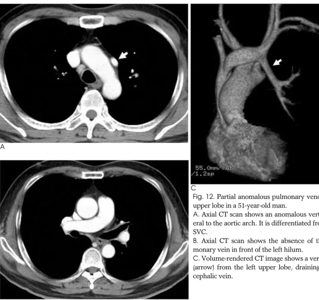
Fig. 13. Partial anomalous pulmonary venous return of the right upper lobe in a 51-year old man.
A. Chest radiograph shows a curvilinear vascular structure (arrow) in the right upper lung directed toward SVC.
B. Axial CT scan shows an abnormal vascular structure (arrow) draining to SVC.
C. Posterior volume-rendered CT image shows an anomalous pulmonary veins (arrows) just above the right pulmonary artery.
한 개의 폐정맥만이 환류이상을 보이면 거의 증상이 없고, 2개 정도이면 심하게 운동할 때 호흡곤란을 호소하게 된다. 정도가 심한 경우에 폐고혈압을 동반할 수도 있다. 성인에서 간혹 볼 수 있으며 심방중격결손과 잘 동반된다.
부분폐정맥환류이상은 약 79%가 좌상엽의 폐정맥에 생기고 (Fig. 12) 약 17%가 우상엽에 생긴다(10) (Fig. 13). 좌상엽 의 폐정맥에 부분폐정맥환류이상이 생기면 대동맥궁의 외측에 이상 혈관으로 보인다. 대동맥궁의 외측에 이상혈관이 보이면 좌상대정맥 혹은 좌상엽의 부분폐정맥환류이상의 가능성을 생 각할 수 있다. 우폐정맥에 생기는 부분폐정맥환류이상에서는 대부분이 우심방과 하대정맥으로 연결되는데 폐 내에서 폐정 맥의 주행이상으로 터키식 칼, 즉 초승달 모양의 특징적인 수 직 음영을 흉부사진에서 보여 쉽게 진단할 수 있다.
참 고 문 헌
1. Demos TC, Posniak HV, Pierce KL, Olson MC, Muscato M.
Venous anomalies of the thorax. AJR Am J Roentgenol 2004;182:
1139-1150
2. Davies M, Guest PJ. Developmental abnormalities of the great ves- sels of the thorax and their embryological basis. Br J Radiol 2003;
76:491-502
3. Padhani AR, Hale HL. Mediastinal venous anomalies: potential pitfalls in cancer diagnosis. Br J Radiol 1998;71:792-798
4. Cole TJ, Henry DA, Jolles H, Proto AV. Normal and abnormal vas- cular structures that simulate neoplasms on chest radiographs:
clues to the diagnosis. Radiographics 1995;15:867-891
5. Whitten CR, Khan S, Munneke GJ, Grubnic S. A diagnostic ap- proach to mediastinal abnormalities. Radiographics 2007;27:657- 671
6. Lawler LP, Corl FM, Fishman EK. Multi-detector row and volume- rendered CT of the normal and accessory flow pathways of the thoracic systemic and pulmonary veins. Radiographics 2002;22:
S45-S60
7. Chen SJ, Liu KL, Chen HY, Chiu IS, Lee WJ, Wu MH, et al.
Anomalous brachiocephalic vein: CT, embryology, and clinical im- plications. AJR Am J Roentgenol 2005;184:1235-1240
8. Hatfield MK, Vyborny CJ, MacMahon H, Chessare JW. Congenital absence of the azygos vein: a cause for aortic nipple enlargement.
AJR Am J Roentgenol 1987;149:273-274
9. Spaggiari L, Solli P, Leo F, Veronesi G, Pastorino U. Anomalous segmental vein for right upper lobe: an unusual anatomical varia- tion. Ann Thorac Surg 2002;74:267
10. Haramati LB, Moche IE, Rivera VT, Patel PV, Heyneman L, McAdams HP, et al. Computed tomography of partial anomalous pulmonary venous connection in adults. J Comput Assist Tomogr 2003;27:743-749
─ 35 ─
J Korean Soc Radiol 2009;60:27-35
Address reprint requests to : Young Tong Kim, M.D., Department of Radiology, Soonchunhyang University Hospital Cheonan 23-20 Bongmyung-dong, Cheonan 330-721, Korea.
Tel. 82-41-570-3515 Fax. 82-41-579-9026 E-mail: [email protected]
Thoracic Venous Anomalies
1Hye Lin Kim, M.D., Young Tong Kim, M.D.2, Sung Shick Jou, M.D.2
1Department of Radiology, Soonchunhyang University Hospital Bucheon
2Department of Radiology, Soonchunhyang University Hospital Cheonan
The radiographic findings of normal and abnormal vascular structures can mimic soft-tissue masses and this can lead to inappropriate diagnostic procedures. An awareness of these anatomical features is required to avoid misinterpretation and then performing unnecessary additional studies. The purpose of this pictorial es- say is to describe the radiologic findings of various anomalies of the pulmonary and systemic venous systems in the thorax.
Index words :Veins Thorax Anatomy
Computed tomography (CT), X-Ray