ISSN: 2233-601X (Print) ISSN: 2093-6516 (Online)
− 216 −
†
This paper was presented at the 19th Gyeonggi-Incheon monthly meeting of the Korean Society for Thoracic and Cardiovascular Surgery.
Received: August 28, 2017, Revised: September 22, 2017, Accepted: September 26, 2017, Published online: June 5, 2018
Corresponding author: Jong Hui Suh, Department of Thoracic and Cardiovascular Surgery, Incheon St. Mary’s Hospital, College of Medicine, The Catholic University of Korea, 56 Dongsu-ro, Bupyeong-gu, Incheon 21431, Korea
(Tel) 82-2-2258-6139 (Fax) 82-2-594-8644 (E-mail) [email protected]
© The Korean Society for Thoracic and Cardiovascular Surgery. 2018. All right reserved.
This is an open access article distributed under the terms of the Creative Commons Attribution Non-Commercial License (http://creativecommons.org/
licenses/by-nc/4.0) which permits unrestricted non-commercial use, distribution, and reproduction in any medium, provided the original work is properly cited.
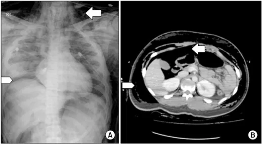
Right Main Bronchus Rupture Presenting with Pneumoperitoneum
Seok Beom Hong, M.D. 1 , Ji Yoon Lee, M.D. 1 , June Lee, M.D. 1 , Kuk Bin Choi, M.D. 2 , Jong Hui Suh, M.D. 3
1