J Korean Soc Radiol 2016;74(1):66-70 http://dx.doi.org/10.3348/jksr.2016.74.1.66
INTRODUCTION
Liposarcomas are the most common primary soft-tissue ma- lignancies of the retroperitoneum. They are divided into five subtypes: well-differentiated, dedifferentiated, myxoid, pleo- morphic, and mixed, according to the 2002 World Health Or- ganization histological classification (1). Well-differentiated ret- roperitoneal liposarcoma is the most common subtype and is considered a low-grade tumor. The dedifferentiated subtype is a transition from well-differentiated liposarcoma to nonlipogenic sarcoma with variable histological grades. Although several jour- nals have reported retroperitoneal liposarcomas presenting as indirect inguinal hernias, dedifferentiated liposarcoma extend- ing into inguinal canal and seemingly presenting as inguinal hernia has been more scarcely described (2, 3). Here we report multidetector computed tomography (CT) and magnetic reso-
neal liposarcoma mimicking a right inguinal hernia.
CASE REPORT
A 37-year-old male visited our hospital with a painless pro- truding mass in the right inguinal region. The patient had no other symptoms or relevant past medical history. Physical ex- amination revealed a palpable mass without tenderness in his right groin. His laboratory findings were unremarkable.
Contrast-enhanced abdominal CT examination using a 128-detector-row CT scanner (Definition AS+, Siemens Medi- cal Solutions, Forchheim, Germany) revealed a predominant fat- density mass with enhancing septa and a few solid nodules measuring approximately 12 × 7.5 cm (Fig. 1). The caudal por- tion of the mass, located in the right inguinal canal, mainly consisted of soft tissue with an enhancement measuring about
Dedifferentiated Retroperitoneal Liposarcoma Presenting as Right Inguinal Hernia: A Case Report
우측 서혜부 탈장으로 나타난 후복막의 역분화 지방육종: 증례 보고
Jung Myung Kim, MD
1, Moon Hyung Choi, MD
2, Su Lim Lee, MD
1, Young Mi Ku, MD
1*
1Department of Radiology, Uijeongbu St. Mary’s Hospital, College of Medicine, The Catholic University of Korea, Uijeongbu, Korea
2Department of Radiology, Seoul St. Mary’s Hospital, College of Medicine, The Catholic University of Korea, Seoul, Korea
Retroperitoneal liposarcomas usually present as painless, slow-growing abdominal masses. When masses grow large enough to compress surrounding structures, symptoms may occur. Retroperitoneal liposarcoma clinically manifesting as inguinal hernia is a very rare entity; only 11 cases have been reported. Herein, we present ra- diographic features of a 37-year-old male with a painless palpable mass in the right groin that was identified as dedifferentiated retroperitoneal liposarcoma herniated through the right inguinal canal.
Index terms Liposarcomas
Retroperitoneal Neoplasms Inguinal Hernia
Multidetector Computed Tomography Magnetic Resonance Imaging
Received June 26, 2015 Revised July 17, 2015 Accepted August 6, 2015
*Corresponding author: Young Mi Ku, MD
Department of Radiology, Uijeongbu St. Mary’s Hospital, College of Medicine, The Catholic University of Korea, 271 Cheonbo-ro, Uijeongbu 11765, Korea.
Tel. 82-31-820-3148 Fax. 82-31-846-3080 E-mail: [email protected]
This is an Open Access article distributed under the terms of the Creative Commons Attribution Non-Commercial License (http://creativecommons.org/licenses/by-nc/3.0) which permits unrestricted non-commercial use, distri- bution, and reproduction in any medium, provided the original work is properly cited.
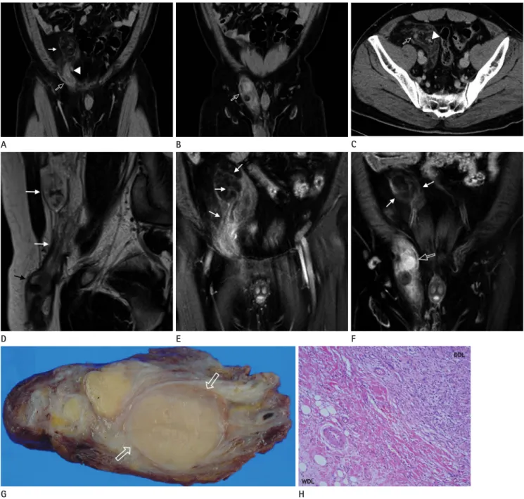
Fig. 1. A 37-year-old male with a retroperitoneal dedifferentiated liposarcoma (DDL) extending into the right inguinal canal.
A, B. Contrast-enhanced computed tomography (CT) with coronal reformatted images shows a large mass with a fatty component in the retro- peritoneum (thin arrows) extending into the right inguinal canal with a solid enhancing component (open arrow). Note the medial displacement of the inferior epigastric vessels (arrowhead).
C. Axial contrast-enhanced CT demonstrates several ill-defined strands and a soft-tissue nodule with homogenous enhancement (open arrow) within the mass. Note the mass adjacent to and extending along the testicular vessels (arrowhead).
D. Sagittal T2-weighted image of the retroperitoneal tumor extending into the right inguinal canal. The retroperitoneal component of the mass shows high signal intensity consistent with mature fatty tissue (white arrows), while the inguinal canal component shows intermediate signal in- tensity consistent with tumor tissue (black arrow).
E, F. Coronal gadolinium-pentetic-acid-enhanced T1-weighted images demonstrate multiple tenuous septa (arrows) and a soft-tissue nodule with marked enhancement (open arrow) within the mass.
G. On cross-section, the surgical specimen taken from the right inguinal canal is solid, with discrete intratumoral nodules of varying size and col- ors ranging from yellow to yellow-tan admixed with firm tan-gray areas corresponding to dedifferentiated foci (open arrows).
H. The tumor shows an abrupt transition between the components of well-differentiated liposarcoma and DDL (hematoxylin & eosin, × 100).
A B
G H
C
D E F
cending colon and mesocolon and extended alongside the right testicular vessels, suggesting an extraperitoneal location of the lesion. Also, the mass passed between the right inferior epi- gastric vessels medially, and the right external iliac vessels later- ally.
Pelvic MRI was performed using a 3.0T system (Magnetom Verio; Siemens Medical Solutions, Erlangen, Germany) with a body phased-array coil, in order to obtain more information. On MR images (Fig. 1), most of the retroperitoneal mass showed high signal intensity on T1-weighted images and intermediate to high signal intensity on T2-weighted images, and demon- strated a signal drop on fat-saturated images. The irregular septa and nodules, which appeared inonly a small part of the retro- peritoneal area, were of low signal intensity on T1-weighted im- ages and were enhanced slightly after administration of gadolin- ium pentetic acid (Gd-DTPA, Magnevist®; Bayer Healthcare Pharmaceuticals, Wayne, NJ, USA). In the right inguinal canal, however, the majority of the mass showed low signal intensity on T1-weighted images and high signal intensity on T2-weight- ed images with profuse contrast enhancement following Gd- DTPA injection.
The patient underwent surgical resection of the mass includ- ing components of both theretroperitoneal and the right ingui- nal canal. Intraoperatively, predominantly fatty masses contain- ing hard, solid lesions with multiple septa were found in the retroperitoneal space. The firm mass in the right inguinal area had clearly originated from the large retroperitoneal mass and broken through the right inguinal canal.
Histopathologically, the surgical specimen was diagnosed as a well-differentiated retroperitoneal liposarcoma with a dedif- ferentiated component. Grossly, the tumor was unencapsulated, well-circumscribed, oval, rubbery, and about 8.0 × 5.0 × 4.0 cm in size. The cut surface of the intracanalicular portion was solid with discrete intratumoral nodules varying in size. Its color ranged from yellow to yellow-tan admixed with tan-gray areas that corresponded to the dedifferentiated foci. There was no ev- idence of hemorrhage or necrosis (Fig. 1G). Viewed microscopi- cally, the tumor had two different histological components sepa- rated by either an abrupt or gradual transition. The well-diff- erentiated area was characterized by proliferating adipocytes varying in size and shape, whereasthe dedifferentiated area was
(Fig. 1H). Most of the dedifferentiated area had the appearance of a high-grade malignant fibrous histiocytoma or a rhabdo- myosarcoma with different degrees of chronic inflammation and fibrosis. The tumor cells showed marked nuclear atypia and pleo- morphism with abundant, finely granular, amphophilic to baso- philic cytoplasm in bipolar, stellate, or polygonal configurations, and vesicular nuclei with one to a few prominent nucleoli. The mitotic count was 1–2 mitotic fields/10 high-power fields in the most mitotically active area. Immunohistochemistry demon- strated strong expression of vimentin and α-1-antitrypsin and negative staining for muscle markers and S100 protein.
DISCUSSION
The inguinal canal is involved in a broad spectrum of patho- logical processes including inguinal hernia, other congenital disease, inflammatory conditions, and even neoplasm, manifest- ing as palpable groin mass. The incidence of malignancy is about 30% in neoplastic disease involving the inguinal canal. The ma- jority of malignant inguinal tumors consist of mesenchymal sar- comas such as liposarcoma (4). Dedifferentiated liposarcoma, one of five liposarcoma subtypes, originates from well-differen- tiated liposarcoma with an abrupt transition to aggressive, high- grade nonlipogenic sarcoma. According to Lahat et al. (5), dedif- ferentiated liposarcomas are likely to recur as local or distant metastatic disease and their treatment accordingly requires in- tensive multimodal approaches. The presence of a nonfatty com- ponent within a predominantly fatty mass or a well-defined fat- ty mass within a well-defined nonfatty mass are often suggestive of dedifferentiated liposarcoma (1, 6). Relative to muscle, these masses are hypointense on T1-weighted MRI, but are heteroge- neously hyperintense on T2-weighted MRI (7). Taking all of this into account, we preoperatively diagnosed the tumor as a retro- peritoneal liposarcoma containing a dedifferentiated component.
The diagnosis of retroperitoneal tumors is often challenging for radiologists. The most important clue is whether the tumor is located within the retroperitoneal space. Displacement of ad- jacent retroperitoneal organs or major vessels in the retroperi- toneal cavity, observed using either CT or MRI, is a useful sign.
Tracing the tumor’s spread and characterizing its contents can also narrow the possible diagnoses (8). In our patient, we con-
because the ascending colon and its mesocolon were displaced anteriorly, and the mass had spread alongside the testicular ves- sels. The retroperitoneum is known to have contact with the pel- vic prevesicular space (9). The anterolateral segments of the tes- ticular vessels travel within this space before entering the internal inguinal ring to become part of the spermatic cord. Prevesicular fat accompanying the testicular vessels is surrounded by the in- ternal spermatic fascia, which arises from the transversalis fascia.
Incarcerated omental inguinal hernias could mimic well-dif- ferentiated or dedifferentiated liposarcomas extending into the inguinal canal not only in clinical manifestation, but also in im- aging features (10). Both of them usually present as irreducible, palpable groin masses with varying degrees of nonfatty or scanty fatty components on imaging studies. However, unlike herniat- ed retroperitoneal liposarcomas, incarcerated omental inguinal hernias originate in omental arteries and have no relation to tes- ticular vessels.
Knowledge of both the imaging findings of dedifferentiated liposarcoma and the retroperitoneal anatomy is critical to cor- rect preoperative diagnosis and treatment planning in cases of retroperitoneal liposarcoma.
REFERENCES
1. Hong SH, Kim KA, Woo OH, Park CM, Kim CH, Kim MJ, et al.
Dedifferentiated liposarcoma of retroperitoneum: spectrum of imaging findings in 15 patients. Clin Imaging 2010;34:
203-210
2. Mizuno Y, Sumi Y, Nachi S, Ito Y, Marui T, Saji S, et al. A case of a large retroperitoneal liposarcoma presenting as an in- carcerated inguinal hernia. Hernia 2006;10:439-442
3. McKinley SK, Abreu N, Patalas E, Chang A. Large Retroper- itoneal Liposarcoma Diagnosed upon Radiological Evalua- tion of Mild Right-Sided Inguinal Hernia. Case Rep Radiol 2013;2013:187957
4. Vagnoni V, Brunocilla E, Schiavina R, Borghesi M, Passaretti G, Gentile G, et al. Inguinal canal tumors of adulthood. An- ticancer Res 2013;33:2361-2368
5. Lahat G, Anaya DA, Wang X, Tuvin D, Lev D, Pollock RE. Re- sectable well-differentiated versus dedifferentiated liposar- comas: two different diseases possibly requiring different treatment approaches. Ann Surg Oncol 2008;15:1585-1593 6. Tateishi U, Hasegawa T, Beppu Y, Satake M, Moriyama N.
Primary dedifferentiated liposarcoma of the retroperitone- um. Prognostic significance of computed tomography and magnetic resonance imaging features. J Comput Assist To- mogr 2003;27:799-804
7. Song T, Shen J, Liang BL, Mai WW, Li Y, Guo HC. Retroperi- toneal liposarcoma: MR characteristics and pathological correlative analysis. Abdom Imaging 2007;32:668-674 8. Nishino M, Hayakawa K, Minami M, Yamamoto A, Ueda H,
Takasu K. Primary retroperitoneal neoplasms: CT and MR imaging findings with anatomic and pathologic diagnostic clues. Radiographics 2003;23:45-57
9. Aikawa H, Tanoue S, Okino Y, Tomonari K, Miyake H. Pelvic extension of retroperitoneal fluid: analysis in vivo. AJR Am J Roentgenol 1998;171:671-677
10. Aguirre DA, Santosa AC, Casola G, Sirlin CB. Abdominal wall hernias: imaging features, complications, and diag- nostic pitfalls at multi-detector row CT. Radiographics 2005;25:1501-1520
우측 서혜부 탈장으로 나타난 후복막의 역분화 지방육종: 증례 보고
김정명
1· 최문형
2· 이수림
1· 구영미
1*
후복막의 지방육종은 서서히 성장하는 무통성의 종괴로 발견되는 경우가 대부분이며 큰 종괴의 경우 주변 구조물을 압박 하여 증상을 일으킬 수 있다. 후복막의 지방육종의 임상 발현이 서혜부 탈장으로 오인되는 경우는 매우 드물며 지금까지 단 11개의 증례가 보고되었다. 본 저자들은 무통성의 촉지되는 우측 서혜부 종괴를 주소로 내원하여 서혜부 탈출을 동반 한 후복막의 역분화 지방육종으로 진단된 37세 남자 환자에 대해 보고하고자 한다.
1가톨릭대학교 의과대학 의정부성모병원 영상의학과, 2가톨릭대학교 의과대학 서울성모병원 영상의학과