ISSN: 2233-601X (Print) ISSN: 2093-6516 (Online)
− 110 −
Received: July 12, 2016, Revised: August 29, 2016, Accepted: August 30, 2016, Published online: April 5, 2017
Corresponding author: Kyung Hwan Kim, Department of Thoracic and Cardiovascular Surgery, Seoul National University Hospital, Seoul National University College of Medicine, 101 Daehak-ro, Jongno-gu, Seoul 03080, Korea
(Tel) 82-2-2072-3971 (Fax) 82-2-764-3664 (E-mail) [email protected]
© The Korean Society for Thoracic and Cardiovascular Surgery. 2017. All right reserved.
This is an open access article distributed under the terms of the Creative Commons Attribution Non-Commercial License (http://creativecommons.org/
licenses/by-nc/4.0) which permits unrestricted non-commercial use, distribution, and reproduction in any medium, provided the original work is properly cited.
Delayed Left Atrial Perforation Associated with Erosion After Device Closure of an Atrial Septal Defect
Ji Seong Kim, M.D., Sang Yoon Yeom, M.D., Sue Hyun Kim, M.D., Jae Woong Choi, M.D., Kyung Hwan Kim, M.D., Ph.D.
Department of Thoracic and Cardiovascular Surgery, Seoul National University Hospital
A 43-year-old man who had had a history of atrial septal defect (ASD) device closure 31 months previously presented with abrupt chest and back pain along with progressive cardiogenic shock and cardiac arrest. After resuscitation, he was diagnosed with cardiac tamponade. Diagnostic and therapeutic surgical exploration re- vealed left atrium (LA) perforation due to LA roof erosion from a deficient aortic rim. Device removal, pri- mary repair of the LA perforation site, and ASD patch closure were performed successfully. The post- operative course was uneventful. The patient was discharged after 6 weeks of empirical antibiotic therapy without any other significant complications.
Key words: 1. Atrial septal defects 2. Atrium
3. Septal occluder device 4. Cardiac tamponade 5. Heart atria
Case report
A 43-year-old man visited the emergency room with complaints of abrupt chest and back pain that began 20 minutes earlier. The patient was previously healthy and did not take any medication, although he had a history of transcatheter atrial septal defect (ASD) device (Amplatzer Septal Occluder; AGA Medical Co. , Golden Valley, MN, USA) closure performed 31 months ago. The initial electrocardiogram demon- strated normal sinus rhythm without ST elevation.
Transthoracic echocardiography (TTE) showed mod- erate pericardial effusion or hematoma. During evalu- ation, the patient complained of severe pain with drowsiness and hypotension. Sudden cardiac arrest followed. The return of spontaneous circulation was
achieved within 1 minute with cardiopulmonary re- suscitation and intubation. Systemic hypotension was improved after urgent pericardiocentesis. Computed tomography (CT) angiography was then performed.
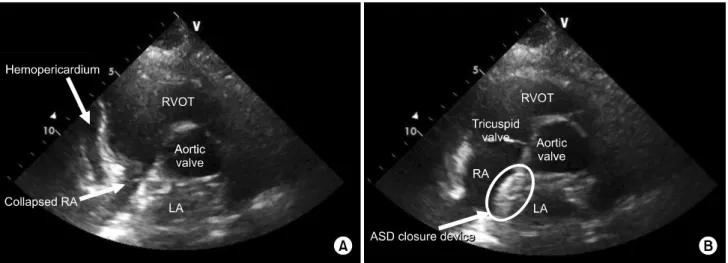
There was no evidence of acute aortic dissection or intramural hematoma. However, the moderate hemo- pericardium extended into the superior recess. The ASD closure device had not migrated (Fig. 1). After the patient’s admission to the intensive care unit, TTE revealed moderate hemopericardium with mild right atrial collapse, suggesting residual tamponade physiology. The ASD closure device was found in situ in the interatrial septum, abutting to the aortic root due to the scant aortic rim (Fig. 2). However, there was no evidence of any residual interatrial shunt, valvular dysfunction, or device erosion. Transeso-
Korean J Thorac Cardiovasc Surg 2017;50:110-113 □ CASE REPORT □
https://doi.org/10.5090/kjtcs.2017.50.2.110
Erosion After Device Closure of Atrial Septal Defect
− 111 −
Fig. 2. In the early systolic phase of the parasternal short-axis view, moderate hemopericardium with mild RA collapse is seen, suggest- ing residual tamponade physiology (A), and after atrial filling, the ASD closure device is seen attached only to the aortic root (B). RVOT, right ventricular outflow tract; LA, left atrium; RA, right atrial; ASD, atrial septum defect.
Fig. 1. Computed tomography angiography showing no evidence of acute aortic dissection or intramural hematoma. The white ar- row depicts the atrial septum defect closure device in situ. The black arrow shows the remaining pericardial effusion, consistent with hemopericardium.
phageal echocardiography showed that the peri- cardial effusion had resolved, and there were no ad- ditional abnormal findings. The patient was ex- tubated 1 day after admission. His left pleural effu- sion was controlled by closed thoracostomy with a 10-Fr mini-tube. Although the patient’s status stabi- lized after appropriate management, we were not able to identify the exact reason for the hemoperi- cardium.
Clinically, we ruled out left atrium (LA) perforation
on the basis of the ASD closure device abutting the aortic root and the LA roof. Elective surgical explora- tion was planned after stabilizing the patient’s vital signs. However, it was difficult to decide to perform surgical exploration for resolved hemopericardium without a definite reason or new symptoms. Although the patient exhibited no abnormal laboratory findings or fever and the culture results were negative, em- pirical antibiotics (vancomycin, gentamicin, and ri- fampicin) were started in accordance with infective endocarditis on the prosthetic valve. Diagnostic and therapeutic surgical exploration was performed on the eighth day after admission. With full median ster- notomy and pericardiotomy, the pericardial space was filled with dark-blood-colored pericardial effusion.
Mild adhesion around the LA roof, aortic root, and the right pulmonary artery was found. However, there was no evidence of active bleeding, organized hematoma, abscess formation, or infective tissue. Right atriotomy was then performed under aorto-bicaval cannulation with antegrade cold crystalloid cardio- plegic arrest. The ASD closure device was located in the appropriate position. However, the aortic rim was nearly absent. The device was removed with electrocautery and dissection using Metzenbaum scis- sors. After inspecting the LA cavity via the ASD, we identified the LA perforation site. A 2-mm probe could pass through the perforation site (Fig. 3). This site was repaired primarily with a 4-0 polypropylene pledgeted buttressed suture. The ASD was closed
Ji Seong Kim, et al
− 112 −
Fig. 3. Intraoperatively, a 2-mm probe (arrow) could pass through the LA roof perforation site (A). Further, with RA wall retraction, the probe was seen in the LA cavity via the ASD (B). LA, left atrium; SVC, superior vena cava; RA, right atrium; ASD, atrial septum defect.
with a glutaraldehyde-fixed autologous pericardial patch. The cardiopulmonary bypass time and the aortic cross clamp time were 108 minutes and 56 minutes, respectively. The patient was extubated 2 hours after surgery and transferred to a general ward on postoperative day 1. Persistent mild sinus tachycardia was controlled with a low dose of oral beta blockers. Postoperative TTE demonstrated nor- mal findings without residual tamponade physiology or pericardial effusion. The rest of the postoperative course was uneventful. The patient was transferred to department of cardiology and maintained with 2 weeks of gentamicin and 6 weeks of vancomycin and rifampicin therapy. He was discharged without any other complications.
Discussion
The closure of secundum ASD without severe irre- versible pulmonary artery hypertension either percu- taneously or surgically is a preferable therapy for adult patients [1]. Usually, no absolute contraindi- cation exists for the closure of ASD using a device.
However, insufficient peridefect rims are considered to be relative contraindications. The Amplatzer Septal Occluder (AGA Medical Co.), which was approved by the US Food and Drug Administration in December 2001, is a safe and effective closure device for chil- dren and adult patients. Although the device has been used widely, several early and late complica- tions have been reported, including device malposi-
tion, device embolization, arrhythmia, pericardial ef- fusion, thrombus formation, and iliac vein dissection [2,3].
Among the complications after transcatheter ASD device closure, cardiac erosion with tamponade is very rare but extremely fatal. Most of the complica- tions from erosion occur within 1 year of device clo- sure, but more than 10% of the cases of erosion oc- curred more than 1 year after device closure [4].
With proper clinical follow-up, early erosion can be treated appropriately [5]. However, as in this case, sudden cardiac tamponade caused by cardiac erosion several years after the procedure is difficult to dis- tinguish from hemopericardium due to other causes.
Therefore, if a patient with a history of device clo- sure of ASD presents with chest pain, syncope, or other shock symptoms, device-related cardiac erosion should be suspected and urgent evaluation should be performed for confirmation. Moreover, long-term reg- ular clinical follow-up is recommended [1].
A deficient retroaortic rim has been considered to be a risk factor for erosion. However, O’Byrne et al.
[5] have argued that it is not associated with an in- creased risk of technical failure or early adverse events. On the other hand, McElhinney et al. [4] have evaluated the relative risk factors and revealed that deficiency of any rim, the ratio of the size of the de- vice to the size of the rim, and oversized devices are associated with erosion. A recent study has shown that absence of the aortic rim, poor posterior rim consistency, septal malalignment, and dynamic mor-
Erosion After Device Closure of Atrial Septal Defect
− 113 − phology of the ASD in echocardiographic findings can significantly increase the risk of erosion [6]. In addi- tion, a case report has described device embolization in the left ventricular outflow tract (LVOT) in a left-to-right shunt ASD patient with an aortic rim de- fect [7].
With a low incidence of early and late complica- tions, interventional cardiologists often do not hesi- tate in using the device. However, given the severity of complications such as LVOT embolization or de- vice erosion, decision-making regarding device clo- sure for ASD patients with some risk factors should be based on a multidisciplinary team approach.
Conflict of interest
No potential conflict of interest relevant to this ar- ticle was reported.
References
1. Warnes CA, Williams RG, Bashore TM, et al. ACC/AHA 2008 guidelines for the management of adults with congenital heart disease: executive summary: a report of the Ameri- can College of Cardiology/American Heart Association
Task Force on practice guidelines (writing committee to develop guidelines for the management of adults with congenital heart disease). Circulation 2008;118:2395-451.
2. Chessa M, Carminati M, Butera G, et al. Early and late complications associated with transcatheter occlusion of secundum atrial septal defect. J Am Coll Cardiol 2002;39:
1061-5.
3. Du ZD, Hijazi ZM, Kleinman CS, Silverman NH, Larntz K;
Amplatzer Investigators. Comparison between trans- catheter and surgical closure of secundum atrial septal defect in children and adults: results of a multicenter nonrandomized trial. J Am Coll Cardiol 2002;39:1836-44.
4. McElhinney DB, Quartermain MD, Kenny D, Alboliras E, Amin Z. Relative risk factors for cardiac erosion following transcatheter closure of atrial septal defects: a case-con- trol study. Circulation 2016;133:1738-46.
5. O’Byrne ML, Gillespie MJ, Kennedy KF, Dori Y, Rome JJ, Glatz AC. The influence of deficient retro-aortic rim on technical success and early adverse events following de- vice closure of secundum atrial septal defects: an analysis of the IMPACT Registry. Catheter Cardiovasc Interv 2017;
89:102-111.
6. Amin Z. Echocardiographic predictors of cardiac erosion after Amplatzer septal occluder placement. Catheter Cardiovasc Interv 2014;83:84-92.
7. Kim YH, Kim H, Kim SJ, et al. Emergent surgical inter- vention for embolization of atrial septal defect closure device. Korean J Thorac Cardiovasc Surg 2012;45:320-2.