INTRODUCTION
Cyclooxygenase (COX) is a bifunctional enzyme with cy- clooxygenase and peroxidase activities. The cyclooxygenase activity of COX is important for the initial conversion of arachidonic acid to prostaglandin (PG) G2, and the peroxi- dase activity of COX then converts it to PG H2. Finally, PG H2 is converted to other PGs by a variety of tissue specific enzymes. These PGs then exert their biologic actions on these tissues (1, 2). There are two isoforms of COX, a constitutive isoform COX-1 and an inducible isoform COX-2. COX-1 is constitutively expressed to fulfill its beneficial housekeep- ing roles. In contrast, COX-2 is frequently undetectable at baseline in most normal tissues, but it is rapidly induced by cytokines, tumor promoters, growth factors and carcinogens (3-5). Increased COX-2 expression has been demonstrated in various human cancers including gastric cancer and has been implicated in carcinogenesis (6-8). Furthermore, selec- tive COX-2 inhibitors have been shown to have antineoplas- tic activities in various cancers (9, 10).
The several mechanisms by which COX-2 may contribute to carcinogenesis are beginning to unravel. These include that COX-2 stimulates tumor cell proliferation, inhibits apopto-
sis, increases the invasiveness of malignant cells, and enhance angiogenesis through the production of angiogenic factors (11-14). We previously reported that COX-2 may play a crit- ical role in carcinogenesis by stimulating tumor angiogenesis in concert with vascular endothelial growth factor (VEGF) in human gastric cancer (15).
Regarding tumor cell proliferation, it is widely accepted that proliferative capacity may influence the clinical course, and hence patient prognosis. Ki-67, a nuclear antigen is ex- pressed in all stages of the cell cycle except G0 and early G1, and it is often used to indicate the proliferative activity of tumors (16).
Tumor development, growth and progression are charac- terized by uncontrolled cell proliferation. This is usually the result of multiple genetic and epigenetic insults to the cell, particularily involving oncogenes and tumor suppressor genes (17). The p53, tumor suppressor gene is one of the most fre- quently mutated genes in human cancers. The p53 tumor suppressor gene is believed to play a pivotal role in prevent- ing the uncontrolled cell proliferation characteristic of can- cer. Recent studies reported that mutation of p53 may con- tribute to the increased COX-2 expression that is observed in malignant tissues (18, 19).
Young-Eun Joo*,�, Ik-Joo Chung*,�, Young-Kyu Park*,�, Yang-Seok Koh�, Jae-Hyuk Lee�, Chang-Hwan Park�, Wan-Sik Lee�, Hyun-Soo Kim�, Sung-Kyu Choi�, Jong-Sun Rew�, Chang-Soo Park�, Sei-Jong Kim�
Gastrointestinal Cancer Research Program*, Departments of Internal Medicine�, Surgery�and Pathology�, Chonnam National University Medical School, Gwangju, Korea
Address for correspondence Young-Eun Joo, M.D.
Gastrointestinal Cancer Research Program, Department of Internal Medicine, Chonnam National University Medical School, 8 Hak-dong, Dong-gu, Gwangju 501-757, Korea
Tel : +82.62-220-6296, Fax : +82.62-225-8578 E-mail : yejoo@jnu.ac.kr
*This study was financially supported by research funds of Chonnam National University in 2003.
871
Expression of Cyclooxygenase-2, p53 and Ki-67 in Gastric Cancer
It has been reported that p53 mutation may contribute to upregulate cyclooxyge- nase (COX)-2 expression that is observed in malignant tissues. These molecules are involved in carcinogenesis by affecting tumor cell proliferation. The aim of this study was to examine the relationship between COX-2 or p53 expression and clinico- pathological characteristics including tumor cell proliferation in gastric cancer. COX-2 and p53 expressions were investigated with immunostaining, in tissue specimens obtained from 119 patients who underwent surgery for gastric cancer. The Ki-67 label- ing index (LI) was counted by Ki-67 immunostaining. COX-2 and p53 expressions correlated significantly with depth of tumor invasion. However, there was no asso- ciation between COX-2 or p53 expression and survival. p53 expression did not cor- relate with COX-2 expression. There was no significant difference in various clini- copathological variables between Ki-67 LI subgroups. The mean Ki-67 LI value of COX-2 positive tumors was significantly higher than that of negative tumors. The mean Ki-67 LI value of p53 positive tumors was not significantly higher than that of negative tumors. The mean Ki-67 LI value of both COX-2 and p53 positive tumors was significantly higher than that of both negative tumors. These results imply that COX-2 expression is associated with tumor cell proliferation of gastric cancer.
Key Words : Cyclooxygenase 2; Tumor Suppressor Protein p53; Cell Proliferation; Stomach Neoplasms; Immu- nohistochemistry
Received : 30 November 2005 Accepted : 13 March 2006
However, the role of increased COX-2 expression and p53 mutation in gastric cancer cell proliferation has not been fully established. The aim of this study was to evaluate the expres- sion of COX-2 and p53 in gastric cancer and to examine the relationship between their expression and various clinicopa- thological characteristics including tumor cell proliferation.
MATERIALS AND METHODS Sample selection
This study was based on an analysis of formalin-fixed, paraf- fin-embedded tissue specimens obtained from 119 patients who had gastric cancer and who underwent surgical resection at Chonnam National University Hospital from July 1994 to June 1995. None of the patients had received preoperative irradiation or chemotherapy before undergoing surgery. The specimens were taken from representative cancerous lesions over their greatest length and included adjacent noncancer- ous areas. Clinicopathological characteristics and survival data were obtained by hospital records, pathologist and physician contact when necessary. The tumors were staged at the time of surgery by the standard criteria for TNM staging using the American Joint Committee on Cancer (AJCC) (20). The mean age was 58.7±10.9 yr (mean±SD) with a range from 28 to 79 yr. Eighty-four patients were male, and 35 were female.
The mean size of tumor was 5.1±2.7 cm (mean±SD) with a range from 0.5 to 15.0 cm. The mean follow-up period was 65.4 months with a range from 1.3 to 119.8 months Immunohistochemistry
All procedures for immunohistochemical staining were done by the Micro-Probe staining system (Fisher Scientific, Pittsburgh, PA, U.S.A.) based on capillary action (21). Forma- lin-fixed, paraffin-embedded tissue blocks were cut to 4- m- thick sections for immunohistochemical staining. A standard avidin-biotin peroxidase complex method was used. Sections were deparaffinized using xylene and transferred to alcohol.
Endogenous peroxidase activity was blocked using the 0.6%
hydrogen peroxide and incubated for 5 min. Antigen retrieval was performed by microwave for 7 min. A monoclonal mouse immunoglobulin antibody to COX-2 (160112; diluted 1:
250; Cayman Chemical Co, Ann Arbor, MI, U.S.A.), p53 (DO-7; diluted 1:100; Dakopatts, Glostrup, Denmark), and Ki-67 (MIB-1; diluted 1:150; Dakopatts, Glostrup, Denmark) were used as primary antibodies. The primary antibodies, in the aforementioned concentrations were diluted in phosphate- buffered saline supplemented with 5% normal horse serum and 1% bovine serum albumin and then incubated with tis- sues for 25, 15 min at 45℃, and 90 min at room tempera- ture, respectively for COX-2, p53 and Ki-67. Anti-mouse immunoglobulin G (Sigma, St. Louis, MO, U.S.A.) labeled
with biotin was used as a secondary antibody for the detec- tion of primary antibodies and slides were incubated for 10 min at 45℃. After multiple rinses with universal buffer, the slides were incubated in streptavidin-horseradish peroxidase solution (Biomeda, Foster, CA, U.S.A.) for 10 min. As the final step, the slides were developed for 10 min with the en- zyme substrate, 3 amino-9-ethyl carbazole (AEC, Sigma, St.
Louis, MO, U.S.A.). The slides were then counterstained with hematoxylin solution for 1 min (Research Genetics, Hunts- ville, AL, U.S.A.). After dehydration, the tissue was sealed with a universal mount (Research Genetics). For negative con- trols, the primary antibody was omitted and replaced with phosphate-buffered saline.
Interpretation of immunohistochemical stains for COX-2, p53 and Ki-67
The immunohistochemical staining was evaluated inde- pendently by two pathologists without knowledge of the clin- ical outcomes, analysing the intensity, area and pattern of im- munohistochemical staining. In case of disagreement, the slides were reviewed and a consensus view achieved. COX-2 immunoreactivity score was calculated as the product of stain- ing intensity and staining area. The staining intensity was arbitrarily graded on a scale of four grades: 0, no staining of cancer cells; 1, weak staining; 2, moderate staining; 3, strong staining. The percentage of staining area was also graded on a scale with four grades: 0, none; 1, <10%; 2, 10-50%; 3, >50
%. Theoretically, the overall scores could range from 0 to 9.
The specimens with a score of more than 4 were regarded as positive expression, and those with a score≤4 as negative expression. p53 immunoreactivity was assessed as being posi- tive only when tumors exhibited intense nuclear staining, and immunoreactivity was categorized into 2 groups: negative expression (less than 10% positive tumor cells) and positive expression (at least 10% positive tumor cells). A distinct nu- clear immunoreactivity for Ki-67 was considered positive. The Ki-67 labeling index (LI) was determined by observing 1000 cancer cell nuclei in areas of the section with highst labeling frequency, and the percentage of Ki-67-labeled nuclei was used for analysis (16).
Statistical analysis
The 2-test and Fisher’s exact test, where appropriate, were used to compare expression of the COX-2, p53 and Ki-67 LI subgroups with various clinicopathological variables. The cor- relation between COX-2 or p53 expression and Ki-67 LI was analyzed for statistical significance using Student’s t test. Actu- arial survival rates of patients were evaluated according to the Kaplan-Meier method and the differences were tested with a log-rank test. The statistical software program used was Sta- tistical Package for the Social Sciences (SPSS/PC+12.0, Chi- cago, IL, U.S.A.). The value of significance was taken as p<0.05.
RESULTS
Expression of COX-2, p53, and Ki-67 in gastric cancer
COX-2 immunoreactivity was cytoplasmic and was almost exclusively restricted to cancerous areas of pathologic specimens (Fig. 1). In some tumors, the COX-2 staining was diffuse, and in other tumors it was localized. In the normal gastric epithelia of non-cancerous area, no COX-2 immunoreactivity was observed. Immunoreactivity specific for p53 protein was evident in the nuclei of cancer cells and heterogenously dis- tributed. Ki-67 immunoreactivity was almost found in the nuclei of cancer cells. Positive cells were frequent in the advanc- ing margin of the tumor. Based on our criteria, expression of COX-2 and p53 in gastric cancer tissues, was demonstrat- ed in 60.5% (72/119) and 34.5% (41/119), respectively.
Correlation between COX-2 or p53 expression and clini- copathological variables
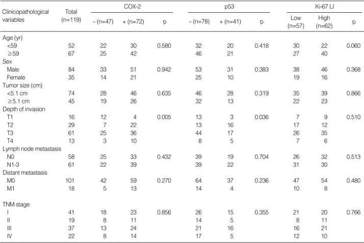
The correlation between COX-2 or p53 expression and clini- copathological variables is shown in Table 1. Expression of COX-2 and p53 correlated significantly with depth of tumor invasion (p=0.005, 0.036 respectively). However, there was
Fig. 1.Immunoreactivity of COX-2 in gastric cancer tissues. COX- 2 immunoreactivity is predominantly detected in the cytoplasm of cancer cells (×200).
Clinicopathological variables
Total (n=119)
COX-2
- (n=47) + (n=72) p
p53
- (n=78) + (n=41) p
Ki-67 LI Low
(n=57)
High
(n=62) p
Age (yr)
<59 52 22 30 0.580 32 20 0.418 30 22 0.060
≥59 67 25 42 46 21 27 40
Sex
Male 84 33 51 0.942 53 31 0.383 38 46 0.368
Female 35 14 21 25 10 19 16
Tumor size (cm)
<5.1 cm 74 28 46 0.635 46 28 0.319 35 39 0.866
≥5.1 cm 45 19 26 32 13 22 23
Depth of invasion
T1 16 12 4 0.005 13 3 0.036 7 9 0.510
T2 29 7 22 13 16 17 12
T3 61 25 36 44 17 26 35
T4 13 3 10 8 5 7 6
Lymph node metastasis
N0 58 25 33 0.432 39 19 0.704 26 32 0.513
N1-3 61 22 39 39 22 31 30
Distant metastasis
M0 101 42 59 0.270 64 37 0.236 47 54 0.480
M1 18 5 13 14 4 10 8
TNM stage
I 41 18 23 0.856 26 15 0.355 21 20 0.766
II 19 8 11 14 5 8 11
III 37 13 24 21 16 16 21
IV 22 8 14 17 5 12 10
Table 1.Correlation between COX-2, p53 expression and Ki-67 labeling index (LI) and clinicopathological variables of gastric cancers
-, Negative; +, Positive; COX-2, Cyclooxygenase-2.
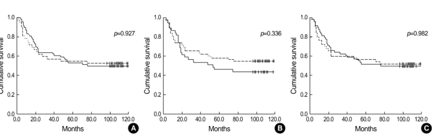
no association between COX-2 or p53 expression and tumor stage, status of lymph node, or distant metastasis. Further- more, COX-2 and p53 expression did not associate with pati- ent survival (p=0.927, 0.336 respectively) (Fig. 2A, B). The expression of p53 did not correlate with COX-2 expression (p=0.939) (Table 2).
Correlation between Ki-67 LI and clinicopathological vari- ables
The Ki-67 LI for 119 tumors ranged from 7.6% to 85.8%
with a mean Ki-67 LI of 49.5±15.5. When a mean Ki-67 LI value of 49.5 was chosen as the cut-off point for discrimi- nation of the 119 patients into two subgroups, 57 patients were categorized as high Ki-67 LI and 62 as low Ki-67 LI.
There was no significant difference in various clinicopatholo- gical variables including survival between the two subgroups (Table 1) (Fig. 2C).
Correlation between COX-2 or p53 expression and Ki-67 LI
The correlation between COX-2 or p53 expression and Ki-67 LI is shown in Table 3. The mean Ki-67 LI value of COX-2 positive tumors was 52.3±14.6 and significantly higher than that of COX-2 negative tumors (p=0.012). The mean Ki-67 LI value of p53 positive tumors was 50.7±14.7 and not significantly higher than that of p53 negative tumors (p=0.525). Combined analysis of COX-2 and p53 status show- ed that the mean Ki-67 LI value of both positive tumors was significantly higher than that of both negative tumors (p=
0.025).
DISCUSSION
Most of antineoplastic effects of aspirin and other non-ste- roidal anti-inflammatory drugs have been linked to their abi- lity to suppress PGs synthesis through inhibition of the activi- ty of the inducible isoform COX-2 (9, 10). Overproduction of COX-2 and PGs has been found to accompany the devel- opment and progression of various human cancers including gastric cancer (6-8). However, the precise mechanisms by which they act is not yet understood. Therefore, understand- ing of underlying mechanisms of COX-2 action in cancer development and progression may lead to a better understand- ing of carcinogenesis. Recent studies have shown that COX-2 and PGs promote carcinogenesis as well as growth and spread of established tumors by stimulating cell proliferation, inhibi- ting apoptosis, increasing the invasiveness of malignant cells, and enhancing the production of VEGF, which promotes an- giogenesis (11-14).
Ki-67 is recognized as a nuclear antigen present in prolif- erating cells but absent in resting quiescent cells. The Ki-67 LI, determined by Ki-67 immunohistochemistry, is a well- known proliferation marker and has been extensively used to
A
Cumulative survival
1.0 0.8 0.6 0.4 0.2
0.00.0 20.0 40.0 60.0 80.0 100.0 120.0 Months
p=0.927
Fig. 2.Kaplan-Meier survival curve correlating disease specific survival with positive (solid line) or negative (dotted line) expression of COX- 2 (A), p53 (B), and high (solid line) or low (dotted line) Ki-67 LI (C).
B
Cumulative survival
1.0 0.8 0.6 0.4 0.2
0.00.0 20.0 40.0 60.0 80.0 100.0 120.0 Months
p=0.336
C
Cumulative survival
1.0 0.8 0.6 0.4 0.2
0.00.0 20.0 40.0 60.0 80.0 100.0 120.0 Months
p=0.982
COX-2 expression p53 expression p-value
Positive (n=41) Negative (n=78)
Positive (n=72) 25 47
Negative (n=47) 16 31 0.939
Table 2.Correlation between COX-2 and p53 expression in gas- tric cancer
COX-2/p53 status Total p-value
(n=119)
Ki-67 LI Mean±SD (%) COX-2
Negative 47 45.1±16.0 0.012
Positive 72 52.3±14.6
p53
Negative 78 48.8±16.0 0.525
Positive 41 50.7±14.7
COX-2/p53
Positive/Positive* 25 54.5±13.0 0.025*
Positive/Negative 47 51.2±15.4 Negative/Positive 16 44.8±15.7 Negative/Negative* 31 45.3±16.4
Table 3.Correlation between Ki-67 labeling index (LI) and sta- tus of COX-2 and p53 in gastric cancer
SD, Standard deviation; COX-2, Cyclooxygenase-2; *Student’s t test.
estimate the growth fraction of tumors (16).
This study is the first to document the possible role of COX- 2 expression for tumor cell proliferation in gastric cancer. In this study, the mean Ki-67 LI value of COX-2 positive tumors was significantly higher than that of COX-2 negative tumors.
This result was concordant with result obtained by Yama- gishi et al. (22). Also, Sawaoka et al. reported that both selec- tive and non-selective COX-2 inhibitors exerted minimal effects on cell proliferation of human gastric cancer cell lines, which expressed lower levels of COX-2, but suppressed cell proliferation of human gastric cancer cell lines that overex- pressed COX-2 (23). These results imply that COX-2 play a critical role in tumor cell proliferation of gastric cancer.
It is known that wild type p53, but not mutant p53, sup- presses a variety of promoters that contain TATA elements (24). A recent in vitro study demonstrated that wild type p53 inhibits the formation of the complex between TATA binding protein and the promoter region of COX-2 gene in a cell-free system (18). Therefore, mutation of p53 may con- tribute to upregulate COX-2 expression, and it may be in- volved in the regulation of tumor cell proliferation.
The second aim of this study was to evaluate the correlation between p53 and COX-2 expression, with special reference to tumor cell proliferation. In this study, the expression of p53 did not correlate with COX-2 expression. In contrast, previous studies reported that tumors with p53 mutation were associated with higher level of COX-2 expression (18, 19).
There are some possible explanations for this discrepancy. First, induction of COX-2 is not dependent on mutation of p53 alone, and regulated by many factors including cytokines, tumor promoters, growth factors, and oncogenes (3-5). Sec- ond, the mutant p53 protein is frequently more stable than wild type, and this stabilized protein product can therefore be detected using immunohistochemistry (25). However, the expression of p53 as revealed by immunohistochemistry might not indicate that the p53 is necessarily mutated and non-func- tional.
Cancer growth and progression is generally regarded as dependent on a high rate of cell proliferation rate and a low rate of apoptosis rate (17). Apoptosis and cell cycle arrest may be reduced when p53 is mutated, allowing cancer growth and progression (18, 19, 25). In this study, the relationship between status of p53 expression and Ki-67 LI was not sta- tistically significant. This result may indicate that p53 muta- tion is predominantly associated with apoptosis, but not cell proliferation. Previous studies reported similar or contrary results (26, 27). The role of p53 mutation in tumor cell pro- liferation has yet been controversial. Combined analysis of p53 and COX-2 status showed that the mean Ki-67 LI value of both positive tumors was significantly higher than that of both negative tumors. This result suggests that molecular and biologic factors including tumor suppressor gene may act in the control of tumor cell proliferation.
The third aim of this study was to examine the expression
of COX-2, p53, and Ki-67 in gastric cancer and their rela- tionship with clinicopathological characteristics including patient prognosis. In this study, expression of COX-2 corre- lated significantly with depth of tumor invasion. However, there was no association between COX-2 expression and sur- vival. There was some agreement, but also several differences between these results and other studies (8, 28, 29). We pre- viously reported that COX-2 expression is associated with well differentiated and intestinal type pathway in gastric car- cinogenesis and not useful for establishing prognoses for gas- tric cancer (8). It is well known that well differentiated and intestinal-type gastric cancer, which less invade and metas- tasize, are associated with a better prognosis than poorly dif- ferentiated and diffuse type cancer. Therefore, the prognostic significance of COX-2 is not established in gastric cancer.
Also, expression of p53 correlated significantly with depth of tumor invasion. However, there was no association between p53 expression and survivial. Although relationship between p53 expression and poorer prognosis is suggested, it is still unclear whether p53 is an independent prognostic factor. Un- derstanding of the molecular and biochemical mechanisms responsible for mutation of p53 is necessary before change of this molecule can be applied in clinical practice as a prognos- tic factor. The Ki-67 LI, an estabilished cell proliferation mar- ker, often correlated to prognosis in gastric cancer (30, 31).
However, in this study, when a mean Ki-67 LI value was cho- sen as the cut-off point for discrimination of the study patients into two subgroups as high Ki-67 LI and low Ki-67 LI, there was no significant difference in various clinicopathological variables including survival between the two subgroups. Liu et al. and Kanai et al. reported that the Ki-67 LI did not in- fluence the prognosis in gastric cancer (32, 33). These con- troversial results may be related to the different score systems and different antibodies used. And because tumor growth and progression result from the imbalance between cell pro- liferation and apoptosis, alterations in the control of apopto- sis can be as important as those of cell proliferation.
In conclusion, COX-2 expression is associated with tumor cell proliferation of gastric cancer. However, tumor cell pro- liferation through the regulation of COX-2 in gastric cancer may not be dependent on p53 status.
REFERENCES
1. DuBois RN, Abramson SB, Crofford L, Gupta RA, Simon LS, Van De Putte LB, Lipsky PE. Cyclooxygenase in biology and disease.
FASEB J 1998; 12: 1063-73.
2. Vane JR, Bakhle YS, Botting RM. Cyclooxygenases 1 and 2. Annu Rev Pharmacol Toxicol 1998; 38: 97-120.
3. Jones DA, Carlton DP, McIntyre TM, Zimmerman GA, Prescott SM.
Molecular cloning of human prostaglandin endoperoxide synthase type II and demonstration of expression in response to cytokines. J Biol Chem 1993; 268: 9049-54.
4. Xie W, Herschman HR. Transcriptional regulation of prostaglandin synthase 2 gene expression by platelet-derived growth factor and serum. J Biol Chem 1996; 271: 31742-8.
5. Sheng H, Williams CS, Shao J, Liang P, DuBois RN, Beauchamp RD. Induction of cyclooxygenase-2 by activated Ha-ras oncogene in Rat-1 fibroblasts and the role of mitogen-activated protein kinase pathway. J Biol Chem 1998; 273: 22120-7.
6. Joo YE, Kim HS, Min SW, Lee WS, Park CH, Park CS, Choi SK, Rew JS, Kim SJ. Expression of cyclooxygenase-2 protein in colorec- tal carcinomas. Int J Gastrointest Cancer 2002; 31: 147-54.
7. Zimmermann KC, Sarbia M, Weber AA, Borchard F, Gabbert HE, Schror K. Cyclooxygenase-2 expression in human esophageal car- cinoma. Cancer Res 1999; 59: 198-204.
8. Joo YE, Oh WT, Rew JS, Park CS, Choi SK, Kim SJ. Cyclooxyge- nase-2 expression is associated with well-differentiated and intestinal- type pathways in gastric carcinogenesis. Digestion 2002; 66: 222-9.
9. Dannenberg AJ, Altorki NK, Boyle JO, Dang C, Howe LR, Weksler BB, Subbaramaiah K. Cyclo-oxygenase 2: a pharmacological target for the prevention of cancer. Lancet Oncol 2001; 2: 544-51.
10. Thun MJ. NSAID use and decreased risk of gastrointestinal cancers.
Gastroenterol Clin North Am 1996; 25: 333-48.
11. Tsujii M, DuBois RN. Alterations in cellular adhesion and apopto- sis in epithelial cells overexpressing prostaglandin endoperoxide synthase 2. Cell 1995; 83: 493-501.
12. Tsujii M, Kawano S, DuBois RN. Cyclooxygenase-2 expression in human colon cancer cells increases metastatic potential. Proc Natl Acad Sci USA 1997; 94: 3336-40.
13. Kambayashi T, Alexander HR, Fong M, Strassmann G. Potential involvement of IL-10 in suppressing tumour-associated macrophages.
Colon-26-derived prostaglandin E2 inhibits TNF-alpha release via a mechanism involving IL-10. J Immunol 1995; 154: 3383-90.
14. Cianchi F, Cortesini C, Bechi P, Fantappie O, Messerini L, Vannacci A, Sardi I, Baroni G, Boddi V, Mazzanti R, Masini E. Up-regulation of cyclooxygenase 2 gene expression correlates with tumor angiogen- esis in human colorectal cancer. Gastroenterology 2001; 121: 1339- 47.
15. Joo YE, Rew JS, Seo YH, Choi SK, Kim YJ, Park CS, Kim SJ. Cy- clooxygenase-2 overexpression correlates with vascular endothelial growth factor expression and tumor angiogenesis in gastric cancer.
J Clin Gastroenterol 2003; 37: 28-33.
16. Weidner N, Moore DH 2nd, Vartanian R. Correlation of Ki-67 anti- gen expression with mitotic figure index and tumor grade in breast carcinomas using the novel ‘‘paraffin’’-reactive MIB1 antibody. Hum Pathol 1994; 25: 337-42.
17. Chan AO, Luk JM, Hui WM, Lam SK. Molecular biology of gas- tric carcinoma: From laboratory to bedside. J Gastroenterol Hepa- tol 1999; 14: 1150-60.
18. Subbaramaiah K, Altorki N, Chung WJ, Mestre JR, Sampat A, Dan- nenberg AJ. Inhibition of cyclooxygenase-2 gene expression by p53.
J Biol Chem 1999; 274: 10911-5.
19. Leung WK, To KF, Ng YP, Lee TL, Lau JY, Chan FK, Ng EK, Chung
SC, Sung JJ. Association between cyclooxygenase-2 overexpression and missense p53 mutations in gastric cancer. Br J Cancer 2001; 84:
335-9.
20. Fleming ID. American Joint Committee on Cancer Classification (AJCC). AJCC cancer staging manual. Philadelphia, Lippincott- Raven, 1997: 71-6.
21. Reed JA, Manahan LJ, Park CS, Brigati DJ. Complete one-hour im- munocytochemistry based on capillary action. Biotechniques 1992;
13: 434-43.
22. Yamagishi M, Noda M, Tatsumi Y, Mukaisho K, Mitsufuji S, Sugi- hara H, Okanoue T, Hattori T. Correlation between cyclooxygenase- 2, proliferative activity, and mucin phenotype in human advanced gastric cancer. J Gastroenterol 2004; 39: 1143-9.
23. Sawaoka H, Kawano S, Tsuji S, Tsujii M, Murata H, Hori M. Effects of NSAIDs on proliferation of gastric cancer cells in vitro: possible implication of cyclooxygenase-2 in cancer development. J Clin Gas- troenterol 1998; 27 (Suppl 1): 47-52.
24. Mack DH, Vartikar J, Pipas JM, Laimins LA. Specific repression of TATA-mediated but not initiator-mediated transcription by wild-type p53. Nature 1993; 363: 281-3.
25. Prives C, Hall PA. The p53 pathway. J Pathol 1999; 187: 112-26.
26. Chen G, Burger MM. p150 overexpression in gastric carcinoma: the association with p53, apoptosis and cell proliferation. Int J Cancer 2004; 112: 393-8.
27. Ishii HH, Gobe GC, Pan W, Yoneyama J, Ebihara Y. Apoptosis and cell proliferation in the development of gastric carcinomas: associ- ations with c-myc and p53 protein expression. J Gastroenterol Hep- atol 2002; 17: 966-72.
28. Murata H, Kawano S, Tsuji S, Tsuji M, Sawaoka H, Kimura Y, Shio- zaki H, Hori M. Cyclooxygenase 2 overexpression enhances lymphatic invasion and metastasis in human gastric carcinoma. Am J Gastroen- terol 1999; 94: 451-5.
29. Lim HY, Joo HJ, Choi JH, Yi JW, Yang MS, Cho DY, Kim HS, Nam DK, Lee KB, Kim HC. Increased expression of cyclooxygenase 2 protein in human gastric carcinoma. Clin Cancer Res 2000; 6: 519- 25.
30. Igarashi N, Takahashi M, Ohkubo H, Omata K, Iida R, Fujimoto S.
Predictive value of Ki-67, p53 protein, and DNA content in the diag- nosis of gastric carcinoma. Cancer 1999; 86: 1449-54.
31. Kakeji Y, Korenaga D, Tsujitani S, Baba H, Anai H, Maehara Y, Sugimachi K. Gastric cancer with p53 overexpression has high poten- tial for metastasising to lymph nodes. Br J Cancer 1993; 67: 589-93.
32. Liu XP, Tsushimi K, Tsushimi M, Kawauchi S, Oga A, Furuya T, Sasaki K. Expression of p21 (WAF1/CIP1) and p53 proteins in gas- tric carcinoma: its relationships with cell proliferation activity and prognosis. Cancer Lett 2001; 170: 183-9.
33. Kanai T, Konno H, Maruyama K, Baba M, Tanaka T, Maruo Y, Ni- shino N, Nakamura S, Baba S, Sugimura H. p53 overexpression and proliferative activity do not correlate with lymph node metastasis in early gastric cancer. Eur Surg Res 1997; 29: 35-41.