췌장 전이암은 원발성 췌장암보다 췌장에 발생하는 빈도가 매우 낮아 암의 2% 미만으로 보고되고 있다. 대부분의 췌장 전이암은 신장암, 유방암, 폐암, 대장암 등이고, 흑색종에 의한 전이는 드물게 보고되고 있다(1-3). Mizushima 등(4)에 의 해 보고된 흑색종의 췌장 전이와 간정맥 혈전을 보인 증례는 두경부에 국한된 전이암으로 수술적 절제를 시행하였다. 저자 들이 경험한 증례는 췌장 전체에 미만성 병변을 동반한 전이암 으로 췌장 전반에 부종 및 췌장주위 조직의 침윤을 동반하고 있어 급성 췌장염이나 림프종으로 오인된 증례로, 비장정맥과 상장간막정맥 내에 혈전을 동반하고 있었으며, 진단 후 급속히 병변이 진행되어 나쁜 예후를 보였던 희귀한 경우로 CT와 MR 영상소견을 보고하고자 한다.
증례 보고
42세 여자가 내원 2주 전부터 시작된 오심과 구토를 동반한 상복부 통증을 주소로 내원하였다. 과거력에서 6개월 전에 우 측 서혜부의 림프절 종대로 절제 생검을 시행 받았으나 전체 림프절의 괴사소견을 보여 병리적 진단을 얻지 못하였다. 그 외 과거력 중 고혈압, 당뇨, 간염 등의 소견은 없었다. 이학적 검사에서 만져지는 종괴는 없었고, 소변검사와 혈청검사에서 는 이상 소견을 보이지 않았다. CA 19-9을 포함한 종양표지자 도 정상이었고 아밀라아제(amylase)와 지질분해효소(lipase) 도 정상이었다. 통증의 원인을 찾기 위해 외래에서 시행한 조 영증강 복부 CT에서 췌장의 미만성 부종과 감소한 밀도를 보 이며 경도의 췌장 주위 조직으로의 침윤을 보였다. 또한, 비장 정맥과 상장간막정맥 내에 혈전이 관찰되었다(Fig. 1A, B).
영상소견으로는 급성 췌장염도 감별해야 했으나 아밀라아제/
지질분해효소 등의 혈액검사 수치가 정상소견을 보여 배제할 수 있었고, 미만성으로 커지고 저밀도 소견을 보인 췌장 때문 에 림프종이나 백혈병 등의 침윤성 암을 먼저 고려하였고, 전 이성 암의 가능성도 감별하였다. MRI는 조영증강을 시행하지 않고 시행하였는데 T1 강조영상에서는 다수의 저신호강도의 결절들이 췌장 두부와 체, 미부에 걸쳐 있었고, T2 강조영상에 서는 이 결절들은 고신호강도를 보였다(Fig. 1C-E). 감별진단 은 CT와 같았다.
췌장에 대한 조직 검사를 위해 입원하여 PET-CT를 촬영하 였는데, PET-CT에서 췌장에 전반적인 평균 4.3 정도의 높은 표준화 섭취계수(standard uptake value: SUV)를 보임과 동시에 왼쪽 겨드랑이 림프절에도 높은 표준화 섭취계수 소견 을 보여, 초음파 유도 하에 겨드랑이 림프절에서 조직 검사결 과 악성 흑색종으로 확진되었다. 그 후 다시 시행한 이학적 검 사에서 환자의 오른쪽 발 뒤꿈치에서 2 cm 크기의 반점성 발 진(malcular rash)이 관찰되어 적출을 시행하여 흑색종으로 확진되었다. 수술 후 항암치료 후 추적 CT에서 정맥 혈전은 점점 진행되었고, 뇌전이가 발생하여 9개월 후 뇌출혈로 사망 하였다.
고 찰
췌장의 전이성 악성 종양은 원발성 췌장 종양과 비교하여 비 교적 드물게 발생한다. 췌장 전이암의 원발 병소로는 신장암 (70.5%)이 가장 많고 다음으로 유방암(6.8%), 폐암(5.9%), 대장암(5.5%) 등으로 알려져 있다(2, 3, 5). 췌장으로의 전이 는 원발 병소의 치료 혹은 진단 후 상당히 병기가 진행된 말기 에 주로 발현되는 것으로 보고되고 있으며, 평균적으로 7년 정 도의 기간이 지난 후 발현되었다. 췌장의 두부에 발생한 경우 가 가장 많아 41.8%를 차지하였고 췌부와 미부에 발생한 경우
─ 325 ─ 대한영상의학회지 2009;61:325-328
비장정맥과 상장간막정맥 혈전을 동반한 Melanoma의 췌장 전이: 증례 보고1
홍성숙∙김정훈∙권귀향∙최득린∙박성태∙김용재∙구동억∙황정화
흑색종의 췌장 전이는 드물고, 더욱이 비장정맥이나 상장간막정맥 내 혈전으로 발현된 증례 는 매우 드물다. 저자들은 췌장 전반에 걸친 미만성 침윤으로 급성 췌장염이나 림프종으로 오인 된 증례로 비장정맥이나 상장간맥정맥 혈전으로 발현되어 나쁜 예후를 보인 흑색종의 췌장 전 이 1예를 CT 및 MRI 소견과 함께 보고하고자 한다.
1순천향대학교병원 영상의학과
이 논문은 2009년 5월 19일 접수하여 2009년 6월 19일에 채택되었음.
는 34.9%, 유두 주위 8.9%, 목 부위 1.6%로 보고 되었다(3).
대부분 임상증상은 췌장에 전이된 위치에 따라 발현되는 증 상이 다른데, 두부의 암은 황달이나 췌장염과 관련된 증상을 나타내고, 미부나 췌부에 전이된 암은 복부 통증이나 만져지는 종괴로 나타나는 경우가 더 흔하다. 220증례를 분석한 보고에
의하면 가장 흔한 임상증상은 황달(25.2%)이고, 복통 (19.7%), 위장관 출혈(11%), 체중 감소(10.2%), 췌장염 (11%) 등의 순이 었다(3). 이 중 췌장염으로 발현되었던 Levine 등(6)이 보고한 증례로는 종양의 괴사(tumor lysis) 에 의하여 췌장염이 발생한 것으로 설명하고 있다. 췌장 전이
─ 326 ─
홍성숙 외: 비장정맥과 상장간막정맥 혈전을 동반한 Melanoma의 췌장 전이
A B
C D
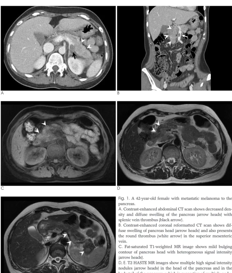
Fig. 1. A 42-year-old female with metastatic melanoma to the pancreas.
A. Contrast-enhanced abdominal CT scan shows decreased den- sity and diffuse swelling of the pancreas (arrow heads) with splenic vein thrombus (black arrow).
B. Contrast-enhanced coronal reformatted CT scan shows dif- fuse swelling of pancreas head (arrow heads) and also presents the round thrombus (white arrow) in the superior mesenteric vein.
C. Fat-saturated T1-weighted MR image shows mild bulging contour of pancreas head with heterogeneous signal intensity (arrow heads).
D, E. T2 HASTE MR images show multiple high signal intensity nodules (arrow heads) in the head of the pancreas and in the body-tail of the pancreas, which is suggestive of multiple nodu- lar metastases.
E
암은 예후가 매우 나쁜데, 최근에는 췌장 전이암도 병소가 국 한된 경우 수술적 치료로 비교적 좋은 결과를 보여주고 있다 (1, 7). 종양이 체부나 미부에 국한되어 있는 경우는 원위부췌 장절제술을 시행하고, 두부에 국한되어 있는 경우는 췌장십이 지장절제술을 시행할 수 있어, 선택된 환자에서는 수술로 생존 율을 증가시킬 수 있다(1, 3, 4, 7). 가장 흔한 전이암인 신장 암의 췌장 전이인 경우는 2년 생존율 78%, 5년 생존율은 65%로 보고 하고 있다(3).
췌장 전이암은 단일 결절로 나타나는 경우가 흔하나, 다발성 결절로 나타나는 경우도 종종 있다. 7증례의 췌장 전이암을 보 고한 Rumancik 등(8)의 보고에 의하면, 다발성 병변은 3증 례로, 2예는 신장암의 전이, 1예는 흑색종 전이였고, 2-4 cm 크기의 다발성으로 경계가 잘 그려지는 결절이었다. 단일 결절 성 전이는 2-9 cm크기로 2예는 유방암의 전이, 2예는 육종의 전이였다(8). 단일 결절로 나타나는 경우는 원발성 췌장암과 의 감별이 필요하고, 다발성 결절로 나타나는 전이암은 미만성 췌장암이나 급성 췌장염, 림프암, 혈액암 등의 가능성을 감별 해야 한다. 병변이 국한되어 있는 단일 결절은 수술적 절제가 생존율을 향상시키는데 도움을 줄 수 있으나, 저자들의 증례와 같이 두부에서 미부까지 미만성으로 침윤된 전이암의 경우는 수술적 절제를 시행할 수 없고, 더욱이 비장정맥 및 상장간막 정맥 혈전을 동반한 증례는 예후가 훨씬 나쁠 것으로 생각된 다.
부검에서 발견된 악성 종양의 췌장 전이는 15%였고, 그 중 흑색종의 췌장 전이는 매우 드물어 0.1%(1/690)였다(5). 더 욱이 비장정맥이나 상장간막정맥 혈전으로 보고된 경우는 매 우 드물다(4). 정맥혈전이 생기는 원인은 종양에 의한 외부성 압박(external compression)이나 이차적인 응고장애 (coagulation disorders)를 일으키기 때문이다(4, 9).
Pestana 등(9)은 신장암의 췌장 전이에서 비장정맥이나 상장 간막정맥 혈전이 있었던 증례에서 원위부췌장절제술(distal pancreatectomy)이나 간정맥과 비장정맥의 혈전제거술 (portosplenic venotomy)을 시행한 후 6개월 추적관찰에서 정상이었다고 보고하였다. 그러나 일반적으로는 비장정맥 혈
전이 있는 환자는 정맥혈전이 없는 환자에 비하여 예후가 더 나쁜 것으로 알려져 있다(9). 저자들이 보고하는 증례도 정맥 혈전이 동반된 전이 환자로 추적관찰 시, 복부에 정맥 혈전이 점점 심해지고 복수가 증가하였으며, 6개월 후 새롭게 뇌전이 가 생겨 9개월 후 뇌출혈로 사망하였다.
이 증례는 미만성 다발성 결절 형태로 췌장에 전이를 일으킨 흑색종 환자로 비장정맥과 상장간막정맥에 혈전이 동반되어 있어 나쁜 예후를 보여 준 증례로, 췌장에 전이된 흑색종의 CT 및 MRI 소견과 정맥 혈전 동반 소견을 보고하고자 한다.
참 고 문 헌
1. Crippa S, Angelini C, Mussi C, Bonardi C, Romano F, Sartori P, et al. Surgical treatment of metastatic tumors to the pancreas: a single center experience and review of the literature. World J Surg 2006;30:1536-1542
2. Roland CF, van Heerden JA. Nonpancreatic primary tumors with metastasis to the pancreas. Surg Gynecol Obstet 1989;168:345-347 3. Sweeney AD, Wu MF, Hilsenbeck SG, Brunicardi FC, Fisher WE.
Value of pancreatic resection for cancer metastatic to the pancreas.
J Surg Res 2009;1:1-10
4. Mizushima T, Tanioka H, Emori Y, Ochi K, Yoshida A, Kiura K, et al. Metastatic pancreatic malignant melanoma: tumor thrombus formed in portal venous system 15 years after initial surgery.
Pancreas 2003;27:201-203
5. Nakamura E, Shimizu M, Itoh T, Manabe T. Secondary tumors of the pancreas: clinicopathological study of 103 autopsy cases of japanese patients. Pathol Int 2001;51:686-690
6. Levine M. Danovitch SH. Metastatic carcinoma to the pancreas.
Another cause of acute pancreatitis. Am J Gastroenterol 1973;60:
290-294
7. Dar FS, Mukherjee S, Bhattacharya S. Surgery for secondary tu- mors of the pancreas. HPB 2008;10:498-500
8. Rumancik WM, Megibow AJ, Bosniak MA, Hilton S. Metastatic disease to the pancreas: evaluation by computed tomography. J Comput Assist Tomogr 1984;8:829-834
9. Pestana IA, David-West G, Livingstone A. Metastatic renal cell car- cinoma to the pancreas with splenic portal vein tumor thrombus.
Am J Surg 2008;74:359-360
─ 327 ─ 대한영상의학회지 2009;61:325-328
─ 328 ─
홍성숙 외: 비장정맥과 상장간막정맥 혈전을 동반한 Melanoma의 췌장 전이
J Korean Soc Radiol 2009;61:325-328
Address reprint requests to : Seong Sook Hong, M.D., Department of Radiology, Soonchunhyang University Hospital 22, Daesakwan-gil, Yongsan-gu, Seoul 140-743, Korea.
Tel. 82-2-709-9396 Fax. 82-2-709-3928 E-mail: [email protected]
Metastatic Melanoma to the Pancreas with a Thrombus in the Splenic Vein and Superior Mesenteric Vein: A Case Report1
Seong Sook Hong, M.D., Jung Hoon Kim, M.D., Kui Hyang Kwon, M.D., Duek Lin Choi, M.D., Sung Tae Park, M.D., Yong Jae Kim, M.D., Dong Erk Goo, M.D., Jung Hwa Hwang, M.D.
1Department of Radiology, Soonchunhyang University Hospital
Metastatic melanoma to the pancreas has been observed infrequently, and the presence of a thrombus in the splenic vein and superior mesenteric vein has rarely been seen for metastatic melanoma. We present the CT and MRI findings of a case of metastatic melanoma to the pancreas, which presented with diffuse multiple pancreatic metastatic nodules and a venous thrombus.
Index words :Pancreatic neoplasms Neoplasm metastasis Melanoma
Venous thrombosis