submit.radiology.or.kr J Korean Soc Radiol 2011;65(5):461-464
461 INTRODUCTION
Marchiafava-Bignami Disease (MBD) is a disease observed in chronic alcoholic patients which results from the symmetric de- myelination of the corpus callosum (1). The clinical diagnosis of MBD can be difficult. Recent magnetic resonance (MR) imaging including diffusion-weighted imaging (DWI) have been helpful in analyzing the distribution of lesions as well as establishing the diagnosis. We report here on a case of MBD with symmetric in- volvement of the corpus callosum, white matter, corticospinal tract, internal capsule, and middle cerebellar peduncle.
CASE REPORT
A 49-year-old man was referred to the emergency depart- ment with dizziness, trembling hands and weakness in both legs for a day. The patient had been an alcoholic with a daily in- take of alcohol. DWI obtained at admission showed symmetri- cal hyperintense lesions involving the corpus callosum, white
matter, corticospinal tract, internal capsule, and middle cere- bellar peduncle (Fig. 1). Fluid-attenuated inversion recovery (FLAIR) and T2 weighted images (T2WI) showed subtle high signal intensity and apparent diffusion coefficient mapping, which showed relatively high hypointensity in those lesions. No further enhancement of the lesions was observed after gadolin- ium. On the basis of the clinical history and imaging findings, MBD was diagnosed. The patient was admitted and given in- travenous thiamine, which resulted in an almost complete re- mission of the symptoms related to the disease. Follow-up MR imaging performed 11 months after the onset showed the dis- appearance of signal-intensity abnormalities on FLAIR, T2WI, and DWI (Fig. 2).
DISCUSSION
MBD is a rare form of toxic demyelination and necrosis of the corpus callosum associated with chronic alcohol con- sumption. This disease is occasionally observed in non-alco-
Case Report
pISSN 1738-2637
J Korean Soc Radiol 2011;65(5):461-464
Received July 12, 2011; Accepted August 24, 2011 Corresponding author: Hae Woong Jeong, MD Department of Radiology, Busan Paik Hospital, Inje University School of Medicine, 633-165 Gaegeum-dong, Busanjin-gu, Busan 614-735, Korea.
Tel. 82-51-890-6549 Fax. 82-51-896-1085 E-mail: [email protected]
Copyrights © 2011 The Korean Society of Radiology
Marchiafava-Bignami disease (MBD) is a rare toxic disorder strongly associated with chronic alcoholism (1-3, 6, 7). It is characterized by progressive demyelination and ne- crosis of the corpus callosum. The process may extend to neighboring white matter and subcortical regions. We report a case of MBD in which fluid-attenuated inversion recovery and diffusion-weighted imaging revealed symmetrical hyperintense lesions with diffuse involvement of the corpus callosum, white matter, corticospinal tract, in- ternal capsule, and middle cerebellar peduncle (3, 4, 8).
Index terms
Marchiafava-Bignami Disease Corpus Callosum
Diffusion-Weighted Imaging
Magnetic Resonance Finding of Acute Marchiafava-Bignami Disease with Diffuse Involvement: A Case Report
광범위한 침범을 보였던 급성 Marchiafava-Bignami 병의 Magnetic Resonance 소견: 1 증례
Young Jin Heo, MD, Hae Woong Jeong, MD, Hyun Sin In, MD
Department of Radiology, Busan Paik Hospital, Inje University School of Medicine, Busan, Korea
MR Finding of Acute Marchiafava-Bignami Disease with Diffuse Involvement
submit.radiology.or.kr
J Korean Soc Radiol 2011;65(5):461-464
462
necrosis. The genu is most frequently involved, but the degen- eration can extend to the entire corpus callosum (5, 6). Other white matter tracts such as the anterior and posterior commi- sures, corticospinal tracts, hemispheric white matter, and middle cerebellar peduncle may also be involved (2). Selective involvement of the entire length of the corpus callosum and chronic alcoholism, other clinical features, and MRI findings support the diagnosis (1). MR imaging is the best technique holic patients as well. It is generally accepted that the cause of
the disease is mainly due to the deficiency of vitamin B com- plex (1, 5, 9). Most patients are male, between 40 and 60 years of age, and have a history of chronic alcoholism with poor oral intake (1, 2).
The main pathologic change associated with MBD is a de- generation of the corpus callosum with different degrees of damage, from demyelination with preservation of axons, to
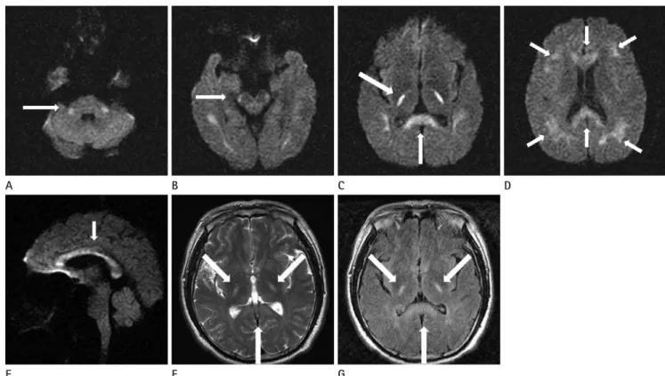
Fig. 1. Axial diffusion-weighted magnetic resonance images obtained upon admission show symmetrical hyperintense lesions involving the middle cerebellar peduncle (A), corticospinal tract (B), posterior limb of internal capsule and the splenium of the corpus callosum (C), as well as the corpus callosum and white matter (D). Sagittal image shows the involvement of the entire corpus callosum (E). Axial T2WI (F) and FLAIR (G) images also showing hyperintense lesions in the posterior limb of the internal capsule and splenium of the corpus callousum.
Note.-FLAIR = fluid-attenuated inversion recovery, T2WI = T2-weighted image
Fig. 2. Follow-up diffusion weighted magnetic resonance images obtained 11 months after the initial study show the disappearance of signal-in- tensity abnormalities including middle cerebellar peduncle (A), corticospinal tract (B), posterior limb of the internal capsule and splenium of the corpus callosum (C), as well as the corpus callosum and white matter (D).
E A
A
F B
B
G C
C
D
D
Young Jin Heo, et al
submit.radiology.or.kr J Korean Soc Radiol 2011;65(5):461-464
463
2. Arbelaez A, Pajon A, Castillo M. Acute Marchiafava-Bigna- mi disease: MR findings in two patients. AJNR Am J Neu- roradiol 2003;24:1955-1957
3. Johkura K, Naito M, Naka T. Cortical involvement in Marchi- afava-Bignami disease. AJNR Am J Neuroradiol 2005;
26:670-673
4. Tung CS, Wu SL, Tsou JC, Hsu SP, Kuo HC, Tsui HW. Marchia- fava-Bignami disease with widespread lesions and complete recovery. AJNR Am J Neuroradiol 2010;31:1506-1507 5. Tuntiyatorn L, Laothamatas J. Acute Marchiafava-Bignami
disease with callosal, cortical, and white matter involve- ment. Emerg Radiol 2008;15:137-140
6. Ihn YK, Hwang SS, Park YH. Acute Marchiafava-Bignami disease: diffusion-weighted MRI in cortical and callosal involvement. Yonsei Med J 2007;48:321-324
7. Yoshizaki T, Hashimoto T, Fujimoto K, Oguchi K. Evolution of Callosal and Cortical Lesions on MRI in Marchiafava- Bignami Disease. Case Rep Neurol 2010;2:19-23
8. Shin HE, Kim JG, Jo SR, Jeong SH, Yoon SJ. Acute Marchi- afava-Bignami disease with typical white matter involve- ment on diffusion weighted MRI. J Korean Neurol Assoc 2008;26:376-378
9. Jang HW. Cortical involvement of Marchiafava-Bignami disease: a case report. J Korean Radiol Soc 2007;56:217-219 for evaluating MBD lesions (3). Characteristic MR imaging
findings are symmetric lesions on the corpus callosum; but, the lesions may be also found in the hemispheric white mat- ter, cortex, middle cerebellar peduncles, and internal capsules (4). In the acute phase, the affected areas are hypointense on T1WI and hyperintense on T2WI, due to edematous change with or without demyelination. However, these lesions do not have mass effect and may show peripheral contrast enhance- ment during the acute stage (2). At 30 hrs after initial imag- ing, hyperintense signals on DWI appeared which was in contrast to the initial image where the signals did not appear.
These findings suggest that the initial change in the corpus callosum was partly attributed to vasogenic edema and that the lesion was then converted into cytotoxic edema. The ne- crotic lesions in the corpus callosum in chronic stage con- firmed the cytotoxic process (7).
Acute MBD is a rare toxic disease, but typical history, clini- cal manifestation, and MR findings with DWI could facilitate its diagnosis.
REFERENCES
1. Kim MJ, Kim JK, Yoo BG, Kim KS, Jo YD. Acute Marchiafa- va-Bignami disease with widespread callosal and cortical lesions. J Korean Med Sci 2007;22:908-911
MR Finding of Acute Marchiafava-Bignami Disease with Diffuse Involvement
submit.radiology.or.kr
J Korean Soc Radiol 2011;65(5):461-464
464
광범위한 침범을 보였던 급성 Marchiafava-Bignami 병의 Magnetic Resonance 소견: 1 증례
허영진 · 정해웅 · 인현신
Marchiafava-Bignami 병은 만성 알코올 중독과 관련성이 많은 드문 독성 장애이다. 이는 뇌들보의 점차적인 탈수초화와 괴사를 나타낸다. 진행과정 중에 인접한 백색실과 피질하의 병변까지 확산될 수 있다. 우리는 액체감쇠역전회복영상과 확 산강조영상에서 뇌들보, 백질, 겉질 척수로, 속섬유막, 양측 소뇌다리 등에 광범위하게 대칭적으로 고신호강도를 보였던 1 증례를 경험하여 보고하고자 한다.
인제대학교 의과대학 부산백병원 영상의학과학교실