INTRODUCTION
Follicular thyroid carcinoma (FTC), which is the second common malignancy of the thyroid gland following papillary thyroid carcinoma (PTC), is usually diagnosed histologically by capsular and/or vascular invasion on permanent section. Mean- while, it is more likely to metastasize to distant organs rather than to regional lymph nodes. The FTC may present with meta- static disease and the diagnosis can be made by histological ex- amination of the metastatic disease (1). It is now well-known that the eggshell calcification of the thyroid nodule occurs in various benign and malignant diseases of the thyroid gland. Our
review of the literature yielded four cases of FTC with eggshell calcification, with three of them presenting with distant metas- tasis and one with thyroid incidentaloma (2, 3).
We report here of a case of FTC in a 74-year-old male, with the interrupted eggshell calcification, presenting with renal and bony metastases along with the findings of primary and meta- static lesions at gray-scale, power Doppler ultrasonography (PD US), and CT scan.
CASE REPORT
A 74-year-old male presented with the poor oral intake and
J Korean Soc Radiol 2014;70(6):403-407 http://dx.doi.org/10.3348/jksr.2014.70.6.403
Received January 27, 2014; Accepted March 15, 2014 Corresponding author: Sang Kwon Lee, MD Department of Radiology, Dongsan Medical Center, Keimyung University School of Medicine, 56 Dalseong-ro, Jung-gu, Daegu 700-712, Korea.
Tel. 82-53-250-7735 Fax. 82-53-250-7766 E-mail: [email protected]
This is an Open Access article distributed under the terms of the Creative Commons Attribution Non-Commercial License (http://creativecommons.org/licenses/by-nc/3.0) which permits unrestricted non-commercial use, distri- bution, and reproduction in any medium, provided the original work is properly cited.
We report a case of follicular thyroid carcinoma (FTC) in a 74-year-old man, with in- terrupted eggshell calcification presenting with renal and bony metastases. The ab- dominal and chest CT showed three homogeneously enhancing masses in the right kidney, second lumbar vertebra, and left fourth rib. The neck CT revealed a mass with interrupted eggshell calcification and homogeneous enhancement in the left thyroid lobe. A thyroid mass with interrupted eggshell calcification and its peripheral and cen- tral vascularity had excellent demonstration on sonography. The pathologic findings of the specimens of the thyroid mass, obtained by sonographically guided fine-needle aspiration biopsy, and the renal mass, obtained by sonographically guided core-needle biopsy, were consistent with follicular neoplasm and metastatic follicular carcinoma, respectively. The interrupted eggshell calcification of FTC in the elderly patients may frequently be associated with the distant metastasis. Thus, this may potentially repre- sent a poor prognostic sign rather than a favorable one.
Index terms Thyroid Gland
Follicular Thyroid Carcinoma Pathologic Calcification Ultrasonography
Tomography, X-Ray Computed
Sonographic and CT Features of Follicular Thyroid Carcinoma with Interrupted Eggshell Calcification Presenting with Renal and Bony Metastases: A Case Report
1신장 및 골전이로 발현한 단절된 계란껍질형 석회화를 가진 여포성 갑상선암의 초음파 및 전산화단층촬영 소견: 1예 보고1
Sang Kwon Lee, MD
1, Sun Young Kwon, MD
2, See Hyung Kim, MD
1, Byung Hak Rho, MD
1Departments of 1Radiology, 2Pathology, Keimyung University School of Medicine, Daegu, Korea
were consistent with follicular neoplasm and metastatic follicu- lar carcinoma, respectively (Fig. 1G, H). He was given dexa- methasone and prednisolone for control of the back pain. He re- fused any further evaluation and management of FTC and metastatic diseases due to financial reasons and was lost to fol- low-up.
DISCUSSION
Our case presented diagnostic dilemma, because the primary lesion causing bony metastases to the vertebra and rib may be a renal mass or a thyroid mass with interrupted eggshell calcifica- tion. The genitourinary radiologist (SHK) interpreted this as a renal cell carcinoma with bony metastasis, while the head and neck radiologist (SKL) strongly suggested FTC with renal and bony metastases. Although the bony lesions of the second lum- bar vertebra and the left fourth rib have not been histopatholog- ically proven, we retrospectively speculated that they might be metastatic lesions from the primary FTC, because the patient had no known primary elsewhere and the enhancement pat- terns of the bony and renal lesions were nearly the same as that of the FTC with interrupted eggshell calcification.
The eggshell calcification on sonography can be defined as a curvilinear echogenic structure along the circumference of a nodule, and interruption of eggshell calcification as focal dis- continuities of a curvilinear echogenic structure. To the best of our knowledge, two case reports concerning FTC with eggshell calcification have been in the literature (2, 3). Cheng et al. (2) re- ported the first case of FTC with eggshell calcification in a 63-year-old woman. They noticed a faint rim-like calcification in the right lower neck on the chest radiograph, which corre- sponded to a cold area of the right lower thyroid on Tc-99m pertechnetate scintigraph, and a focal osteolytic lesion involving frontal bone with epidural extension on the cranial CT. Ultra- sound-guided fine-needle aspiration biopsy of the thyroid nod- ule and biopsy of the skull lesion proved to be follicular neo- plasm and metastatic follicular carcinoma, respectively. However, they did not mention sonographic features of the thyroid mass.
We previously reported three cases of FTC with eggshell calci- fication (3). All three cases of FTC were spherical in shape and had interrupted eggshell calcification, and two had intranodular vacularity on PD US. Among them, two cases presented with the pain, recently being aggravated, in the back and in both of
the lower legs, which he had for 7 months following a traffic ac- cident. The magnetic resonance imaging (MRI) of the lumbar spine, performed at another hospital 7 months prior, had re- vealed a suspicious metastatic lesion at the second lumbar verte- bra and an enhancing mass in the lower pole of the right kidney.
He had refused further examination, and had medicated at the local medical clinic for relief of pain. Otherwise, the physical ex- amination was unremarkable. No remarkable abnormalities were noted on the routine laboratory tests, including thyroid function test.
He underwent CT of the abdomen, chest, and neck by using SOMATOM Definition Flash scanner (Siemens Healthcare, Forchheim, Germany), for investigation of the unknown possi- ble metastatic lesions of the second lumbar vertebra and the right kidney detected by MRI at another hospital. The CT of the abdomen and chest showed approximately 35 × 34 × 39 mm in size, exophytically grown, homogeneously enhancing mass at the lower pole of the right kidney, and homogeneously enhanc- ing masses with bony destruction at the body, right pedicle of the second lumbar vertebra, and left fourth rib (Fig. 1A, B). The CT of the neck revealed a mass with interrupted eggshell calcifi- cation in the lower pole of the left thyroid lobe, which enhanced homogeneously following intravenous administration of con- trast material (Fig. 1C, D). The sonographic examination, per- formed by using Acuson Sequoia 512 scanner (Siemens Medical Solutions, Mountain View, CA, USA) equipped with a 8- to 15- MHz linear array transducer, showed an approximately 17 × 20
× 23 mm-sized solid mass with interrupted eggshell calcification (Fig. 1E). The central and peripheral color signals were noted on PD US (Fig. 1F). He underwent sonographically guided fine- needle aspiration biopsy of the left thyroid mass with interrupt- ed eggshell calcification, using 25-gauge needle attached to a 10- mL disposable syringe, which in turn was held by a reusable syringe holder. Three samples were obtained from the mass. We cautiously performed sonographically guided core-needle biop- sy of the mass in the lower pole of the right kidney, by using a freehand technique with a 18-gauge needle (Bard Peripheral Technologies, Covington, GA, USA) and spring-loaded biopsy gun (Pro-Mag 2.2; Manan Medical Products, Northbrook, IL, USA). Two samples were obtained from the mass. The cytologi- cal and histological findings of the thyroid and renal masses
the eggshell calcification, which corresponded to the tumor in- vasion through (or over) eggshell calcification on pathologic ex- distant metastasis.
Kim et al. (4) found that a hypoechoic halo and disruption of
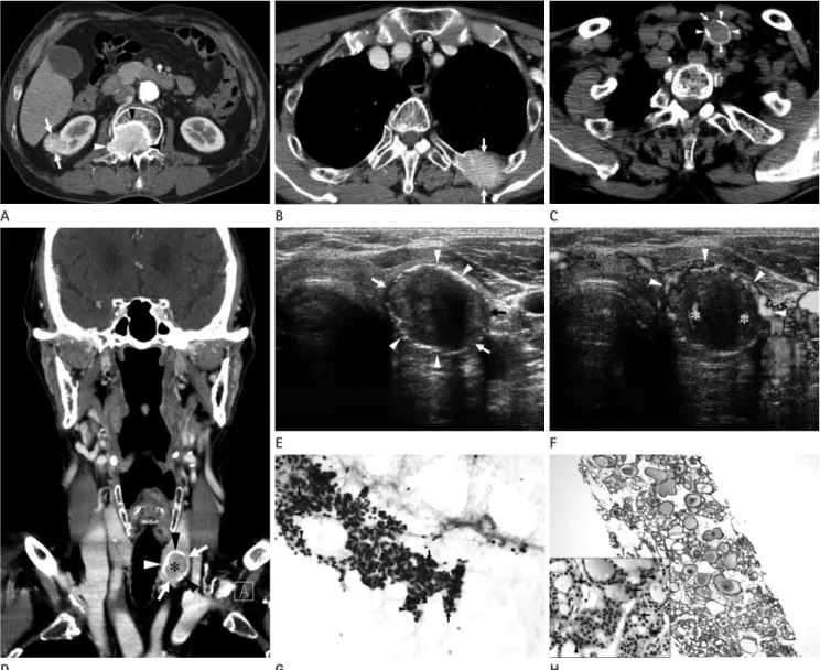
Fig. 1. CT and sonographic features of follicular thyroid carcinoma with interrupted eggshell calcification and metastatic lesions of the kidney and bones in a 74-year-old man.
A, B. Contrast-enhanced axial CT image of the abdomen (A) shows an exophytically grown, homogeneously enhancing mass at the lower pole of the right kidney (arrows) and a homogeneously enhancing mass with bony destruction at the body and right pedicle of the second lumbar vertebra (arrowheads). A contrast-enhanced axial CT image of the chest (B) shows a homogeneously enhancing mass with bony destruction at the left fourth rib (arrows).
C, D. A non-enhanced axial CT image of the neck (C) reveals a mass with eggshell calcification (arrowheads) that has an area of interruption (ar- row). Contrast-enhanced coronal reformatted CT image (D) shows homogeneous enhancement of the mass (asterisk), and eggshell calcification (arrowheads) that has areas of interruption (arrows).
E, F. Transverse gray-scale sonographic image of the thyroid gland (E) shows an ovoid, solid mass with eggshell calcification (arrowheads) that has areas of interruption (arrows) in the lower pole of the left thyroid lobe. Transverse power Doppler sonography (F) reveals color signals within (asterisks) and around the mass (arrowheads).
G, H. Photomicrograph of the cytological examination of the specimen obtained by sonographically guided fine-needle aspiration biopsy of the left thyroid mass with interrupted eggshell calcification (G) demonstrates sheets of atypical follicular cells (arrowheads) and scanty colloid (as- terisks). Follicular cells are arranged to form microfollicles (arrows) (Papanicolaou stain, × 400). Photomicrograph of the histological examination of the specimen obtained by sonographically guided core-needle biopsy of the right renal mass (H) shows numerous, variable sized follicles filled with colloid (asterisks) (hematoxylin & eosin stain, × 100). Tumor cells show mild cytologic atypia (arrowheads) and form microfollicles (arrows) (inset: hematoxylin & eosin stain, × 400).
H E
B
G D
A
F C
the tumor.
In summary, FTC should be included in the differential diag- nosis of a thyroid mass with interrupted eggshell calcification.
Furthermore, interrupted eggshell calcification itself in the el- derly patients with FTC may frequently be associated with the distant metastasis. Thus, it may potentially represent poor prog- nostic sign rather than a favorable one. Further investigation with a large number of cases is anticipated, and intensive diag- nostic investigations including surgical exploration may be war- ranted in elderly patients with cytologically suggested follicular neoplasm that has interrupted eggshell calcification.
REFERENCES
1. Xu H, Zeng W, Tang Y. Metastatic thyroid follicular carci- noma presenting as a primary renal tumor. Intern Med 2012;51:2193-2196
2. Cheng SP, Lee JJ, Lin J, Liu CL. Eggshell calcification in fol- licular thyroid carcinoma. Eur Radiol 2005;15:1773-1774 3. Lee SK, Rho BH. Follicular thyroid carcinoma with an egg-
shell calcification: report of 3 cases. J Ultrasound Med 2009;28:801-806
4. Kim BM, Kim MJ, Kim EK, Kwak JY, Hong SW, Son EJ, et al.
Sonographic differentiation of thyroid nodules with egg- shell calcifications. J Ultrasound Med 2008;27:1425-1430 5. Lee J, Soh EY. Differentiated thyroid carcinoma presenting
with distant metastasis at initial diagnosis clinical out- comes and prognostic factors. Ann Surg 2010;251:114-119 6. Ito Y, Hirokawa M, Masuoka H, Yabuta T, Fukushima M, Ki- hara M, et al. Distant metastasis at diagnosis and large tu- mor size are significant prognostic factors of widely inva- sive follicular thyroid carcinoma. Endocr J 2013;60:829- 833
aminations, may be more useful as sonographic predictors of PTC than known sonographic criteria including hypoecho- genicity, irregular or microlobulated margins, and a taller-than- wide shape.
While the eggshell calcification may be lined inside of, within, or outside of the tumor capsule, the disruption of the eggshell calcification in the follicular neoplasm may be a useful finding that predicts capsular invasion. However, the patient refused to undergo surgical exploration and we could not obtain thyroid- ectomy specimen to verify sonographic-histologic correlation.
Distant metastasis is a major cause of thyroid cancer-specific mortality (5). Ito et al. (6) reported that distant metastasis at di- agnosis is the strongest prognostic factor for cause-specific sur- vival of the patients with widely invasive FTC, and the tumor size larger than 4 cm significantly affects the disease-free surviv- al and cause-specific survival of the patients who did not show distant metastasis at diagnosis. The sizes of 3 cases, with FTC with interrupted eggshell calcification and distant metastasis, re- ported in the literature were 20 mm (2), 28.5 mm, and 10.3 mm (3) in the greatest diameter, and that of the present case was 23 mm. The size of a FTC with interrupted eggshell calcification that did not metastasize was 24.4 mm (3). It should be noted that the sizes of the masses of all FTC with interrupted eggshell calcification presenting with distant metastasis in the literature and present case were all less than 30 mm in the greatest diame- ter (range, 10.3--28.5 mm; average, 20.5 mm) (2, 3). Regarding the age of four patients with FTC with eggshell calcification pre- senting with distant metastasis, in the literature and present case, the average age was 71.8 years (range, 63--75 years) (2, 3).
The age of a patient who had FTC with interrupted eggshell cal- cification, but without distant metastasis was 54 years (3). As such, interrupted eggshell calcification of FTC in elderly pa- tients may be a poor prognostic sign, irrespective of the size of
신장 및 골전이로 발현한 단절된 계란껍질형 석회화를 가진 여포성 갑상선암의 초음파 및 전산화단층촬영 소견: 1예 보고1
이상권
1· 권선영
2· 김시형
1· 노병학
174세 남성에서 신장과 골전이로 발현한 단절된 계란껍질형 석회화를 가진 여포성 갑상선암 1예를 보고하고자 한다. 복부 및 흉부 CT에서 우측 신장, 제2요추, 좌측 4번늑골에 균질한 조영증강을 보이는 종괴가 보였으며, 경부 CT에서 좌측 갑 상선엽에서 단절된 계란껍질형 석회화를 가지며, 균질한 조영증강을 보이는 종괴가 관찰되었다. 단절된 계란껍질형 석회 화를 가진 갑상선 종괴와 종괴의 주변부 및 중심부 혈류는 초음파검사에서 잘 관찰되었다. 갑상선 종괴와 신장 종괴의 초 음파유도하 세침흡인생검 및 핵생검에 의한 검체의 병리학적 소견은 각각 여포성 종양 및 전이성 여포암에 합당하였다. 여 포성 갑상선암에 있어서 단절된 계란껍질형 석회화는 고연령군에서는 원격전이와 흔히 연관되어 있을 수 있으며, 따라서 잠재적으로 나쁜 예후를 나타내는 징후일 수 있다.
계명대학교 의과대학 1영상의학과학교실, 2병리과학교실