74 https://e-jcvi.org
A 68-year-old male presented with the history of a recent anterior wall myocardial infarction 1-month ago which was managed by streptokinase thrombolysis. The patient stabilised temporarily with medical management but complained of progressive dyspnoea on exertion for the past one week. The patient was a chronic smoker with the history of long-standing diabetes mellitus controlled on oral hypoglycaemic agents. Cardiovascular examination revealed a double apical impulse and auscultation identified a gallop rhythm (S3).
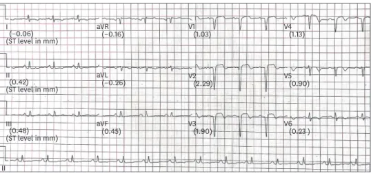
Electrocardiogram was suggestive of recent anterior wall myocardial infarction (Figure 1).
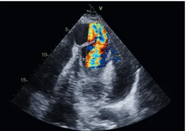
Transthoracic echocardiography revealed a giant apical pseudoaneurysm with a narrow neck and thin wall (Figure 2, Movie 1). Colour Doppler evaluation revealed bidirectional shunt (Figure 3, Movie 2, 3). There was a large pericardial effusion with tamponade physiology as evidenced by right ventricular diastolic collapse. Left ventricular ejection fraction was 25%-30%. The patient was advised urgent surgery and pericardial drainage. Pseudoaneurysm excision followed by left ventricular remodelling with triple layered suture was performed. The patient had an uneventful post-operative period and was discharged on day 7.
Left ventricular aneurysm are of two basic types: true aneurysm and pseudoaneurysm.1) On echocardiography, pseudoaneurysm produces an echo-free space with a narrow neck that J Cardiovasc Imaging. 2020 Jan;28(1):74-76
https://doi.org/10.4250/jcvi.2019.0082 pISSN 2586-7210·eISSN 2586-7296
Images in
Cardiovascular Disease
Received: Aug 24, 2019 Revised: Aug 30, 2019 Accepted: Sep 8, 2019 Address for Correspondence:
Pradyot Tiwari, MD
Department of Cardiology, Apex Heart Institute, G-L, Mondeal Business Park, S.G.
Highway, Ahmedabad 380059, Gujarat, India.
E-mail: [email protected] Copyright © 2020 Korean Society of Echocardiography
This is an Open Access article distributed under the terms of the Creative Commons Attribution Non-Commercial License (https://
creativecommons.org/licenses/by-nc/4.0/) which permits unrestricted non-commercial use, distribution, and reproduction in any medium, provided the original work is properly cited.
ORCID iDs Pradyot Tiwari
https://orcid.org/0000-0002-0461-9607 Tejas Patel
https://orcid.org/0000-0001-9433-6493 Munish Dev
https://orcid.org/0000-0003-0727-0428 Conflict of Interest
The authors have no financial conflicts of interest.
Pradyot Tiwari , MD, Tejas Patel , MD, Sanjay Shah, MD, and Munish Dev , MD
Department of Cardiology, Apex Heart Institute, Ahmedabad, Gujarat, India
Left Ventricular Apical
Pseudoaneurysm with Cardiac Tamponade
I(−0.06) (−0.16)
(ST level in mm)
II
II (0.42) (ST level in mm)
III(0.48) (ST level in mm)
aVR V1(1.03)
(2.29) V2
(1.90) V3
(1.13) V4
(0.90) V5
(0.23 ) V6 (−0.26)
aVL
(0.45) aVF
Figure 1. Electrocardiogram shows QS complexes in V1-V4 with persistent ST segment elevation and T wave inversion in precordial leads.
communicates with the left ventricular cavity. In contrast, true aneurysm results in local bulging and dilatation of the left ventricular wall with a wide neck.2)
Our patient is an unusual survivor of anterior wall myocardial resulting in apical free wall rupture leading to pericardial effusion and tamponade which was contained in time by pericardial inflammation and adhesions. Thrombolysis has been shown to increase the rate of free wall rupture3) and this might be a contributing factor in our case. Urgent surgery, as performed, is necessary in order to prevent sudden death due to re-rupture of the contained rupture.4)
75 https://e-jcvi.org https://doi.org/10.4250/jcvi.2019.0082
Left Ventricular Apical Pseudoaneurysm with Cardiac Tamponade
LV
PE
Pseudoaneurysm
Figure 2. Modified apical 4 chambered view showing a large apical pseudoaneurysm originating from the LV with a narrow neck and thin walls. Massive PE can be seen surround the LV. LV: left ventricle, PE: pericardial effusion.
Figure 3. Modified apical 4 chambered view with colour Doppler demonstrating bidirectional shunt.
SUPPLEMENTARY MATERIALS
Movie 1
Modified apical 4 chambered view showing a large apical pseudoaneurysm originating from the left ventricle (LV) with a narrow neck and thin walls. Massive pericardial effusion can be seen surround the LV.
Click here to view Movie 2
Modified apical 4 chambered view with colour Doppler demonstrating bidirectional shunt.
Click here to view Movie 3
Simultaneous view demonstrating the same pseudoaneurysm with and without colour Doppler.
Click here to view
REFERENCES
1. Brown SL, Gropler RJ, Harris KM. Distinguishing left ventricular aneurysm from pseudoaneurysm. A review of the literature. Chest 1997;111:1403-9.
PUBMED | CROSSREF
2. Gatewood RP Jr, Nanda NC. Differentiation of left ventricular pseudoaneurysm from true aneurysm with two dimensional echocardiography. Am J Cardiol 1980;46:869-78.
PUBMED | CROSSREF
3. Kawakami Y, Hirose K, Watanabe Y, et al. Myocardial free wall rupture and thrombolytic therapy in acute myocardial infarction. Kokyu To Junkan 1989.37:1109-12.
PUBMED
4. Vlodaver Z, Coe JI, Edwards JE. True and false left ventricular aneurysms. Propensity for the altter to rupture. Circulation 1975;51:567-72.
PUBMED | CROSSREF
76 https://e-jcvi.org https://doi.org/10.4250/jcvi.2019.0082
Left Ventricular Apical Pseudoaneurysm with Cardiac Tamponade