서 론
유도객담검사를 시행하는 목적은 천식 및 기타 호흡기 질환에서 기도 염증의 특징을 확인하기 위한 것이며, 객담이 자연적으로 생 성될 수 없는 하부 기도에서 적절한 검체를 수집하기 위하여 객담 유도를 하게 된다.
객담분석은 지난 수세기 동안 중요한 진단 기법으로 사용되었 고, 최근까지 다양한 질환에서 이들의 결과가 이용되고 있다. 천식, 만성폐쇄성폐질환, 호산구성 기관지염, 간질성폐질환에서 기도의
염증세포의 분포를 확인하여, 진단 및 치료에 이용되며, 악성 세포 확인을 통한 폐암 진단 및 폐결핵이나 면역저하 환자에서의 원인균 을 확인하는 데 도움이 되기도 한다. 하기도에서 저절로 가래가 많 이 배출되지 않는 환자들에 대하여 객담유도 방법이 이용되며, 적 절한 분비를 유도하기 위해 등장성 혹은 고장성 용액을 흡입하는 과정이 필요하다. 이 방법은 1958년 처음으로 폐암 진단에 적용시 킨 방법으로 Bickerman에 의해 처음 기술되었다.1
유도객담은 기도 염증을 측정하는 직접적, 상대적으로 비침습적 이며, 유효하고 반복 가능한 방법이다.2 이를 통한 기도의 염증 분
Allergy Asthma Respir Dis 5(6):307-311, November 2017 https://doi.org/10.4168/aard.2017.5.6.307 REVIEW
Correspondence to: Jae-Woo Jung https://orcid.org/0000-0002-3411-735X
Department of Internal Medicine, Chung-Ang University Hospital, Chung-Ang University College of Medicine, 102 Heukseok-ro, Dongjak-gu, Seoul 06973, Korea
Tel: +82-2- 6299-1437, Fax: +82-2-825-7571, E-mail: [email protected]
• This research was supported by a fund (A092076) by Research of Korea Centers for Disease Control and Prevention.
Received: March 17, 2017 Revised: September 6, 2017 Accepted: September 8, 2017
© 2017 The Korean Academy of Pediatric Allergy and Respiratory Disease The Korean Academy of Asthma, Allergy and Clinical Immunology This is an Open Access article distributed under the terms of the Creative Commons Attribution Non-Commercial License
(Allergy Asthma Respir Dis 2017;5:307-311)
유도객담검사의 시행과 해석: 전문가 의견서
김민혜,1 김미영,2 임경환,3 양민석,4 송우정,5 이정민,6 서동인,7 신유섭,8 권재우,9 김세훈,10 김상헌,11 이병재,12 조상헌,5 정재우,13 대한천식알레르기학회 표준화위원회
1이화여자대학교 의과대학 내과학교실, 2인제대학교 부산백병원 내과, 3국군수도병원 내과, 4서울대학교 보라매병원 내과, 5서울대학교 의과대학 내과학교실,
6연세대학교 원주 의과대학 소아청소년과학교실, 7서울대학교 의과대학 소아청소년과학교실, 8아주대학교 의과대학 알레르기내과학교실, 9강원대학교 의과대학
내과학교실, 10서울대학교 분당병원 내과, 11한양대학교 의과대학 내과학교실, 12성균관대학교 의과대학 내과학교실, 13중앙대학교 의과대학 내과학교실
KAAACI Standardization Committee report on the procedure and application of induced sputum examination
Min-Hye Kim,1 Mi-Yeong Kim,2 Kyung-Hwan Lim,3 Min-Suk Yang,4 Woo-Jung Song,5 Jeongmin Lee,6 Dong In Suh,7 Yoo Seob Shin,8 Jae-Woo Kwon,9 Sae-Hoon Kim,10 Sang-Heon Kim,11 Byung-Jae Lee,12 Sang-Heon Cho,5 Jae-Woo Jung,13 Korean Academy of Asthma, Allergy and Clinical Immunology Standardization Committee
1Department of Internal Medicine, Ewha Womans University School of Medicine, Seoul; 2Department of Internal Medicine, Inje University Busan Paik Hospital, Inje University College of Medicine, Busan; 3Department of Internal Medicine, Armed Forces Capital Hospital, Seongnam; 4Department of Internal Medicine, Seoul Metropolitan Government - Seoul National University Boramae Medical Center, Seoul; 5Department of Internal Medicine, Seoul National University College of Medicine, Seoul; 6Department of Pediatrics, Yonsei University Wonju College of Medicine, Wonju; 7Department of Pediatrics, Seoul National University College of Medicine, Seoul; 8Department of Allergy and Clinical Immunology, Ajou University School of Medicine, Suwon; 9Department of Internal Medicine, Kangwon National University School of Medicine, Chuncheon; 10Department of Internal Medicine, Seoul National University Bundang Hospital, Seongnam; 11Department of Internal Medicine, Hanyang University College of Medicine, Seoul; 12Division of Allergy, Department of Medicine, Samsung Medical Center, Sungkyunkwan University School of Medicine, Seoul; 13Department of Internal Medicine, Chung-Ang University College of Medicine, Seoul, Korea
Induced sputum and sputum cell count analysis is a test for the diagnosis of various respiratory diseases. In particular, it has long been used as an important biomarker in the diagnosis or characterization of asthma or eosinophilic bronchitis. Despite a relatively long history of this test, there has been no consensus report for conducting and interpreting the analyses in Korea. Based on this awareness and necessity, the Korean Academy of Asthma, Allergy and Clinical Immunology launched the Standardization Commit- tee to review the international guidelines and the literature and to develop a consensus report on the diagnostic procedure and in- terpretation of the sputum induction test. (Allergy Asthma Respir Dis 2017;5:307-311)
Keywords: Sputum, Induced sputum, Methods, Analysis
석 결과는 기관지세척검사(bronchial washing) 결과와 상관관계가 높으나, 기관지 생검(bronchial biopsy)이나 기관지폐포세척술 (bronchoalveolar lavage, BAL) 결과와는 일관되지 않은 상관관계 를 보이고 있다.3 이는 유도객담과 기관지세척은 주로 중심부기관지 의 분비물을 대변하고, BAL은 말초 폐포를, 기관지 생검은 중심기 도벽을 대상으로 하기 때문이다. 객담 내 호산구분율은 혈액 내 호 산구나 eosinophilic cationic protein,4 호기산화질소보다 천식에 대하여 더 민감도와 특이도가 높기 때문에 진단적 가치가 높다.5
하지만 현재까지 국내에서는 유도객담검사법에 대한 고찰이나 진료지침이 제대로 마련되지 않은 실정으로 이에 대한 필요성이 있 어 대한천식알레르기학회의 기술정보위원회 내 표준화위원회에서 는 국제 가이드라인 및 문헌고찰, 전문가 의견 교환을 통해 유도객 담검사법에 대한 전문가 의견을 정리하였다.
검사법 개요
유도객담검사에는 객담을 유도하는 과정과 유도된 객담에 대하 여 염증세포를 확인하기 위해 객담을 처리하는 과정이 포함된다.
객담유도는 주로 폐기능검사실에서 시행되며 폐기능검사 기사에 의해 시행되는 경우가 많다. 이들은 의사에 의해 감독되어야 한다.
검사는 총 30분 정도 소요된다. 유도된 객담의 처리는 주로 실험실 에서 진행되며, 백혈구분류에 능숙한 전문가에 의해 시행되어야 한다. 이들 모두 숙련된 검사자에 의하여 시행되어야 하고, 정기적 인 질 관리가 필요하다.
장비 및 시설
환자들은 별도의 장소에서 가래를 수집하는 것이 좋고, 이는 객 담 처리실과 인접한 것이 좋다. 유도객담검사를 하는 장소에는 필 요 시 산소 포화도를 모니터링하고, 산소를 즉각적으로 공급할 수 있는 장비가 갖추어져 있어야 한다. 만약의 경우를 대비하여, 인공 호흡장비, 심폐소생술장비를 갖추거나, 이들에 쉽게 접근할 수 있 는 곳에 위치하는 것이 좋다. 생리식염수 에어로졸의 충분한 출력 을 위해 초음파 네뷸라이저(ultrasonic nebulizer)를 추천한다. 기관 지 수축 여부를 모니터링하는 데 있어 폐기능측정기(spirometer) 를 통한 1초간강제호기량(forced expiratory volume in 1 second, FEV1)이 높은 민감도를 보이기 때문에, FEV1이 최대유량계(peak expiratory flow)보다 더 추천된다. 필요 장비는 Table 1과 같다.
주의사항
식염수를 흡입하는 시간은 검체의 세포 조성에 영향을 줄 수 있 으므로, 총 식염수 흡입 시간을 기록하여야 한다. 폐기능 측정이나
가래를 뱉는 데 소요된 시간은 여기서 제외하여야 한다. 유도된 객 담은 4°C에서 보관하여야 하고, 2시간 내에 객담 처리를 시작하여 야 한다. 유도객담검사는 가장 침습적 검사이므로, 호기산화질소 측정검사, 폐기능검사, 기관지천식유발시험 등을 먼저 시행하고, 객 담유도를 가장 마지막에 시행하는 것이 좋다. 객담유도를 2회 이상 시행하여야 하는 경우 객담유도 간 최소 24시간 간격을 두는 것이 염증세포 결과에 있어, 재현성을 높일 수 있다.
검사 시행
1. 객담유도
객담의 유도는 기화된 멸균식염수(등장성 혹은 고장성)를 흡입 하고 이후 기침을 하여 기도의 분비물을 뱉는 과정으로 진행된다.
식염수 흡입은 기관지 수축을 유발할 수 있기 때문에 객담유도 전 기저 폐기능과 전 처치로 기관지확장제인 살부타몰(salbutamol) 200 μg 흡입 10분 후 폐기능을 측정하여야 하며, 객담 유도 과정 동 안 폐기능을 모니터링하여야 한다. 낮은 기저 FEV1 결과가 객담유 도 동안의 기관지 수축 발생을 예측할 수 있는 것은 아니다. 객담유 도에 사용되는 식염수는 멸균된 상태로 환자 당 새로이 준비되어야 한다.6 일반적으로 4.5%의 생리식염수가 권장된다. 고장성 식염수 를 사용하는 경우 더 많은 양의 객담을 유도할 수 있으며, 객담 내 세포 조성에는 등장성 식염수와 차이는 없다.7 적절한 기화를 위하 여, 초음파 네뷸라이져가 추천되며 1 mL/min 이상의 출력이 성공 적인 객담 수집에 필요하다. 일반적으로 식염수를 흡입하는 시간은 15–20분 정도 소요되며, 5분 간격으로 폐기능을 측정하고, 이와 함 께 기침을 하여 객담을 뱉도록 요청하는 것이 필요하다. 이 과정 중 FEV1 값이 검사 직전에 비하여 20% 이상 감소한 경우 객담유도를 중단하여야 한다. 또한 악화된 상태의 천식 환자와 같은 고위험군 의 경우에는 환자의 상태에 따라 검사 중간에 폐기능을 더 자주 측 정해 볼 수 있다(Table 2).
2. 유도객담의 처리 1) 전체 객담 샘플 이용 시
유도객담 처리는 세포 수 측정 목적의 경우 실온에서 시행 가능 Table 1. Equipment for sputum induction
Oxygen saturation monitor, oxygen supply device Salbutamol inhaler (200–400 μg)
Spirometry or peak expiratory flow meter, nose clip Ultrasonic nebulizer, tube, lids
Sterile hypertonic (3.0% or 4.5%) or isotonic (0.9%) saline Glass of water, petri dish for sample collection Worksheet, calculator
Clock
하다. 하지만 다른 연구 목적이라면 각각의 목적에 따라 다를 수 있 다. 먼저 객담 샘플의 부피와 무게를 기록한다. 이후 0.1%의 dithiot- reitol (DTT) 또는 dithioerythritol (DTE)이 포함된 phosphate- buffered 생리식염수(phosphate-buffered saline, PBS)를 같은 부피 로 넣어 10–30분간 실온에서 섞어준다. 이때 균질화를 위해 vortex 를 이용한다. 그리고 나서 슬라이드 질을 높이기 위해 나일론 mesh (48 μm)에 여과한다. 세포 생존 여부(viability)를 평가하기 위해 10 μL의 세포 현탁액과 10 μL의 tryptan blue를 섞어서 염색을 하고 혈구계(hemocytometer counting chamber)를 이용하여 총 세포의 개수를 센다. 총 세포 수에 대한 생존 세포(cell viability) 수의 분율 은 %로 보고한다. 이 작업의 경우 신뢰할 수 있는 결과를 얻기 위해 수작업으로 진행할 것을 권장한다.8 세포와 상층액(supernatant) 을 분리하기 위해 원심분리(300–1,500×g, 5–10분; 보통 400×g, 10분)를 하고, 상층액은 이후의 분석을 위해 따로 분리하여 -80°C 에서 보관할 수 있다. 여기에서 얻은 세포 침전물을 balanced salt solution (혹은 buffer)에 다시 녹이며, 이때 세포 농도는 1.0×10⁶ cells/mL로 맞춘다. 이 후 각각의 cytospin에 대략 40–65 μL의 샘플 (450–650×103 cells)을 분주한다. 이때 cytospin 원심 속도는 10–51×g으로, 6분간 시행할 것을 권장한다.6 이후 cytospin 슬라 이드 염색을 위해 Giemsa 또는 Wright 염색을 이용할 수 있으며 다 른 염색 방법도 가능하다. 감별 세포 계산(differential cell count)을 위해 400–500개의 비편평세포(nonsquamous cells)를 세면서, 이 들 중 호산구, 호중구, 대식세포, 림프구, 기관지 상피세포의 분율을 퍼센트로 기록한다. 편평세포의 분율도 따로 기록해야 하며, 전체 세포 중 편평세포 분율이 높을 경우(>20%), 즉 비편평세포가 80%
미만일 경우 감별 세포 계산의 재현성이 낮아지기 때문에 부적절 한 검체로 생각해야 하며, 결과 해석에 주의가 필요하다.6 정상 객담 호산구 분율은 3% 미만이다.9
2) 객담 점액 plug 샘플만 이용 시
유도객담의 처리에 객담 점액 plug 샘플만 이용할 때에는, 침의 오 염을 최소화하기 위해 전체 객담 샘플을 세포 배양 접시에 붓고 고밀 도의 끈끈한 객담 덩어리만(침이 섞이지 않은 진한 부위) 골라낸다.
객담 샘플의 부피와 무게를 기록하고 0.1%의 DTT 또는 DTE가 포함 된 PBS를 샘플과 4:1 비율의 부피로 넣어 10–30분간 희석한다. 다음 과정은 전체 객담 샘플 이용 시 객담 처리 방법과 동일하다(Table 3).
결과 기록
유도객담의 세포 분율 기록은 다음과 같이 한다. 총 세포 개수 (상피세포와 염증세포 수의 합)를 적고 그 안에 포함되는 각각의 세 포의 수와 이를 통한 분율을 %로 계산하여 작성한다. 편평세포의 분율이 높은 경우 결과 해석에 주의가 필요하다(Table 4, Fig. 1).
결과 해석
객담 내 염증세포 분율은 기도 염증의 표현형의 지표로 흔히 이 용된다. 일반 정상인의 객담 내 호산구 세포 분율은 3% 미만으로, 호산구가 3% 이상일 때 증가되어 있다고 볼 수 있으며, 이는 한국인 을 대상으로 한 연구에서도 증명되었다.10,11 객담 호산구 수가 기도 염증의 표현형의 구분 및 이에 따른 맞춤형 치료법 선택에 중요한 지표로 이용된다는 것이 여러 연구들을 통해 증명되었다.12-14 현재 천식 가이드라인에서도 객담 호산구는 기도 염증을 평가하는 지표 이며 스테로이드 반응을 예측하거나 평가하는 데 유용한 지표로 자리잡았다.15,16
유도객담은 재현성이 좋으며, 이 방법을 모니터링에 이용하였을 때 천식 조절이 향상된다고 알려져 있다. 이에 American Thoracic Society와 European Respiratory Society 천식 가이드라인에서도 유도객담 호산구를 모니터링 지표로 이용하는 것을 포함하고 있 다.15 예를 들어, 치료중인 천식 환자에서 객담 내 호산구가 증가해 있다면 약제 순응도가 떨어지거나, 알레르겐이나 직업적인 천식유 발 물질에 지속적으로 노출되고 있는 것은 아닌지,17 스테로이드 치 료가 불충분한지에 대한 고려가 필요하고, 호산구 증가 없이 호중 구 증가가 관찰될 경우에는 오진의 가능성, 관련된 다른 질환, 스테 로이드 불응성 천식 등을 고려해야 한다.18 또한 천식 환자에서 스 테로이드 치료 용량의 결정이나 치료 반응성 예측, 추가 치료에 대 Table 2. Process of sputum induction
① Explain the test method to the patient in detail (rinsing the mouth before the test, inhalation with tidal breathing, and collecting sputum by coughing every 5 minutes)
② Set the nebulizer output (≥1 mL/min) and sterile saline preparation (usually hypertonic saline, 4.5%)
③ Measure baseline FEV1 (or PEF)
④ FEV1 measurement after 10 minutes of inhalation of pre-treatment Salbutamol (200 μg)
⑤ Start inhalation of saline with tidal breathing through a nebulizer. The sputum is collected every 5 minutes by coughing, during 15 to 20 minutes in total. (The time required for cough and sputum excretion was excluded from the total examination time and the total sputum induction time is recorded). However, if the patient wants to spit out,
they can collect sputum at any time.
⑥ Measure FEV1 (or PEF) every 5 minutes after the start of the test. Examination should be stopped when symptoms occur or FEV1 is reduced by more than 20% of postbron chodilator FEV1.
FEV1, forced expiratory volume in 1 second; PEF, peak expiratory flow.
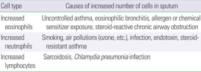
한 결정에도 도움을 줄 수 있다.18 심한 객담 호중구 증가가 있다면 감염을 고려해야 하며 이 경우 항생제 치료가 도움이 될 수도 있 다.18 뿐만 아니라, 만성폐쇄성폐질환 환자에서도 스테로이드 반응 성을 결정하는 데 이용될 수 있다.19 또한 폐암, 간질성폐질환, 결핵, 면역결핍 환자에서 기회 감염 등의 질병을 진단하고, 원인균을 감 별하는데 이용될 수 있다(Table 5).18,20-23
만성기침 환자에서 유도 객담 내 호산구 증가는 호산구성 기관 지염의 진단에 사용될 수 있으며, 총 세포 수와 함께 호중구가 증가 해있다면 감염성기관지염, 기관지확장증, 낭성섬유증 등을 의심해 볼 수 있겠다.18 객담 세포 수가 정상 범위이거나, 전체 세포 수는 정 상이면서 호중구 분율이 증가해있다면 위식도 역류증이나 습관성 기침을 고려해야 한다.18
기타 고려 사항
표준 유도객담검사 및 처리 과정에는 반드시 재현성을 보장하기 위해 각각의 객담 실험실의 질 관리 방법이 포함되어야 한다. 이러 Table 3. Process of induced sputum preparation
When using whole sputum samples
① Put total sputum sample in a polystyrene tube and record volume and weight.
② Mix PBS with 0.1% DTT or DTE in a 1:1 volume ratio with the sputum sample.
③ Mix the sample with pipet and mix more with vortex.
④ Mix the sample using rocker or water-bath at room temperature (22°C).
⑤ The mixture is filtered and the weight is measured.
⑥ Cell viability and total cell count is measured.
⑦ Calculate as total number of cells per mL.
⑧ Cells and supernatant are separated by centrifugation (300–1,500×g, 5–10 minutes; usually 400×g, 10 minutes).
⑨ The cell precipitate is resuspended in a salt solution (or buffer) (concentration 1.0×10⁶ cells/mL). Use approximately 40–65 μL of sample for each cytospin.
⑩ Cytospin is centrifugated at 10–51×g for 6 minutes.
⑪ Giemsa or Wright staining of cytospin slides.
⑫ Differential cell count: Count 400–500 nonsquamous cells and record the percentage of eosinophils, neutrophils, macrophages, lymphocytes, and bronchial epithelial cells. Record the fraction of squamous cells.
When using only sputum mucus plug samples
① Only a thick sputum mucus plug without saliva is selected and put into a polystyrene tube. Record volume and weight.
② Dilute sample with PBS with 0.1% DTT or DTE in a 1:4 volume ratio.
③ The following procedure is the same as the treatment step for the whole sputum sample.
PBS, phosphate-buffered saline; DTT, dithiotreitol; DTE, dithioerythritol.
Table 4. Report form of induced sputum processing
Cell counts %
Bronchial epithelial cells Macrophages Neutrophils Eosinophils Lymphocytes
Total cell counts 400 (500)* 100
*For differential cell count, count 400–500 nonsquamous cells, and then record per- centage of eosinophils, neutrophils, macrophages, lymphocytes, and bronchial epi- thelial cells.
Fig. 1. Image of the sputum cells (× 200). M, macrophage; N, neutrophil; E, eo- sinophil; EP, epithelial cell.
Table 5. Causes of increase in specific cell type of induced sputum examination Cell type Causes of increased number of cells in sputum Increased
eosinophils
Uncontrolled asthma, eosinophilic bronchitis, allergen or chemical sensitizer exposure, steroid-reactive chronic airway obstruction Increased
neutrophils
Smoking, air pollutions (ozone, etc.), infection, endotoxin, steroid- resistant asthma
Increased
lymphocytes Sarcoidosis, Chlamydia pneumonia infection
한 방법에는 정기적인 장비 보정과 매달 내부적으로 슬라이드 해석 에 대한 확인이 필요하다.6
또한 객담 내 세포 감별 계산처럼 잘 정립된 표지자 외에도, 유도 객담 샘플을 이용한 다른 검사들도 가능하다. 예를 들어, 분자미생 물학, 바이러스나 세균 동정, polymerase chain reaction, chip 기술, proteomics, lipidomics, functional assays, 객담 내 액체에서 사이 토카인이나 케모카인을 측정할 수 있는 기술—enzyme-linked immunosorbent assay나 fluorescent bead-based multiplex sand- wich immunoassays, high performance liquid chromatography, microRNA—의 적용이 가능하다. 또한 유도 객담을 통해 얻은 대 부분의 세포들이 살아있는 상태이기 때문에 세포 배양이나 West- ern blot 등의 다른 기술의 적용도 가능하다.6
결 론
유도객담검사는 호흡기 질환의 기도 염증을 반영하는 대표적인 검사법이다. 유도객담 내 호산구 수는 천식의 매우 유용한 지표 중 하나이며, 천식의 표현형이나 치료 반응의 예측에도 도움을 주고, 호산구성기관지염 진단의 가장 중요한 지표로 사용되기도 한다. 그 동안 유도객담검사법을 많이 사용하면서도 이에 대한 국내 가이드 라인이 없어, 문헌고찰 및 국내 전문가들의 의견을 정리한 의견서 를 발표하는 바이다. 하지만 아직까지 유도객담 내의 세포 분율의 기준은 연구마다 다르게 사용하고 있어 명확한 기준을 제시하기는 어렵다. 이번에 정리된 검사법을 기반으로 하여 이러한 세포 분율 의 기준에 대해서도 더 연구가 필요하며 향후 이에 대한 공통된 합 의가 이루어져야 할 것이다.
REFERENCES
1. Bickerman HA, Sproul EE, Barach AL. An aerosol method of producing bronchial secretions in human subjects: a clinical technic for the detec- tion of lung cancer. Dis Chest 1958;33:347-62.
2. Pin I, Gibson PG, Kolendowicz R, Girgis-Gabardo A, Denburg JA, Harg- reave FE, et al. Use of induced sputum cell counts to investigate airway inflammation in asthma. Thorax 1992;47:25-9.
3. Fahy JV, Wong H, Liu J, Boushey HA. Comparison of samples collected by sputum induction and bronchoscopy from asthmatic and healthy subjects. Am J Respir Crit Care Med 1995;152:53-8.
4. Pizzichini E, Pizzichini MM, Efthimiadis A, Dolovich J, Hargreave FE.
Measuring airway inflammation in asthma: eosinophils and eosinophilic cationic protein in induced sputum compared with peripheral blood. J Allergy Clin Immunol 1997;99:539-44.
5. al-Ali MK, Howarth PH. Nitric oxide and the respiratory system in
health and disease. Respir Med 1998;92:701-15.
6. Weiszhar Z, Horvath I. Induced sputum analysis: step by step. Breathe 2013;9:300-6.
7. Cataldo D, Foidart JM, Lau L, Bartsch P, Djukanovic R, Louis R. Induced sputum: comparison between isotonic and hypertonic saline solution in- halation in patients with asthma. Chest 2001;120:1815-21.
8. Efthimiadis A, Spanevello A, Hamid Q, Kelly MM, Linden M, Louis R, et al. Methods of sputum processing for cell counts, immunocytochemistry and in situ hybridisation. Eur Respir J Suppl 2002;37:19s-23s.
9. Spanevello A, Confalonieri M, Sulotto F, Romano F, Balzano G, Migliori GB, et al. Induced sputum cellularity. Reference values and distribution in normal volunteers. Am J Respir Crit Care Med 2000;162(3 Pt 1):1172-4.
10. Belda J, Leigh R, Parameswaran K, O'Byrne PM, Sears MR, Hargreave FE. Induced sputum cell counts in healthy adults. Am J Respir Crit Care Med 2000;161(2 Pt 1):475-8.
11. Kim MY, Jo EJ, Lee SE, Lee SY, Song WJ, Kim TW, et al. Reference ranges for induced sputum eosinophil counts in Korean adult population. Asia Pac Allergy 2014;4:149-55.
12. Jatakanon A, Lim S, Barnes PJ. Changes in sputum eosinophils predict loss of asthma control. Am J Respir Crit Care Med 2000;161:64-72.
13. Pavord ID, Brightling CE, Woltmann G, Wardlaw AJ. Non-eosinophilic corticosteroid unresponsive asthma. Lancet 1999;353:2213-4.
14. Green RH, Brightling CE, McKenna S, Hargadon B, Parker D, Bradding P, et al. Asthma exacerbations and sputum eosinophil counts: a randomised controlled trial. Lancet 2002;360:1715-21.
15. Reddel HK, Taylor DR, Bateman ED, Boulet LP, Boushey HA, Busse WW, et al. An official American Thoracic Society/European Respiratory Society statement: asthma control and exacerbations: standardizing end- points for clinical asthma trials and clinical practice. Am J Respir Crit Care Med 2009;180:59-99.
16. Lougheed MD, Lemiere C, Ducharme FM, Licskai C, Dell SD, Rowe BH, et al. Canadian Thoracic Society 2012 guideline update: diagnosis and management of asthma in preschoolers, children and adults. Can Respir J 2012;19:127-64.
17. Maestrelli P, Calcagni PG, Saetta M, Di Stefano A, Hosselet JJ, Santonas- taso A, et al. Sputum eosinophilia after asthmatic responses induced by isocyanates in sensitized subjects. Clin Exp Allergy 1994;24:29-34.
18. Jayaram L, Parameswaran K, Sears MR, Hargreave FE. Induced sputum cell counts: their usefulness in clinical practice. Eur Respir J 2000;16:150- 19. Eltboli O, Brightling CE. Eosinophils as diagnostic tools in chronic lung 8.
disease. Expert Rev Respir Med 2013;7:33-42.
20. D'Urso V, Doneddu V, Marchesi I, Collodoro A, Pirina P, Giordano A, et al. Sputum analysis: non-invasive early lung cancer detection. J Cell Physiol 2013;228:945-51.
21. Olivieri D, D'Ippolito R, Chetta A. Induced sputum: diagnostic value in interstitial lung disease. Curr Opin Pulm Med 2000;6:411-4.
22. Bothamley GH, Ditiu L, Migliori GB, Lange C; TBNET contributors. Ac- tive case finding of tuberculosis in Europe: a Tuberculosis Network Euro- pean Trials Group (TBNET) survey. Eur Respir J 2008;32:1023-30.
23. LaRocque RC, Katz JT, Perruzzi P, Baden LR. The utility of sputum in- duction for diagnosis of Pneumocystis pneumonia in immunocompro- mised patients without human immunodeficiency virus. Clin Infect Dis 2003;37:1380-3.