DOI : 10.3341/jkos.2008.49.5.845
베스트병의 빛간섭단층촬영 소견
김현경1․노창래2․이원기1․김기석3
가톨릭대학교 의과대학 안과학교실1, 해군 3함대 의무대2, 새빛안과병원3
목적 : 베스트병을 가진 환자 3명에서 병의 단계에 따른 빛간섭단층촬영(optical coherence tomography, OCT) 소견을 보고하고자 한다
증례요약 : 베스트병으로 진단된 3명 6안의 병력기록을 통하여 안저 검사, 눈전위도, 빛간섭단층촬영 등을 후향적으로 조사하였다. 베스트병 환자 6안 안저에서 노른자모양단계, 가성전방축농단계, 계란스크램블 단계등 특징적인 단계를 구 분할 수 있었고 이때 빛간섭단층촬영에서 바깥 망막 맥락막 복합체(outer retina-choroid complex, ORCC)가 낮 은 반사도 영역에 의해 두 층으로 나뉘어지는 형태이거나 또는 낮은 반사도 영역위로 융기된 후 두꺼워지는 형태로 나 타났다.
고찰 : 베스트병에 있어서 진행 단계에 따라 특징적인 OCT 소견을 보였다. 특히 가성전방 축농단계는 감각망막박리 혹은 망막색소상피박리 모양의 형태를 나타낸다고 알려져 있는데 빛간섭단층촬영을 통해 두 가지 경우 모두를 관찰할 수 있었다. 이때 병변의 위치는 베스트로핀에 의한 유체이동장애의 상대적 정도에 따라 정해지는 것으로 생각된다.
<한안지 49(5):845-852, 2008>
<접수일 : 2006년 5월 4일, 심사통과일 : 2007년 11월 27일>
통신저자 : 김 기 석
경기도 고양시 일산구 백석동 1334-3 새빛안과병원
Tel: 031-900-7700, Fax: 031-900-7788 E-mail: kiseok-kim@daum.net
* 본 논문의 요지는 2004년 대한안과학회 제91회 춘계학술대회 에서 포스터로 발표되었음.
베스트병은 염색체 우성으로 유전되는 망막 질환으 로 다양한 표현형으로 나타난다. 이 질환은 초기에 특 징적으로 양안에서 난황모양의 황반 병변을 나타내며 이 병변은 시간이 지날수록 파괴되거나 흉터를 남기게 된다.1 베스트병 환자의 망막기능을 검사해보면 망막전 위도(electroretinogram, ERG) 소견은 정상이지 만, 눈전위도(eletro-oculogram, EOG) 소견은 아 덴비(Arden ratio)가 1.5 이하로 감소하는 특징적인 변화를 보인다. 형광안저혈관조영술에서는 난황 모양에 일치하는 영역에 차단 형광을 보인다. 이 질환을 조직 병리학적으로 연구한 결과에 따르면 망막색소상피내, 망막하 공간 또는 맥락막층에 지방갈색소(lipofuscin) 이나 이에 유사한 물질이 침착되는 것으로 밝혀져있 다.2 이와 같이 베스트병은 매우 특징적인 임상양상을 가지고 있기 때문에 소아에서 발견될 경우 진단자체가
어렵지는 않다. 하지만 황반의 병변이 진행하여 위축되 는 시기까지는 시력이 매우 양호하므로3 성인이 되어서 야 뒤늦게 발견되는 경우가 빈번하다.
최근 망막분야에서 널리 사용되는 빛간섭단층촬영 (optical coherence tomography, OCT)은 조직내 다른 깊이에서 반사되는 빛의 시간차이를 광학적 간섭 계를 사용하여 고해상도의 횡단 영상을 나타내는 새로 운 기술이다.4 기존의 안저사진과 혈관조영술 등으로 알 수 있는 병변의 2차원적 형태에 이 기술을 함께 이 용하면 형태가 비슷한 질병의 3차원 형태를 예상할 수 있어서 망막, 특히 황반부 질환의 진단에 많은 도움을 주고 있다. 따라서 저자들은 아직까지 국내에 보고되지 않은 베스트병 환자들의 황반 빛간섭단층촬영 소견들을 보고하고자 한다.
증례보고
베스트병으로 진단된 3명 6안의 병력기록을 통하여 안저검사, 눈전위도, OCT 등을 후향적으로 조사하였 다. 진단은 특징적인 안저소견과 비정상적으로 낮은 아 덴비를 보이는 눈전위도검사에 기초하였고 추가적으로 OCT를 시행하였다. OCT는 STRATUS OCT (Carl Zeiss Meditec Inc. San Leandro, CA)를 사용하였 다. 저자는 베스트 노른자모양 황반 이영양증을 전노른 자모양 단계(previtelliform stage), 노른자모양 단계
= 증례보고 =
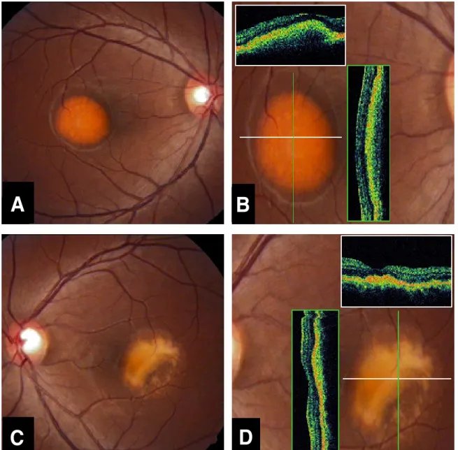
Figure 1. Case 1. Fundus photographs and optical coherence tomography (OCT) findings of the right eye (A, B) and the left eye (C, D) in a patient with Best disease. Each color-framed OCT finding represents a sectioned view of the retina along the axis of the corresponding color-coded line. (A) Fundus photograph shows a 1.5 disc diameter (DD)-sized homogeneous, round, elevated, well- demarcated, yellowish lesion. (B) Vertical and horizontal central macular OCT illustrates a diffuse thickened ORCC*
elevation by underlying hyporeflective area. (C) Fundus photograph shows a 1.5 DD-sized round, elevated, yellowish lesion with an irregular margin. (D) Vertical and horizontal central macular OCT illustrates irregularly thickened ORCC* elevation.
* ORCC = outer retina-choroid complex.
A B
C D
(vitelliform stage), 가성전방축농 단계(pseudohy- popyon stage), 계란스크램블 단계(scrambled egg (vitelliruptive) stage), 후기/반흔단계(late/fibrotic stage)의 다섯 단계로 구분하였다.4
증례 1
28세 건강한 남자가 2개월 동안의 좌안 시력 감소를
주소로 내원하였다. 과거력에서 특별한 병력이나 가족 력은 없었다. 시력은 우안 20/20, 좌안 20/400에서 교정되지 않았으며 전안부 검사에서 특이소견은 없었 다. 안저 검사에서 우안 황반부에는 1.5 유두직경 크기 의 잘 경계지어진 균일한 황색의 둥근 모양의 융기된 병변이 관찰되었고(Fig. 1A), 좌안 황반부에는 1.5 유 두직경 크기의 불규칙한 경계를 가진 황색의 융기된 병 변이 관찰되었다(Fig. 1C). 눈전위도 검사에서 아덴비
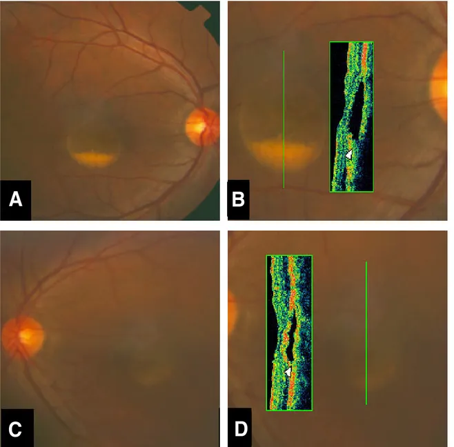
Figure 2. Case 2. Fundus photographs and optical coherence tomography (OCT) findings of the right eye (A, B) and the left eye (C, D) in a patient with Best disease. (A) Fundus photograph shows a 1 DD-sized round lesion within which a pseudohypopyon is forming a fluid level. (B) Vertical central macular OCT illustrates a splitting of ORCC* by hyporeflective area. The broadened, moderate reflective signal (arrowhead) corresponds to the accumulation of subretinal material. (C) Fundus photograph shows s 1 DD-sized round lesion within which a pseudohypopyon is forming a fluid level. (D) Vertical central macular OCT illustrates a splitting of ORCC* by hyporeflective area. The broadened, moderate reflective signal (arrowhead) corresponds to the accumulation of subretinal material.
* ORCC = outer retina-choroid complex.
A B
C D
(Arden ratio)는 우안 1.13, 좌안 0.95로 감소되어 있었다. 검사 결과에 의하여 저자들은 베스트병으로 진 단하고 빛간섭단층촬영을 시행하였다. 빛간섭단층촬영 검사에서 우안의 병변은 높은 반사도를 가지는 바깥 망 막 맥락막 복합체가 두꺼워지면서 바깥 망막 맥락막 복 합체보다 바깥쪽에 위치한 낮은 반사도 영역에 의해 융 기된 소견이었다(Fig. 1B). 같은 검사에서 좌안의 병 변은 전반적으로 높은 반사도를 가지는 바깥 망막 맥락 막 복합체가 불규칙하게 두꺼워져 있는 소견이었다
(Fig. 1D).
증례 2
34세 건강한 남자가 수개월 동안의 양안 시력 감소를 주소로 내원하였다. 과거력에서 특별한 병력이나 가족 력은 없었다. 시력은 우안 20/32, 좌안 20/63에서 교 정되지 않았으며 전안부 검사에서 특이소견은 없었다.
안저 검사에서 양안 황반부에는 1 유두직경 크기의 황
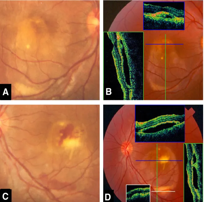
Figure 3. Case 3. Fundus photograph and optical coherence tomography (OCT) findings of the right eye (A, B) and the left eye (C, D) in a patient with Best disease. Each color-framed OCT finding represents a sectioned view of the retina along the axis of the corresponding color-coded line. (A) Fundus photograph shows a 1 DD-sized round, elevated, yellowish lesion. Inferior to this lesion, note a well demarcated 3 DD-sized RPE detachment with yellowish precipitate at the inferior margin (B) Vertical central macular OCT (green-coded) illustrates a well defined sub-RPE detachment and moderately reflective (seen as green) tissue corresponding to the yellow precipitate. Horizontal central macular OCT (blue-coded) shows a moderate reflective signal corresponding to the accumulation of sub-RPE material. (C) Fundus photograph shows a 1 DD-sized round, elevated, yellowish lesion with underlying submacular hemorrhage. There is subretinal hemorrhage over the inferior yellowish precipitate at the inferior margin of the 3 DD-sized RPE detachment. (D) Vertical central macular OCT (green-coded) illustrates a well-defined sub-RPE detachment and moderately reflective (seen as green) tissue corresponding to the yellow precipitate. Horizontal inferior OCT (white-coded) shows a moderate reflective signal corresponding to the accumulation of sub-RPE material.
A B
C D
색의 둥근 모양의 융기된 병변이 관찰되었는데 우안의 병변이 좌안의 병변보다 뚜렷하였다. 이와 함께 둥근 병변 아래쪽 부위에는 수면상을 보이는 반달모양의 노 란 침착물이 관찰되었다(Fig. 2A, C). 눈전위도 검사 에서 아덴비는 우안 0.98, 좌안 1.15로 감소되어 있었
다. 검사 결과에 의하여 저자들은 베스트병으로 진단하 고 빛간섭단층촬영을 시행하였다. 빛간섭단층촬영 검사 에서 양안의 병변 부위와 일치하는 위치에서 잘 경계지 어진 낮은 반사도 영역이 바깥 망막 맥락막 복합체를 두 층으로 나누는 듯한 소견을 관찰할 수 있었다. 반달
Case 1 Case 2 Case 3
Right Left Right Left Right Left
Visual acuity 20/20 20/400 20/32 20/63 20/20 20/20
Stage of Best disease Vitelliform Scrambled egg Pseudo -hypopyon
Pseudo -hypopyon
Pseudo -hypopyon
Pseudo -hypopyon ORCC pathology in OCT Elevation Thickening Splitting Splitting Elevation Elevation Table 1. Summary of visual acuity, stage of Best disease and ORCC pathology in OCT
모양의 침착물에 해당하는 부위는 다소 높은 반사도 (moderate reflectivity)를 나타냈다(Fig. 2B, D).
증례 3
20세 건강한 남자가 정기적인 외래 경과관찰을 위해 내원하였으며 양안 시력은 20/20이었다. 환자는 4년 전 좌안의 급격한 중심 시력 감소를 주소로 처음 내원 하셨으며 당시 우안시력은 20/20, 좌안 시력은 20/80 으로 교정되지 않았고 저자는 우안은 베스트병, 좌안은 베스트병에 동반된 맥락막혈관신생으로 진단하고 특별 한 치료 없이 경과관찰 중이었다. 안저검사에서 우안은 황반부위에 1 유두직경 크기의 황색의 둥근 모양의 융 기된 부위와 그 아래쪽으로 약 3 유두직경 크기의 잘 경계된 망막색소상피 박리소견이 관찰되었다. 망막색소 상피 박리의 아래쪽 경계부위에는 노란 침착물이 보였 다(Fig. 3A). 좌안의 안저 검사는 황반하 출혈 및 망 막색소상피 박리 부분 아래쪽으로 모인 혈액 소견이외 에는 우안과 동일한 소견을 나타내었다(Fig. 3C). 눈 전위도 검사에서 아덴비(Arden ratio)는 우안 1.04, 좌안 0.99로 감소되어있었다. 빛간섭단층촬영 검사에 서 양안의 바깥 망막 맥락막 복합체는 낮은 반사도 영 역에 의해 융기되어 있었고, 병변 아래쪽의 침착물은 초록색으로 나타났다. 이 물질은 노른자 모양 단계에서 보이던 노른자 물질로서 망막색소상피를 뚫고 나온 뒤 중력에 의해 쌓이면서 수면상을 보였다. 그리고 좌안에 서 이전에 맥락막혈관신생이 있었던 부위로서 현재는 반흔형태를 보이는 병변은 피라미드 모양의 다소 높은 반사도인 초록색으로 나타났는데 이 구조는 낮은 반사 도 영역안에 있으면서 망막색소상피층에 연접해있었다.
증례 1의 우안은 안저소견상 노른자모양 단계로서 빛 간섭단층촬영에서는 안저검사에서 관찰된 융기된 병변 위치와 일치하여 바깥 망막 맥락막 복합체가 융기되어 관찰되고 그 밑에 낮은 반사도의 낭형공간이 위치하고 있다. 증례 1의 좌안은 계란스크램블 단계로 빛간섭단 층촬영에서 반사도가 낮은 공간은 존재하지 않으면서 바깥 망막 맥락막 복합체가 불규칙하게 두꺼워져 있다.
증례 2는 안저소견상 양안 모두 가성전방축농 단계로서
빛간섭단층촬영에서는 바깥 망막 맥락막 복합체가 낮은 반사도의 낭형 공간에 의해 두 층으로 나뉘면서 침착물 이 병변 아래쪽에 쌓여있었다. 낭형 공간은 망막색소상 피 위에 존재하였다. 증례 3은 안저소견상 양안 모두 가성전방축농 단계로서 비전형적으로 크고 타원형인 병 변을 가진 형태이며 좌안은 맥락막혈관신생이 동반된 이후 생긴 반흔형태를 함께 보여준다. 증례 2와 달리 증례 3의 빛간섭단층촬영에서는 낭형 공간이 망막색소 상피 밑으로 존재하였다(Table 1).
고 찰
베스트병의 병리 소견은 명확하지 않다. O’Gorman et al2은 망막색소상피하 공간에 침착물이 존재한다고 보고한 반면 Weingeist et al6은 망막하 공간에 존재 한다고 보고하였다. 본 증례의 경우 베스트병을 가진 환자의 황반부 빛간섭단층촬영 소견에서 병변의 위치는 6안 모두 바깥 망막 맥락막 복합체(outer retina- choroid complex, ORCC)에 인접하여 위치하고 있 었다. 바깥 망막 맥락막 복합체는 정상의 망막 빛간섭 단층촬영에서 적색 한 줄로 나타나지만 조직학적으로는 일부의 광수용세포, 망막색소상피, 부르흐씨막 그리고 맥락막 혈관계 등의 높은 반사도를 나타내는 조직의 다 층구조물로 알려져 있다.7 그러므로 이 구조물 사이사 이에 병변이 존재하는 질병의 경우 병변의 정확한 위치 해석에 어려움이 있다. 특히 베스트병과 같이 병인이 세포의 기능이상에 의한 침착물의 증가인 경우 침착위 치와 세포변성의 정도에 따라 빛간섭단층촬영 영상에 차이가 있다.
본 예에 있어서 증례 1의 우안은 안저소견상 노른자 모양 단계이며 증례 2의 양안과 증례 3의 양안은 가성 전방축농 단계이고 증례 1의 좌안은 계란스크램블 단계 였다. 증례 1, 2, 3의 빛간섭단층촬영 소견을 종합해 볼 때 베스트병은 노른자 모양단계에 진입한 후 병변이 진행함에 따라 낮은 반사도의 낭형공간이 생기며 이에 의해 황색의 둥근 병변이 융기되어 보인다. 이후 노란 색 물질의 침착을 보이는 가성전방축농 단계로 접어들 고 병변이 더욱 진행하면 반사도가 낮은 공간이 사라지
면서 전반적으로 바깥 망막 맥락막 복합체가 두꺼워 진 다고 생각할 수 있다.
베스트병이 가성전방축농 단계일 때 빛간섭단층촬영 에서 물질이 어디에 축적하는 지에 대해 Vedantham et al8은 망막색소상피 밑에 존재할 수 있다고 보고한 반면 Men et al9은 물질이 감각망막과 망막색소상피 사이에 존재한다고 보고하였다. 또 안저 소견을 바탕으 로 Kraushar et al10은 가성전방축농 단계의 두 명의 환자에서 중력에 따른 액체 이동을 관찰하여 노른자 모 양 물질이 감각망막과 망막색소상피 사이인 망막하 공 간에 위치하는 것으로 보고하였다. 하지만 가성전방축 농 단계의 환자의 다수가 정상의 시력을 보인다고 알려 져 있으며 본 증례 3에서도 양안 시력은 20/20 이었다.
따라서 정상의 시력을 보이는 대부분의 경우에는 낭형 공간이 망막색소상피밑에 발생하고 안저검사에서 관찰 되는 노란색 물질이 망막색소 상피 밑에 존재한다고 생 각하는 것이 더 합리적일 수 있다.8 어떤 물질이든지 망 막하 공간에 축적된다면 황반 기능에 큰 영향을 주었을 것이기 때문이다.
본 증례 2, 3은 안저소견상 같은 가성전방축농 단계 임에도 빛간섭단층촬영에서는 바깥 망막 맥락막 복합체 의 분열이 서로 다른 위치에서 발생함을 보여주며 이것 은 병인과 연관지어서 생각해 볼 수 있다. 베스트병은 염색체 11q13의 VMD2 유전자가 만드는 베스트로핀 의 이상으로 알려져 있다.11 베스트로핀은 망막색소상 피의 기저바깥막에 배타적으로 위치하며 형질막의 Ca2+/Cl- 이동에 관여한다고 한다.12,13
베스트로핀 변이에 의한 베스트병의 발생은 크게 유 체 이동의 장애와 망막색소상피의 소화기능 장애로 나 누어 기전을 추론할 수 있다. 망막색소상피를 통한 유 체의 이동에는 여러 인자가 영향을 주지만, 그 중에서 도 용질에 연결된 유체의 흡수가 주된 기전이다. 첨단 부를 통한 유체의 유입은 첨단부에 위치한 Na+, K+, 2Cl- 공동이동체(cotransporter)에 의하고 유체의 배출은 기저바깥막에 위치한 Cl- (염소이온) 채널에 의 한다.14 따라서 망막색소상피를 통한 유체의 이동은 망 막색소상피를 가로지르는 Cl-의 운송과 연관된다. 베스 트로핀은 Ca2+/Cl- 채널의 조절 성분 역할을 할 것으 로 생각되며 베스트로핀은 중합하여 작용하기 때문에15 유전자 변이는 올바른 중합의 상호작용을 다양한 범위 에서 방해할 것이다. 결국 베스트병에서 기저바깥막의 비정상적인 Cl- 이동은 망막색소상피를 통한 유체이동 의 장애를 가져오고 망막색소상피의 첨단부 및 기저바 깥막 양쪽 모두에 비정상을 가져온다. 이때 유체나 세 포 부스러기 등이 망막색소상피와 광수용체 사이에 축 적되면 장액성 망막박리와 유사한 형태의 감각망막박리
가 생길수 있고 같은 물질이 망막색소상피와 부루흐씨 막 사이에 축적되면 망막색소상피박리를 가져오게 될 것이다.16 한편 Cl- (염소이온) 채널은 흥분 및 비흥분 세포에서 다양한 역할을 담당하는데 망막색소상피와 같 은 비흥분세포에서는 엔도솜(endosome)의 산성도에 영향을 미쳐서 산성도에 민감한 효소의 적절한 환경을 유지해 주는 기능을 한다. 따라서 베스트로핀은 망막색 소상피가 레티노이드(retinoid)를 소화시키는 데에 영 향을 미친다. 결국 이 단백질의 변이는 리포푹신 축적 을 증가를 통해 베스트병을 유도할 수 있다.17
베스트병의 황반 안저소견은 매우 특이하고 대부분 전형적이지만 형광안저촬영이나 인도사이아닌그린혈관 조영술 소견은 시기에 따라 다양하게 나타난다.18 빛간 섭단층촬영은 망막혈관촬영 소견에서 확인하기 어려운 횡단면상의 병의 조직병리학적 특성을 알려주기 때문에 질병의 특성을 이해하는데 많은 도움을 준다. 특히 가 성전방축농 단계에서는 낭형공간의 위치에 따라 시력에 큰 차이를 보일 수 있다. 따라서 횡단면에서 낭형공간 의 위치를 정확히 알려주는 빛간섭단층촬영은 그 의미 가 크다고 생각한다.
참고문헌
1) Bard LA, Cross HE. Genetic counseling of families with Best macular dystrophy. Trans Sect Ophthalmol Am Acad Ophthalmol Otolaryngol 1975;79:865-73.
2) O’Gorman S, Flaherty WA, Fishman GA, Berson EL.
Histopathologic findings in Best’s vitelliform macular dystrophy. Arch Ophthalmol 1988;106:1261-8.
3) François J, De Rouck A, Fernandez-Sasso D. Electro- oculography in vitelliform degeneration of the macula. Arch Ophthalmol 1967;77:726-33.
4) Gass JD. Steroscopic atlas of macular disease-diagnosis and treatment, 4th ed. Vol. 4. St.Louis: Mosby, 1997;303-436.
5) Hee MR, Izatt JA, Swanson EA, et al. Optical coherence tomography of the human retina. Arch Ophthalmol 1995;113:325-32.
6) Weingeist TA, Kobrin JL, Watzke RC. Histopathology of Best’s macular dystrophy. Arch Ophthalmol 1982;100:1108-14.
7) Huang Y, Cideciyan AV, Aleman TS, et al. Optical coherence tomography (OCT) abnormalities in rhodopsin mutant transgenic swine with retinal degeneration. Exp Eye Res 2000;70:247-51.
8) Vedantham V, Ramasamy K. Optical coherence tomography in Best’s disease: An observational case report. Am J Ophthalmol 2005;139:351-3.
9) Men G, Batioğlu F, Ozkan SS, et al. Best’s vitelliform macular dystrophy with pseudohypopyon: An Optical coherence tomography study. Am J Ophthalmol 2004;137:963-5.
10) Kraushar MF, Margolis S, Morse PH, Nugent ME.
Pseudohypopyon in Best’s vitelliform macular dystrophy. Am J Ophthalmol 1982;94:30-7.
11) Petrukhin K, Koisti MJ, Bakall B, et al. Identification of the gene responsible for Best macular dystrophy. Nat Genet 1998;19:241-7.
12) Marmorstein AD, Marmorstein LY, Rayborn M, et al.
Bestrophin, the product of the Best vitelliform macular dystrophy gene (VMD2), localizes to the basolateral membrane of the retinal pigment epithelium. Proc Natl Acad Sci U S A 2000;97:12758-63.
13) Qu Z, Wei RW, Mann W, Hartzell HC. Two bestrophins cloned from Xenopus laevis oocytes express Ca (2+) activated Cl (-) currents. J Biol Chem 2003;278:49563-72.
14) Hughes BA, Gallemore RP, Miller SS. Transport mechanisms in the retinal pigment epithelium. In : Marmor MF,
Wolfensberger TJ, eds. The retinal pigment epithelium:
Function and disease, 1st ed. New York: Oxford University Press, 1998; chap. 6.
15) Tsunenari T, Sun H, Williams J, et al. Structure-function analysis of the bestophin family of anion channels. J Biol Chem 2003;278:41114-25.
16) Pianta MJ, Aleman TS, Cideciyan AV, et al. In vivo micropathology of Best macular dystrophy with optical coherence tomography. Exp Eye Res 2003;79:203-11.
17) Wabbels B, Presing MN, Kretschmann U, et al. Genotype- phenotpye correlation and longitudinal course in ten families with Best vitelliform macular dystrophy. Graefes Arch Clin Exp Ophthalmol 2006;244:1453-66.
18) Kim JY, Lee SY, Kim IT. Angiographic findings in patients with vitelliform macular dystrophy. J Korean Ophthalmol Soc 2004;45:1917-26.
=ABSTRACT=
Optical Coherence Tomography Findings in Best Disease
Hyun Kyung Kim, M.D.1, Chang Rae Rho, M.D.2, Won Ki Lee, M.D.1, Ki Seok Kim, M.D.3
Department of Ophthalmology, The Catholic University of Korea, College of Medicine1, Seoul, Korea Medical Corps, Navy 3rd Fleet2,
Saevit Eye Hospital3, Gyeonggi, Korea
Purpose: To report the optical coherence tomography (OCT) findings of three cases in various stages of juvenile-onset vitelliform macular dystrophy (Best disease).
Case summary: Medical records of six eyes from three patients diagnosed with Best disease were reviewed retrospectively. We evaluated the clinical features of the fundus, the electro-oculogram, and the optical coherence tomography (OCT) results. In the fundi of the three patients with Best disease, the characteristic stages of vitelliform, pseudohypopyon, and scrambled egg appearance were identified. Optical coherence tomography findings in the eyes of the patients with Best disease showed two types of outer retina-choroid complex (ORCC) changes, including splitting with intervening hyporeflective areas and elevation over hyporeflective area.
Conclusions: The OCT findings showed variable patterns according to the progression of Best disease. In the pseudohypopyon stage, both neurosensory detachment and retinal pigment epithelial detachment appearance were identified. The exact location of the resulting lesions seems to depend on the relative impediment of fluid movement caused by the mutation of bestrophin.
J Korean Ophthalmol Soc 49(5):845-852, 2008
Key Words: Best disease, Optical coherence tomography
Address reprint requests to Ki Seok Kim, M.D.
Saevit Eye Hospital
#1334-3 Baekseok-dong, Ilsandong-gu, Goyang-si, Gyeonggi-do 410-360 Korea Tel: 82-31-900-7700, Fax: 82-31-900-7788, E-mail: kiseok-kim@daum.net