© 2020 The Korean Ophthalmological Society
This is an Open Access article distributed under the terms of the Creative Commons Attribution Non-Commercial License (http://creativecommons.org/licenses /by-nc/3.0/) which permits unrestricted non-commercial use, distribution, and reproduction in any medium, provided the original work is properly cited.
Original Article
Effect of 0.1% Bromfenac for Preventing Macular Edema after Cataract Surgery in Patients with Diabetes
Seok Hyeon Song, Seung Kook Baek, Min Woo Lee, Young Hoon Lee
Department of Ophthalmology, Konyang University College of Medicine, Daejeon, Korea
Purpose: To investigate the effect of 0.1% bromfenac sodium hydrate ophthalmic solution for prevention of mac- ular edema after cataract surgery in patients with diabetes.
Methods: A retrospective analysis of 75 patients with diabetes who underwent cataract surgery was performed.
Thirty-eight patients (52 eyes) were instilled with 0.1% bromfenac solution (bromfenac group) and 37 patients (46 eyes) were not (control group).
Results: There were no significant preoperative between-group differences. Compared to the control group, at 1 month after surgery, the bromfenac group showed slightly better best-corrected visual acuity (0.12 ± 0.12 vs. 0.32 ± 0.42, p = 0.142), lower central macular thickness (265.58 ± 31.28 vs. 314.15 ± 76.11 µm, p < 0.001), and lower macular volume (8.46 ± 0.60 vs. 9.14 ± 1.53 mm3, p = 0.022). There were no significant differences between the two groups at 4 and 6 months postoperatively (p > 0.05). Mean changes in central macular thick- ness showed significant differences at 1 and 4 months postoperatively (-1.44 ± 11.72 and 10.44 ± 22.48 μm in bromfenac group vs. 47.19 ± 70.24 and 31.69 ± 48.04 μm in control group, p < 0.001 and p = 0.016) and mean changes in macular volume showed a significant difference at 1 month postoperatively (-0.08 ± 0.47 mm3 in bromfenac group vs. 0.58 ± 1.28 mm3 in control group, p < 0.001). There were no significant differences there- after (p > 0.05).
Conclusions: Treatment with 0.1% bromfenac sodium hydrate ophthalmic solution showed good efficacy for preventing cystoid macular edema early after cataract surgery in patients with diabetes.
Key Words: Bromfenac, Cataract, Diabetes mellitus, Macular edema, Nonsteroidal anti-inflammatory agents
Postoperative macular edema is a major cause of im- paired vision following cataract surgery. Macular edema
usually develops 4 to 6 weeks after surgery in 1% to 4% of patients [1-3]. Although the etiology of postoperative mac- ular edema is unclear, it is thought that inflammation caused by manipulation of intraocular structures during surgery increases prostaglandin synthesis, which in turn increases retinal vascular permeability [4-7]. Diabetes, hy- pertension, old age, uveitis, and retinal vein occlusions are risk factors for macular edema that weaken the retinal vas- cular walls. In particular, diabetes-related factors such as
Received: April 19, 2019 Final revision: September 30, 2019 Accepted: October 8, 2019
Corresponding Author: Young Hoon Lee, MD. Department of Ophthal- mology, Konyang University College of Medicine, 158 Gwanjeodong-ro, Seo-gu, Daejeon 35365, Korea. Tel: 82-10-3410-0329, Fax: 82-42-600- 9176, E-mail: [email protected]
severity of diabetic retinopathy are related to the develop- ment of macular edema following cataract surgery [8,9].
The most common treatments for macular edema after cataract surgery are corticosteroids, nonsteroidal anti-in- flammatory drugs (NSAIDs), carbonic anhydrase inhibi- tors, and anti-vascular endothelial growth factor agents;
these drugs are topics of many ongoing studies [5].
NSAIDs inhibit cyclo-oxygenase (COX) and therefore decrease inflammation by blocking prostaglandin synthe- sis [10-12]. NSAIDs are used to treat allergic conjunctivitis and scleritis and are effective for maintaining mydriasis during surgery, alleviating postoperative pain, controlling inflammation, and preventing macular edema when used before and after cataract surgery [13-17]. Koh and Chung [18] reported that the use of pranoprofen ophthalmic solu- tion after cataract surgery in patients with diabetes signifi- cantly limited increases in central macular thickness (CMT) and macular volume (MV), thereby preventing macular edema. Chun et al. [19] reported that use of topical diclofenac and pranoprofen prevented postoperative macu- lar edema in a cohort of patients without diabetes. Addi- tionally, intravitreal injection of anti-vascular endothelial growth factor agents used together with topical NSAIDs decreased macular edema in retinal vein occlusion, diabet- ic retinopathy, and age-related macular degeneration [20- 23]. A body of literature indicates that, compared to steroid ophthalmic solutions, NSAIDs are more effective for pre- venting inflammation and macular edema following cata- ract surgery. In addition, NSAIDs are associated with low- er rates of steroid-related side effects such as increased intraocular pressure (IOP), infection, and delayed corneal epithelial wound healing [3,16,17,20-31].
The most common NSAID ophthalmic solutions used in South Korea are 0.1% diclofenac sodium, 0.45% ketorolac tromethamine, 0.1% pranoprofen, and 0.1% bromfenac.
Jeong et al. [32] compared the utility of 1% prednisolone and 0.1% bromfenac for preventing cystoid macular edema following cataract surgery and reported no significant dif- ference in efficacy. In addition, there were a number of studies comparing the efficacy of preventing macular ede- ma between NSAID ophthalmic solutions and comparing NSAID ophthalmic solution versus steroid ophthalmic solution for preventing macular edema after cataract sur- gery. A number of studies have shown that NSAID oph- thalmic solution can help prevent macular edema after cat- aract surgery. However, there has been no report on using
bromfenac for prevention of macular edema after cataract surgery in Korean patients with diabetes.
We studied the preventive effect of 0.1% bromfenac so- dium hydrate ophthalmic solution on macular edema after cataract surgery in patients with diabetes by analyzing ef- fects on best-corrected visual acuity (BCVA), CMT, and MV.
Materials and Methods
A retrospective cohort study was conducted. The study protocol adhered to the tenets of the Declaration of Helsin- ki and was approved by the institutional review board of Konyang University Hospital of Korea (2019-04-010). In- formed consent was waived due to the retrospective nature of the study. The study included 98 eyes from 75 patients with diabetes who underwent cataract surgery from De- cember 2015 to February 2017 and were followed-up for more than 6 months. All patients underwent a comprehen- sive ophthalmic examination, including review of their medical history, visual acuity testing, slit-lamp examina- tion, IOP measurement, and dilated funduscopic examina- tion. Optical coherence tomography (OCT) was conducted using Heidelberg Spectralis (Heidelberg Engineering, Hei- delberg, Germany) prior to surgery. The exclusion criteria included preoperative CMT >300 μm, glaucoma, retinal diseases other than diabetic retinopathy, and history of pri- or intraocular surgery or intravitreal injection.
The severity of diabetic retinopathy was classified by a single ophthalmologist as “no diabetic retinopathy,”
“non-proliferative diabetic retinopathy (NPDR),” or “pro- liferative diabetic retinopathy (PDR)” as per the Early Treatment Diabetic Retinopathy Study criteria. Ophthal- mic examinations were performed at 1 day, 1 week, 1 month, 4 months, and 6 months postoperatively to regular- ly monitor BCVA and IOP. OCT was conducted at 1, 4, and 6 months postoperatively to compare to pre- and postoper- ative CMT and MV values. CMT was defined as the aver- age value of macular thickness < 1.0 mm in diameter with the foveal centralis at the center. BCVA was measured us- ing the Snellen chart and was converted to logarithm of minimum angle of resolution (logMAR) visual acuity for analysis. IOP was measured using a Goldmann applanation tonometer.
All surgeries were performed by the same surgeon after
retrobulbar and nadbath anesthesia using 2% lidocaine. A clear corneal incision of approximately 2.75 mm was made at 11 o’clock, and the anterior chamber was stabilized with 1% sodium hyaluronate (Microvisc; Bohus Bio Tech AB, Stromstad, Sweden) to protect the corneal endothelium during surgery. A 5.5-mm continuous curvilinear capsu- lorhexis was performed using a cystotome and capsulor- hexis forceps. After phacoemulsification using a Constel- lation vision system (Alcon, Fort Worth, TX, USA), a one- piece aspheric foldable soft artificial prosthetic intraocular lens (Tecnis ZCB00; Abbott Medical Optics, Santa Ana, CA, USA) was implanted into the posterior chamber.
In the bromfenac group, 0.1% bromfenac sodium hydrate ophthalmic solution (Bronuck; Taejoon, Seoul, Korea) was applied twice daily from 3 days before cataract surgery to 1 month postoperatively. Both groups applied levofloxacin ophthalmic solution (Cravit; Santen, Osaka, Japan) four times daily for 3 days before cataract surgery. After sur- gery, both groups applied moxifloxacin (Vigamox, Alcon) and 1% prednisolone acetate ophthalmic solution (Pred- forte; Allergan, Irvine, CA, USA) four times daily for
1 week followed by levofloxacin and 0.1% fluorometholone ophthalmic solution (Fluorometholone, Taejoon) for 3 weeks.
Statistical analyses were performed using PASW Statis- tics ver. 18.0 (SPSS Inc., Chicago, IL, USA). Pearson’s chi- squared tests and Mann-Whitney U-tests were used for be- tween-group comparisons. Wilcoxon signed-rank tests were used to compare preoperative and postoperative val- ues within each group. In all cases, the threshold for sig- nificance was p < 0.05.
Results
Of 98 eyes from 75 patients with diabetes, 52 eyes (38 patients) were assigned to the bromfenac group and 46 eyes (37 patients) were assigned to the control group. There were no significant between-group differences in terms of age, sex, duration of diabetes mellitus, hemoglobin A1C within 2 months before surgery, severity of preoperative diabetic retinopathy, or history of panretinal photocoagu-
Table 1. Patient demographics and preoperative clinical characteristics
Characteristics Control (n = 46) Bromfenac (n = 52) p-value
Age (yr) 61.80 ± 7.53 62.85 ± 9.24 0.270*
Sex
Male 14 (30.4) 17 (32.7) 0.810†
Female 32 (69.6) 35 (67.3)
BCVA (logMAR) 0.47 ± 0.35 0.49 ± 0.34 0.657*
IOP (mmHg) 15.13 ± 3.12 14.31 ± 3.08 0.090*
Hypertension 25 (54.3) 28 (53.8) 0.960†
CMT (μm) 267.98 ± 22.66 267.23 ± 26.26 0.724*
MV (mm3) 8.50 ± 0.63 8.55 ± 0.75 0.591*
Duration of DM (yr) 13.39 ± 8.69 13.23 ± 9.07 0.651*
Hemoglobin A1C (%) 7.40 ± 0.97 7.33 ± 1.24 0.350*
Severity of DM
NDR 11 (23.9) 13 (25.0) 0.517†
NPDR 16 (34.8) 23 (44.2)
PDR 19 (41.3) 16 (30.8)
Panretinal photocoagulation 24 (52.2) 19 (36.5) 0.120†
Values are presented as mean ± standard deviation or number (%).
BCVA = best-corrected visual acuity; logMAR = logarithm of the minimum angle of resolution; IOP = intraocular pressure; CMT = central macular thickness; MV = macular volume; DM = diabetes mellitus; NDR = no diabetic retinopathy; NPDR = non-proliferative diabetic retinopathy; PDR = proliferative diabetic retinopathy.
*Mann Whitney U-test; †Pearson’s chi-squared test.
lation laser treatment. Similarly, there were no significant differences in preoperative BCVA (logMAR), CMT, or MV between groups (Table 1).
Visual outcomes
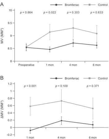
Both groups showed improvements in mean BCVA at 1, 4, and 6 months postoperatively compared to preoperative status ( p < 0.001). At 1 month postoperatively, mean BCVA was 0.12 ± 0.12 in the bromfenac group and 0.32 ± 0.42 in the control group. Although the bromfenac group showed better BCVA than the control group, this differ- ence was not statistically significant (p = 0.142). At 4 and 6 months postoperatively, mean BCVA was 0.15 ± 0.12 and 0.16 ± 0.12 in the bromfenac group, whereas it was 0.28 ± 0.36 and 0.25 ± 0.35 in the control group (p = 0.305 and 0.824) (Table 2 and Fig. 1).
Anatomic outcomes
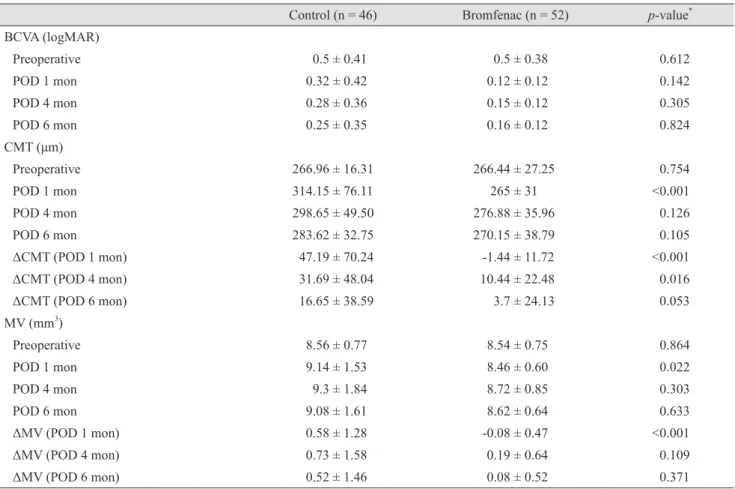
Mean CMT in the bromfenac group was 265.58 ± 31.28, 276.88 ± 35.96, and 270.15 ± 38.79 μm at 1, 4, and 6 months, respectively, while in the control group it was 314.15 ± 76.11, 298.65 ± 49.50, and 283.62 ± 32.75 μm, respectively.
In the bromfenac group, there was no significant differ- ence in CMT between preoperative status and at 1 and 6 months postoperatively (p = 0.52 and 0.089), but there was a significant increase at 4 months compared to the preoperative status (p = 0.001). In the control group, there were significant increases in CMT between preoperative status and at 1, 4, and 6 months postoperatively (p < 0.001, 0.002, and 0.012). Additionally, there were significant be- tween-group differences in CMT at 1 month postopera- tively (p < 0.001), but not at 4 or 6 months postoperatively (p = 0.126 and 0.105). Mean changes in CMT in the brom-
Table 2. Between-group comparisons of postoperative clinical outcomes
Control (n = 46) Bromfenac (n = 52) p-value* BCVA (logMAR)
Preoperative 0.5 ± 0.41 0.5 ± 0.38 0.612
POD 1 mon 0.32 ± 0.42 0.12 ± 0.12 0.142
POD 4 mon 0.28 ± 0.36 0.15 ± 0.12 0.305
POD 6 mon 0.25 ± 0.35 0.16 ± 0.12 0.824
CMT (μm)
Preoperative 266.96 ± 16.31 266.44 ± 27.25 0.754
POD 1 mon 314.15 ± 76.11 265 ± 31 <0.001
POD 4 mon 298.65 ± 49.50 276.88 ± 35.96 0.126
POD 6 mon 283.62 ± 32.75 270.15 ± 38.79 0.105
ΔCMT (POD 1 mon) 47.19 ± 70.24 -1.44 ± 11.72 <0.001
ΔCMT (POD 4 mon) 31.69 ± 48.04 10.44 ± 22.48 0.016
ΔCMT (POD 6 mon) 16.65 ± 38.59 3.7 ± 24.13 0.053
MV (mm3)
Preoperative 8.56 ± 0.77 8.54 ± 0.75 0.864
POD 1 mon 9.14 ± 1.53 8.46 ± 0.60 0.022
POD 4 mon 9.3 ± 1.84 8.72 ± 0.85 0.303
POD 6 mon 9.08 ± 1.61 8.62 ± 0.64 0.633
ΔMV (POD 1 mon) 0.58 ± 1.28 -0.08 ± 0.47 <0.001
ΔMV (POD 4 mon) 0.73 ± 1.58 0.19 ± 0.64 0.109
ΔMV (POD 6 mon) 0.52 ± 1.46 0.08 ± 0.52 0.371
Values are presented as mean ± standard deviation.
BCVA = best-corrected visual acuity; logMAR = logarithm of the minimum angle of resolution; POD = postoperative day; CMT = cen- tral macular thickness; MV = macular volume.
*Mann-Whitney U-test.
fenac group were -1.44 ± 11.72, 10.44 ± 22.48, and 3.70 ± 24.13 μm at 1, 4, and 6 months postoperatively, respective- ly, while those in the control group were 47.19 ± 70.24, 31.69 ± 48.04, and 16.65 ± 38.59 μm, respectively. There were significant between-group differences in mean change at 1 (p < 0.001) and 4 months postoperatively (p = 0.016), but there was no significant difference at 6 months postoperatively (p = 0.053) (Table 2 and Fig. 2A, 2B).
MV showed similar trends to CMT. Mean MVs in the bromfenac group were 8.46 ± 0.60, 8.72 ± 0.85,and 8.62 ± 0.64 mm3 at 1, 4, and 6 months postoperatively, respective- ly, while those in the control group were 9.14 ± 1.53, 9.30 ± 1.84,and 9.08 ± 1.61 mm3, respectively. In the bromfenac group, there was no significant difference in MV between preoperative status and 1 month postoperatively ( p = 0.421), but there was a significant increase at 4 and 6 months postoperatively compared to preoperative status (p = 0.009 and 0.015). In the control group, there were sig- nificant increases in MV between preoperative status and status at 1, 4, and 6 months postoperatively (p = 0.006, 0.011, and 0.083). Mean changes in MV in the bromfenac group were -0.08 ± 0.47, 0.19 ± 0.64, and 0.08 ± 0.52 mm3 at 1, 4, and 6 months postoperatively, respectively, while those in the control group were 0.58 ± 1.28, 0.73 ± 1.58, and 0.52 ± 1.46 mm3, respectively. There were significant be- tween-group differences in mean change at 1 month post- operatively (p < 0.001) (Table 2 and Fig. 3A, 3B).
There were eight eyes (15.4%) with CMT >300 μm in the bromfenac group and 15 eyes (32.6%) with CMT >300 μm in the control group during follow-up. Only one eye in the control group exhibited an increase in CMT >150 μm com- pared to preoperative status. The patient’s BCVA (log-
MAR) was 1.30 at 1 month postoperatively and showed immediate improvement after treatment with intravitreal bevacizumab injection to 0.30. The patient’s BCVA was then 0.20 at 3 months after injection (4 months postopera- tively). There were no significant between-group differ- ences in IOP at 1 day, 1 week, 1 month, 4 months, or 6 months postoperatively (p > 0.05).
Analysis by severity of diabetic retinopathy
In patients without diabetic retinopathy, mean change in CMT was significantly smaller in the bromfenac group than in the control group at 1 month postoperatively (p = 0.038), but not mean change in MV (p = 0.571). There were no significant between-group differences in mean change in CMT or MV at 4 and 6 months postoperatively.
In patients with NPDR, mean changes in CMT and MV were significantly smaller in the bromfenac group than in the control group at 1 month postoperatively (p = 0.014,
BCVA (logMAR)
0.3 0.4 0.5 0.6 0.7
0.2 0.1 0
Bromfenac Control
Preoperative 1 mon 4 mon 6 mon
p = 0.612 p = 0.142 p = 0.305 p = 0.824
Fig. 1. Between-group comparison of best-corrected visual acuity (BCVA). logMAR = logarithm of the minimum angle of resolu- tion.
A
p = 0.754 p < 0.001 p = 0.126 p = 0.105
Preoperative 1 mon 4 mon 6 mon
CMT (μm)
300 320 340
280 260 240
Bromfenac Control
1 mon p < 0.001
4 mon p = 0.016
6 mon p = 0.053
ΔCMT (μm) 4050
70 60
30 10 20
B
-10 0
Bromfenac Control
Fig. 2. Between-group comparison of (A) mean central macular thickness (CMT) and (B) mean change in CMT from preopera- tive status between two groups over time (p < 0.05, Mann-Whit- ney U-test).
0.043, respectively), but not at 4 months postoperatively. At 6 months postoperatively, there was a significant be- tween-group difference in mean change in CMT (p = 0.034), but not in MV (p = 0.196). In patients with PDR, mean changes in CMT and MV were not significant- ly different between the two groups (Table 3). Serious bromfenac-associated complications such as toxic keratop- athy, corneal perforation, corneal melting, or corneal ul- ceration were not identified. In addition, subjective irrita- tion or discomfort were not identified.
Discussion
Previous studies have associated the presence of diabetes mellitus and severity of diabetic retinopathy with macular edema after cataract surgery. Accordingly, patients with diabetes have an increased risk of macular edema due to weak retinal vascular walls [8,9,33]. Macular edema can be diagnosed on OCT and fluorescein angiography. While
conventional fluorescence angiography is invasive, OCT is a non-invasive technique that images the retinal layer structure and quantifies retinal thickness and volume in a highly reproducible manner [2,9,34].
Several studies have reported the incidence of macular edema after cataract surgery. The previously reported inci- dence of cystoid macular edema affecting visual acuity is 1% to 4% [2,3]. The present study only identified decreased visual acuity and macular edema with CMT exceeding 300 μm in 1 eye at 1 month postoperatively, consistent with an incidence of 1.02% (1 / 98).
Prostaglandin, which is produced during inflammatory reactions, is an inflammatory mediator that causes vasodi- lation, increased vascular permeability, destruction of the retinal vascular wall, pain, and miosis [35]. NSAIDs are COX inhibitors that inhibit synthesis of prostaglandin. Ac- cordingly, NSAIDs have anti-inflammatory, antipyretic, analgesic, and anticoagulant effects; they are used to treat allergic conjunctivitis, scleritis, uveitis, mydriasis during cataract surgery, postoperative pain, and inflammation, as well as to prevent macular edema when taken before and after cataract surgery [10-17]. COX-1 and COX-2 are in- volved in prostaglandin synthesis, and COX-2 is the main mediator of inflammatory responses in the eye. Bromfenac is more selective for COX-2 than COX-1, whereas NSAID ophthalmic solutions such as pranoprofen, diclofenac, ke- torolac, nepafenac, and amfenac act on both COX-1 and COX-2. Bromfenac has a half-maximal inhibitory concen- tration of 0.0066–0.0075 μmol/L for COX-2, which is 3 to 4 times more selective for COX-2 than other NSAIDs. Bro- mine present in the chemical structure of bromfenac en- hances the lipophilicity of the molecule and facilitates its penetration into the cornea, vitreous fluid, and intraocular tissue; another study demonstrated that bromination at the four position of the phenyl ring increased the duration of the compound’s analgesic and anti-inflammatory activity [7,35-41]. Kida et al. [42] demonstrated that bromfenac is superior to diclofenac, nepafenac, and amefenac for pene- tration into retinochoroidal tissues, and Baklayan et al. [43]
reported that bromfenac penetrates deeper and persists longer in the retina than nefapenac. Because of these struc- tural features, bromfenac has high penetration into intra- ocular tissues and a longer duration compared to other NSAID ophthalmic solutions–may result in faster and more potent anti-inflammatory and analgesic effects and better macular edema prevention than other NSAID oph-
p = 0.864 p = 0.022 p = 0.303 p = 0.633
Preoperative 1 mon 4 mon 6 mon
MV (MM3) 9 9.5 10
8.5
A
8
Bromfenac Control
p < 0.001 p = 0.109 p = 0.371
1 mon 4 mon 6 mon
ΔMV (MM3) 0.6 0.8 1.2 1
0.4 0.2
B
-0.2 0
Bromfenac Control
Fig. 3. Between-group comparison of (A) macular volume (MV) and (B) mean change in MV from preoperative status (p < 0.05, Mann-Whitney U-test).
thalmic solutions. Compared with other NSAID ophthal- mic solutions, bromfenac can improve a patient’s compli- ance by reducing the number of drops to twice a day with a comfortable sensation.
Previous studies have indicated that the use of NSAIDs alone or in combination with a steroid ophthalmic solution is more effective for preventing macular edema after cata- ract surgery than use of a steroid ophthalmic solution alone. Moreover, studies suggest that use of a steroid oph- thalmic solution alone after cataract surgery is insufficient
to prevent macular edema in patients with diabetes and should be supplemented with NSAIDs [3,16,17,24-31].Endo et al. [44] reported that bromfenac was more effective for managing inflammation and changes in perifoveal thick- ness after cataract surgery than steroids in patients with non-proliferative diabetic retinopathy, a high-risk group for cystoid macular edema. In the present study, clinical outcome of diabetic retinopathy according to severity was significantly reduced in patients with NPDR who were treated with bromfenac at 1 month postoperatively. How- Table 3. Between-group comparisons of postoperative clinical outcomes according to severity of diabetic retinopathy
Control (n = 46) Bromfenac (n = 52) p-value* Preoperative
CMT (μm) NDR 272.75 ± 14.84 256.14 ± 28.93 0.217
NPDR 261.91 ± 14.91 263.76 ± 25.73 0.981
PDR 269.91 ± 18.04 278.20 ± 27.19 0.459
MV (mm3) NDR 7.67 ± 0.63 8.31 ± 1.03 0.257
NPDR 8.46 ± 0.64 8.54 ± 0.79 0.495
PDR 8.99 ± 0.63 8.69 ± 0.39 0.231
POD 1 mon
CMT (μm) NDR 329.75 ± 44.69 259.57 ± 41.10 0.038
NPDR 333.82 ± 110.49 260.24 ± 26.62 0.014
PDR 288.82 ± 21.08 276.90 ± 30.24 0.275
MV (mm3) NDR 7.74 ± 1.11 8.17 ± 0.86 0.571
NPDR 9.46 ± 1.90 8.42 ± 0.58 0.043
PDR 9.32 ± 0.95 8.74 ± 0.25 0.067
POD 4 mon
CMT (μm) NDR 270.25 ± 27.51 269.43 ± 48.53 0.571
NPDR 311.27 ± 61.10 277.00 ± 28.26 0.151
PDR 296.36 ± 40.75 281.90 ± 41.00 0.672
MV (mm3) NDR 6.93 ± 1.21 8.24 ± 0.91 0.059
NPDR 9.89 ± 2.00 8.72 ± 0.97 0.269
PDR 9.57 ± 1.14 9.06 ± 0.31 0.217
POD 6 mon
CMT (μm) NDR 281.75 ± 29.55 266.00 ± 51.16 0.345
NPDR 296.45 ± 40.26 268.29 ± 33.82 0.034
PDR 271.45 ± 21.30 276.20 ± 40.97 0.597
MV (mm3) NDR 7.58 ± 1.45 8.23 ± 0.87 0.507
NPDR 9.76 ± 1.84 8.59 ± 0.62 0.196
PDR 8.95 ± 1.01 8.93 ± 0.33 0.481
Values are presented as mean ± standard deviation unless otherwise indicated.
CMT = central macular thickness; NDR = no diabetic retinopathy; NPDR = non-proliferative diabetic retinopathy; PDR = proliferative diabetic retinopathy; MV = macular volume; POD = postoperative day.
*Mann-Whitney U-test.
ever, there was no difference in the PDR group, similar to results of a previous study [44].
This study demonstrated the utility of 0.1% bromfenac sodium hydrate ophthalmic solution for preventing macu- lar edema, as indicated by changes in CMT and MV after cataract surgery in patients with diabetes. At 1 month postoperatively, CMT and MV were significantly lower in the bromfenac group than in the control group; moreover, changes between preoperative status and 1 month were also lower in the bromfenac group. While the bromfenac group did not show any significant changes from preoper- ative status at 1 month, the control group exhibited signifi- cant increases in CMT and MV, suggesting an increased likelihood of developing macular edema in the control group. At 4 and 6 months after surgery, the groups were equivalent in terms of BCVA, CMT, and MV; yet, both groups showed significant increases in CMT and MV compared to preoperative status. These observations are consistent with those of Kim et al. [45], who reported that prophylactic NSAIDs decrease the incidence of cystoid macular edema and accelerate recovery from cystoid mac- ular edema as well as visual acuity within 3 months post- operatively, but do not affect macular thickness or visual acuity beyond 3 months postoperatively. In the present study, bromfenac was applied for 1 month postoperatively;
thus, macular edema may have been promoted by weak vascular retinal walls due to diabetes.
Side effects associated with ophthalmic NSAIDs include temporary stinging, burning sensation, conjunctival hy- peremia, toxic keratopathy, corneal perforation, corneal melting, and corneal ulceration [46,47]. Serious corneal complications such as toxic keratopathy, corneal perfora- tion, and corneal melting have been reported but are very rare. We did not observe any serious complications in this study; however, NSAIDs should be used carefully in pa- tients with corneal injury or underlying corneal disease.
The present study had some limitations. First, this study was retrospective in design. Given a limited number of subjects and short follow-up period, it was difficult to compare long-term visual prognoses or the incidence of macular edema between the bromfenac group and control group. Therefore, future long-term prospective studies are necessary to investigate whether there is a difference in long-term visual prognosis and incidence of macular ede- ma according to duration and type of ophthalmic solution use. In addition, bromfenac was used for only one month
after surgery in this study. The effect of prolonged use of bromfenac should be investigated in subsequent studies.
Analysis was grouped into no diabetic retinopathy, NPDR, and PDR, but the limited number of subjects in each sub- group was insufficient to analyze the results. Further stud- ies in more subjects are needed to investigate the effect of bromfenac on diabetic retinopathy severity.
Nonetheless, this study was the first to report the pre- ventive effect of bromfenac on macular edema after cata- ract surgery in diabetic patients in Korea. This is the first study to independently evaluate the effect of 0.1% brom- fenac on CMT and MV after cataract surgery in diabetic patients. Our findings suggest that 0.1% bromfenac is safe and effective for preventing macular edema early after cataract surgery in patients with diabetes in Korea. More- over, it may increase postoperative satisfaction and reduce anxiety regarding the possibility of pseudophakic CME.
Conflict of Interest
No potential conflict of interest relevant to this article was reported.
References
1. Yonekawa Y, Kim IK. Pseudophakic cystoid macular ede- ma. Curr Opin Ophthalmol 2012;23:26-32.
2. Kim SJ, Bressler NM. Optical coherence tomography and cataract surgery. Curr Opin Ophthalmol 2009;20:46-51.
3. Daien V, Papinaud L, Domerg C, et al. Incidence and char- acteristics of cystoid macular edema after cataract surgery.
Ophthalmology 2016;123:663-4.
4. Rossetti L, Chaudhuri J, Dickersin K. Medical prophylaxis and treatment of cystoid macular edema after cataract sur- gery. The results of a meta-analysis. Ophthalmology 1998;105:397-405.
5. Flach AJ. The incidence, pathogenesis and treatment of cystoid macular edema following cataract surgery. Trans Am Ophthalmol Soc 1998;96:557-634.
6. Miyake K, Ibaraki N. Prostaglandins and cystoid macular edema. Surv Ophthalmol 2002;47:S203-18.
7. Kim SJ, Flach AJ, Jampol LM. Nonsteroidal anti-inflam- matory drugs in ophthalmology. Surv Ophthalmol 2010;55:108-33.
8. Almeida DR, Johnson D, Hollands H, et al. Effect of pro- phylactic nonsteroidal antiinflammatory drugs on cystoid macular edema assessed using optical coherence tomogra- phy quantification of total macular volume after cataract surgery. J Cataract Refract Surg 2008;34:64-9.
9. Kim SJ, Equi R, Bressler NM. Analysis of macular edema after cataract surgery in patients with diabetes using opti- cal coherence tomography. Ophthalmology 2007;114:881-9.
10. Brown RM, Roberts CW. Preoperative and postoperative use of nonsteroidal antiinflammatory drugs in cataract sur- gery. Insight 1996;21:13-6.
11. Schmidl B, Mester U, Diestelhorst M, Konen W. Laser flare measurement with 3 different nonsteroidal anti-inflamma- tory drugs after phacoemulsification with posterior cham- ber lens implantation. Ophthalmologe 1997;94:33-7.
12. Notivol R, Martinez M, Bergamini MV. Treatment of chronic nonbacterial conjunctivitis with a cyclo-oxygenase inhibitor or a corticosteroid. Pranoprofen Study Group. Am J Ophthalmol 1994;117:651-6.
13. Kraff MC, Sanders DR, McGuigan L, Raanan MG. Inhibi- tion of blood-aqueous humor barrier breakdown with di- clofenac. A fluorophotometric study. Arch Ophthalmol 1990;108:380-3.
14. Henderson BA, Gayton JL, Chandler SP, et al. Safety and efficacy of bromfenac ophthalmic solution (Bromday) dosed once daily for postoperative ocular inflammation and pain. Ophthalmology 2011;118:2120-7.
15. Silverstein SM, Cable MG, Sadri E, et al. Once daily dos- ing of bromfenac ophthalmic solution 0.09% for postopera- tive ocular inflammation and pain. Curr Med Res Opin 2011;27:1693-703.
16. Russo A, Costagliola C, Delcassi L, et al. Topical nonsteroi- dal anti-inflammatory drugs for macular edema. Mediators Inflamm 2013;2013:476525.
17. Schoenberger SD, Kim SJ. Nonsteroidal anti-inflammatory drugs for retinal disease. Int J Inflam 2013;2013:281981.
18. Koh CH, Chung SK. Effect of non-steroidal anti-inflamma- tory drugs on cystoid macular edema in diabetic patients af- ter cataract surgery. J Korean Ophthalmol Soc 2013;54:427- 31.
19. Chun BY, Kang SY, Song JS, Kim HM. Comparison of the effects of prophylactic nonsteroidal anti-inflammatory drugs on macular edema after cataract surgery. J Korean Ophthalmol Soc 2010;51:935-40
20. Shimura M, Yasuda K. Topical bromfenac reduces the fre- quency of intravitreal bevacizumab in patients with branch
retinal vein occlusion. Br J Ophthalmol 2015;99:215-9.
21. Flaxel C, Schain MB, Hamon SC, Francis PJ. Prospective randomized controlled trial of combination ranibizumab (Lucentis) and bromfenac (Xibrom) for neovascular age-re- lated macular degeneration: a pilot study. Retina 2012;32:417-23.
22. Gomi F, Sawa M, Tsujikawa M, Nishida K. Topical brom- fenac as an adjunctive treatment with intravitreal ranibi- zumab for exudative age-related macular degeneration.
Retina 2012;32:1804-10.
23. Zweifel SA, Engelbert M, Khan S, Freund KB. Retrospec- tive review of the efficacy of topical bromfenac (0.09%) as an adjunctive therapy for patients with neovascular age-re- lated macular degeneration. Retina 2009;29:1527-31.
24. Heier JS, Topping TM, Baumann W, et al. Ketorolac versus prednisolone versus combination therapy in the treatment of acute pseudophakic cystoid macular edema. Ophthal- mology 2000;107:2034-8.
25. Cho H, Wolf KJ, Wolf EJ. Management of ocular inflam- mation and pain following cataract surgery: focus on bromfenac ophthalmic solution. Clin Ophthalmol 2009;3:199-210.
26. Perry HD, Donnenfeld ED. An update on the use of oph- thalmic ketorolac tromethamine 0.4%. Expert Opin Phar- macother 2006;7:99-107.
27. McGhee CN, Dean S, Danesh-Meyer H. Locally adminis- tered ocular corticosteroids: benefits and risks. Drug Saf 2002;25:33-55.
28. Jones J, Francis P. Ophthalmic utility of topical bromfenac, a twice-daily nonsteroidal anti-inflammatory agent. Expert Opin Pharmacother 2009;10:2379-85.
29. Wielders LH, Lambermont VA, Schouten JS, et al. Preven- tion of cystoid macular edema after cataract surgery in nondiabetic and diabetic patients: a systematic review and meta-analysis. Am J Ophthalmol 2015;160:968-81.
30. Shorstein NH, Liu L, Waxman MD, Herrinton LJ. Com- parative effectiveness of three prophylactic strategies to prevent clinical macular edema after phacoemulsification surgery. Ophthalmology 2015;122:2450-6.
31. Kessel L, Tendal B, Jorgensen KJ, et al. Post-cataract pre- vention of inflammation and macular edema by steroid and nonsteroidal anti-inflammatory eye drops: a systematic re- view. Ophthalmology 2014;121:1915-24.
32. Jeong HK, Shin WB, Seo KY, Lee JH. Comparison of 1%
prednisolone and 0.1% bromfenac solutions for preventing macular edema after cataract surgery. J Korean Ophthal-
mol Soc 2016;57:1834-9.
33. Kato S, Fukada Y, Hori S, et al. Influence of phacoemulsi- fication and intraocular lens implantation on the course of diabetic retinopathy. J Cataract Refract Surg 1999;25:788- 93.
34. Baumal CR. Clinical applications of optical coherence to- mography. Curr Opin Ophthalmol 1999;10:182-8.
35. Ahuja M, Dhake AS, Sharma SK, Majumdar DK. Topical ocular delivery of NSAIDs. AAPS J 2008;10:229-41.
36. Flach AJ. Topical nonsteroidal antiinflammatory drugs in ophthalmology. Int Ophthalmol Clin 2002;42:1-11.
37. Lindstrom R. The pharmacologic and pathophysiologic ra- tionale for using NSAIDs in ocular inflammatory disease and ocular surgery. Int Ophthalmol Clin 2006;46:7-11.
38. Farah AE, Rosenberg F. Potential therapeutic applications of aspirin and other cyclo-oxygenase inhibitors. Br J Clin Pharmacol 1980;10:261S-78S.
39. Oka T, Shearer T, Azuma M. Involvement of cyclooxygen- ase-2 in rat models of conjunctivitis. Curr Eye Res 2004;29:27- 34.
40. Sancilio LF, Nolan JC, Wagner LE, Ward JW. The analgesic and anti-inflammatory activity and pharmacologic proper- ties of bromfenac. Arzneimittelforschung 1987;37:513-9.
41. Waterbury LD, Silliman D, Jolas T. Comparison of cycloo- xygenase inhibitory activity and ocular anti-inflammatory effects of ketorolac tromethamine and bromfenac sodium.
Curr Med Res Opin 2006;22:1133-40.
42. Kida T, Kozai S, Takahashi H, et al. Pharmacokinetics and efficacy of topically applied nonsteroidal anti-inflammato- ry drugs in retinochoroidal tissues in rabbits. PLoS One 2014;9:e96481.
43. Baklayan GA, Patterson HM, Song CK, et al. 24-hour eval- uation of the ocular distribution of (14)C-labeled bromfenac following topical instillation into the eyes of New Zealand White rabbits. J Ocul Pharmacol Ther 2008;24:392-8.
44. Endo N, Kato S, Haruyama K, et al. Efficacy of bromfenac sodium ophthalmic solution in preventing cystoid macular oedema after cataract surgery in patients with diabetes.
Acta Ophthalmol 2010;88:896-900.
45. Kim SJ, Schoenberger SD, Thorne JE, et al. Topical non- steroidal anti-inflammatory drugs and cataract surgery: a report by the American Academy of Ophthalmology. Oph- thalmology 2015;122:2159-68.
46. Congdon NG, Schein OD, von Kulajta P, et al. Corneal complications associated with topical ophthalmic use of nonsteroidal antiinflammatory drugs. J Cataract Refract Surg 2001;27:622-31.
47. Lin JC, Rapuano CJ, Laibson PR, et al. Corneal melting as- sociated with use of topical nonsteroidal anti-inflammatory drugs after ocular surgery. Arch Ophthalmol 2000;118:1129- 32.