망막전막 환자의 유리체절제술 후 낭포황반부종의 발생에 따른 맥락막두께 비교
Comparison of Choroidal Thickness in Epiretinal Membrane According to Cystoid Macular Edema after Vitrectomy
방종욱, 채수혜, 이민수, 전혜민, 윤희성
Jong Wook Bang, Sue Hey Chae, Min Soo Lee, Hey Min Jeon, Hee Seong Yoon 성모안과병원
Sungmo Eye Hostipal
Purpose: To compare choroidal thickness in patients with epiretinal membrane according to existence of cystoid macular edema after vitrectomy.
Methods: This retrospective, observational study included patients diagnosed with epiretinal membrane treated with vitrectomy and membrane peeling. Twenty eyes with cystoid macular edema after vitrectomy (group 1) and 112 eyes without cystoid macular edema after vitrectomy (group 2) were compared. At the 6 months follow-up, subfoveal choroidal thickness (SFCT), central foveal thickness (CFT), and changes in best corrected visual acuity (BCVA) were evaluated.
Results: The mean occurrence period for cystoid macular edema was 2.89 months in group 1 patients. There were no significant dif- ferences in SFCT preoperatively, and at postoperative 1 week, 1 month, and 6 months, but SFCT significantly differed at postoperative 3 month (group 1, 253.0 μm; group 2, 179.3 μm; p = 0.036). There were no significance differences in preoperative and postoperative CFT and BCVA.
Conclusions: At the time that cystoid macular edema occurs after vitrectomy and membrane peeling, SFCT increased and there was no correlation between CFT and BCVA.
Keywords: Choroidal thickness; Epiretinal membrane; Macular edema; Macular thickness; Vitrectomy
서론
낭포황반부종은 잘 알려진 초음파유화술 및 유리체절제술 후 발생할 수 있는 합병증이며, 이를 설명하는 가설은 다양한데, 염증 반응과 사이토카인의 분비로 인한 혈액 방수 장벽의 파괴 [1-6] 또는 맥락막혈류 감소로 인해 발생한 망막 저산소증으로
증가된 혈관내피성장인자가 혈액 방수 장벽을 파괴함으로써 낭 포황반부종이 발생한다고 설명한다[7,8].
빛간섭단층활영으로 고급깊이 모드(enhanced depth imaging, EDI) 촬영법을 통해 맥락막두께 측정이 가능해졌고, 안내 수술 전후 맥락막두께의 변화를 비교하기 용이해졌다[9,10]. 이를 통 해 초음파유화술 및 유리체절제술 후 맥락막두께의 변화를 파
Address reprint requests to Hee Seoung Yoon, MD, PhD Sungmo Eye Hostipal, #409 Haeun-daero, Haeundae-gu, Busan 48064, Korea Tel: 82-51-743-0775, Fax: 82-51-743-0776
E-mail: [email protected]
Received: 2019. 7. 25 Revised: 2019. 9. 22 Accepted: 2019. 10. 21
악할 수 있게 되었고, 초음파유화술 후 낭포황반부종이 발생하 였을 때의 맥락막두께의 변화를 비교해 본 연구도 보고되고 있 다[11,12].
망막전막 환자에서 유리체절제술 및 초음파유화술을 함께 또 는 단독으로 시행한 연구에서 낭포황반부종의 발생과 시력의 예후는 수술 전 중심황반두께와 관련이 있다는 연구[13]가 있었 으나, 이때 중심와 아래 맥락막두께의 변화를 비교해 본 연구는 아직 보고되지 않아 본 연구를 시행하게 되었다.
대상과 방법
2016년 1월부터 2018년 12월까지 본원 안과 외래에서 망막전막 으로 진단받고 유리체절제술 및 막제거술을 시행받은 환자 중 6개월 이상 경과 관찰이 가능했던 망막전막 환자 830안을 대상 으로 후향적으로 의무 기록을 분석하였다. 망막 및 맥락막염, 망막혈관폐쇄, 안구외상, 망막박리, 포도막염, 녹내장, 당뇨병 성망막병증, 고도근시(spherical equivalent>-6D or axial length
>26.0 mm)안은 연구에서 제외하여 특발성 망막전막 환자 132안 중 유리체절제술 및 막제거술 후 낭포성황반부종이 발생한 20안 에 대하여 연구를 진행하였다. 특발성 망막전막 환자 중 수술 후 낭포황반부종이 발생하지 않은 112안을 대조군으로 설정하 여 두 군을 비교하였다. 빛간섭단층촬영에서 형태적으로 망막 내 낭포 변화가 있는 경우를 낭포황반부종으로 정의하여 연구 를 진행하였다. 본 연구는 헬싱키선언을 준수하였으며, 본원 임 상시험심사위원회(institutional review board, IRB)의 승인을 받 아 진행하였다(승인 번호: 2019-04-BM-03).
모든 환자는 수술 전 병력 청취를 통해 안내 수술 병력, 안 내 질환 및 전신 질환 유무를 확인하고, 최대교정시력, 세극등 검사, 안저검사, 빛간섭단층활영을 시행하였다. 최대교정시력은 스넬렌시력표로 측정 후 logarithm of the minimum angle of resolution (logMAR)로 변환하였다. 빛간섭단층촬영은 Cirrus HD-OCT 5000 with AngioPlex OCT Angiography (Carl Zeiss Meditec, Dublin, CA, USA)를 이용하였다. 최대교정시력(best corrected visual acuity) 측정은 수술 전, 수술 후 1, 3, 6개월에 시 행하였으며 빛간섭단층촬영을 수술 전, 수술 후 1주, 1, 3, 6개 월에 시행하였다.
중심황반두께는 optical coherence tomography에서 fast macular scan mode로 황반부 단면상을 얻은 후 retinal thickness analysis 로 자동으로 측정된 값을 이용하였으며 1,500 μm를 최대 측정 기준으로 설정하였다. 중심와 아래 맥락막두께는 빛간섭단층 촬영의 고급깊이 모드(EDI)를 이용하여 황반부에서 단면 영상 을 얻은 후 망막색소상피인 고반사선의 외측에서 맥락막-공막 의 경계선까지의 수직 거리를 내장된 캘리퍼를 이용하여 측정 하였다[14].
수술은 한 명의 술자(Y.H.S)에 의해 이루어졌으며, 백내장이 존재하는 눈의 경우 유리체절제술로 인해 백내장의 진행 속도 가 빨라질 것으로 예상되는 경우, Lens Opacities Classification System III상 2단계 이상의 노년 백내장에서 수정체 초음파유화 술 및 인공수정체삽입술을 시행하였다. 모든 수술은 섬모체평 면부 공막창을 통한 3-port 유리체절제술을 시행하였으며, 망 막으로부터 뒤유리체피질을 제거하고, 트리암시놀론을 도포하 여 눈속집게를 이용하여 망막전막 및 내경계막을 제거하였다.
통계학적 분석은 IBM SPSS ver. 20.0 for Windows (IBM Corp., Armonk, NY, USA)을 사용하였다. 두 군의 중심황반 두께와 중심와 아래 맥락막두께, 최대교정시력의 비교에는 student t-test를 이용하였다. 0.05 미만의 p값을 통계적으로 유 의한 것으로 정의하였다.
결과
본 연구에 포함된 환자 총 132명 132안 중 성별은 남자 56명(56안), 여자 76명(76안)이었고, 초진시 환자의 평균 연령은 69.3 ± 8.7 세였다. 특발성 망막전막 환자에서 유리체절제술 및 막제거술 을 시행한 후 경과 관찰과정에서 낭포황반부종이 발생한 20안 (I군)과 낭포황반부종이 발생하지 않은 112안(II군)을 비교하였 다(Table 1).
I군에서는 14안(70%), II군에서는 74안(66%)에서 백내장 수술 과 유리체절제술을 병합하여 시행되었으며, I군에서 수술 후 낭포황반부종이 발생한 평균 시점은 2.89 ± 0.34개월이었다.
I군 낭포황반부종의 치료를 위해 국소 비스테로이드성소염제 (Bromfenac eye drop)를 하루 두 번 점안하였다.
중심황반두께는 I군에서 수술 전, 수술 후 1주, 수술 후 1개
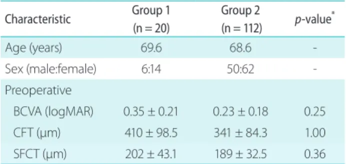
Table 1. Baseline characteristics Characteristic Group 1
(n = 20)
Group 2
(n = 112) p-value*
Age (years) 69.6 68.6 -
Sex (male:female) 6:14 50:62 -
Preoperative
BCVA (logMAR) 0.35 ± 0.21 0.23 ± 0.18 0.25
CFT (μm) 410 ± 98.5 341 ± 84.3 1.00
SFCT (μm) 202 ± 43.1 189 ± 32.5 0.36
Values are presented as mean ± standard deviation unless other- wise indicated.
BCVA = best corrected visual acuity; logMAR = logarithm of mini- mal angle of resolution; CFT = central foveal thickness; SFCT = sub- foveal choroidal thickness.
*t-test.
월, 수술 후 3개월, 수술 후 6개월에 410.5 ± 131.5 μm, 395.0 ± 107.7 μm, 395.0 ± 102.5 μm, 533.5 ± 181.5 μm, 498.5 ± 100.2 μm로 수술 후 3개월째 낭포황반부종으로 가장 두껍게 측정되 었다. 낭포황반부종이 발생하지 않은 II군과 비교하였을 때 낭 포황반부종이 발생한 수술 후 3개월에 통계적으로 유의한 차이 가 있었다(p = 0.036) (Table 2, Fig. 1).
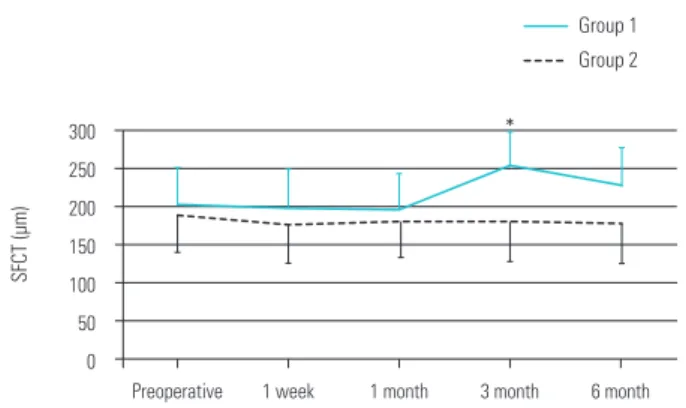
중심와 아래 맥락막두께는 I군에서 수술 전, 수술 후 1주, 수 술 후 1개월, 수술 후 3개월, 수술 후 6개월에 202.0 ± 92.5 μm, 198.5 ± 82.7 μm, 194.5 ± 87.5 μm, 253.0 ± 91.5 μm, 226.5 ± 88.2 μm로 수술 후 3개월째 낭포황반부종이 발생한 시점에 가장 두껍게 측정되었다. 낭포황반부종이 발생한 수술 후 3개월째에 중심와 아래 맥락막두께가 낭포황반부종이 발생하지 않은 II군 과 비교하였을 때 통계적으로 유의한 차이가 있었다(p = 0.036) (Table 3, Fig. 2).
최대교정시력(logMAR)은 I군에서 수술 전, 수술 후 1개월, 수 술 후 3개월, 수술 후 6개월에 0.354 ± 0.5, 0.328 ± 0.3, 0.283
± 0.5, 0.242 ± 0.3으로 수술 후 6개월까지 점차로 개선되는 양 상을 보이고 있었으며, II군에서도 최대교정시력은 수술 후 시 간이 지남에 따라 점차 호전되었다(Table 4, Fig. 3). 낭포황반부 종이 발생한 3개월째에 최대교정시력은 지속 호전을 보여, 최대
교정시력의 변화와 중심황반두께의 변화 및 중심와 아래 맥락 막두께의 변화 양상은 일치하지 않았다.
600 550 500 450 400 350 300 250 200
*
Preoperative 410.5
341
1 week 395 389.7
1 month 395 383.3
3 month 533.5 369.2
6 month 498.5 350.1
CFT (µm)
Group 2 Group 1
Figure 1. Pattern of central foveal thickness (μm) at preoperative and in the postoperative period, in both groups. CFT = central foveal thickness. *p = 0.036 at postoperative 3 month by student t-test.
0.7 0.6 0.5 0.4 0.3 0.2 0.1 0
Preoperative 0.354 0.277
1 month 0.328
0.22
3 month 0.283 0.157
6 month 0.242 0.087
BCVA (logMAR)
Group 2 Group 1
Figure 3. Pattern of mean best corrected visual acuity (BCVA logMAR) at preoperative and in the postoperative period, in both groups. BCVA = best corrected visual acuity; logMAR = logarithm of the minimum angle of resolution.
300 250 200 150 100 50 0
*
Preoperative 202 189
1 week 198.5 176.7
1 month 194.5 181.9
3 month 253 179.3
6 month 226.5 176.8
SFCT (µm)
Group 2 Group 1
Figure 2. Pattern of subfoveal choroidal thickness (μm) at preoper- ative and in the postoperative period, in both groups. SFCT = sub- foveal choroidal thickness. *p = 0.036 at postoperative 3 month by student t-test.
Table 2. Pattern of central foveal thickness (μm) at preoperative and in the postoperative period, in both groups
Group Preoperative 1 week 1 month 3 months 6 months
1 410.5 395.0 395.0 533.5 498.5
2 341.0 389.7 383.3 369.2 350.1
Table 3. Pattern of subfoveal choroidal thickness (μm) at preopera- tive and in the postoperative period, in both groups
Group Preoperative 1 week 1 month 3 months 6 months
1 202.0 198.5 194.5 253.0 226.5
2 189.0 176.7 181.9 179.3 176.8
Table 4. Pattern of mean best corrected visual acuity (BCVA logMAR) at preoperative and in the postoperative period, in both groups
Group Preoperative 1 month 3 months 6 months
1 0.354 0.328 0.283 0.242
2 0.277 0.220 0.157 0.087
BCVA = best corrected visual acuity; logMAR = logarithm of the minimum angle of resolution.
고찰
특발성 망막전막은 후유리체박리가 섬유세포 증식을 유도하여 막을 형성하는 것으로 알려져 있으며, 망막전막의 형성에의 맥 락막의 역할에 대해서는 명확히 알려진 바는 없다. 망막전막에 서 유리체절제술이 맥락막두께에 미치는 영향에 대한 연구들이 많이 진행되고 있으며, 결과는 망막전막의 형태에 따라 상이하 게 나타난다[14-18]. 백내장 수술 이후 위수정체 낭포황반부종 이 발생하였을 때 맥락막의 두께에 대한 연구도 진행되고 있으 며 Odrobina et al. [12]은 위수정체 낭포황반부종 환자에서 맥락 막두께가 대조군에 비해 얇게 측정되었다고 보고하였으며, 얇은 맥락막에서는 혈류 흐름이 감소하여 망막 저산소증을 유발하고 이로 인해 혈관내피세포성장인자(VEGF)의 농도를 올려 혈관- 망막 장벽을 파괴하여 낭포황반부종을 유발한다고 설명하였다.
Fleissig et al. [11]은 대조군에 비해 맥락막두께가 두껍게 측정 되었으며, 이는 안내 수술 후 발생하는 염증 과정에서 나타난 반응으로 낭포황반부종이 발생하며 이에 따른 맥락막혈관의 확 장으로 맥락막두께 증가를 유발한다고 설명하였고, 낭포황반부 종이 감소함에 따라 맥락막두께도 함께 감소함을 보고하였다.
본 연구는 유리체절제술 및 막제거술 단독 또는 백내장 수술 과 병합 시행한 수술 이후 발생한 낭포황반부종에서의 맥락막 두께의 변화를 알고자 하는 연구이며, Frisina et al. [13]은 유 리체절제술 단독과 백내장 수술 병합의 비교에서 수술 후 낭포 황반부종의 발생은 두 군에서 통계적으로 유의한 차이를 보이 지는 않는다고 보고하였다. 본 연구에서 I군의 경우 유리체절제 술 및 막제거술을 단독 시행한 후 낭포황반부종이 발생한 경우 는 6안으로, 대상안의 숫자가 적어 이를 비교하는 연구는 이루 어지지 못했다.
유리체절제술 및 막제거술 이후 낭포황반부종이 발생하는 원 인으로 망막전막과 내경계막을 제거할 때 발생하는 힘으로 뮬 러세포의 기계적인 손상을 유도하고, 망막내 낭종이 존재하는 경우 광수용체층 및 광수용체 내외층경계부(ellipsoid zone)가 더 쉽게 손상되어 혈관-망막 장벽이 파괴되고 결과적으로 위수 정체 낭포황반부종이 발생한다고 설명한다. 또 다른 기전으로 Irvine-Gass syndrome으로 알려진 안내 수술 후 발생하는 염 증유도인자로 인해 혈관-망막 장벽이 파괴되어 낭포황반부종을 일으킨다는 가설도 있다[13].
망막전막에 작용하는 힘은 접선 방향과 전후 방향의 견인력 이며 망막내층을 전방으로 끌어당기는 힘에 대한 반작용이 후 방으로 작용하여 맥락막두께의 증가가 발생하며, 견인력으로 인 하여 망막혈관 흐름이 변화가 생긴다면 망막에 산소와 영양을 공급하기 위해 맥락막혈관이 확장되고 맥락막이 두꺼워질 가능 성을 생각해 볼 수 있다[19]. 망막전막의 유리체절제술 및 막제 거술 이후 발생한 낭포황반부종에서의 맥락막두께에 대한 보고
는 아직 이루어지지 않고 있지만 유리체절제술 및 막제거술 시 에 발생하는 견인력과 함께 추후 동반되는 염증 반응으로 인해 혈관-망막 장벽이 파괴되어 낭포황반부종이 발생하면 기계적, 염증 과정으로 인한 반응으로 맥락막두께 또한 증가할 수 있음 을 설명할 수 있겠다.
타 연구에서 망막전막에서 유리체절제술 및 막제거술 후 낭포 황반부종이 발생하는 시점은 수술 후 3개월로 보고하고 있으며 [13], 본 연구에서는 평균 수술 후 2.89개월로 일치하는 결과를 보였다.망막전막의 수술 과정에서의 조작으로 인한 망막과 맥 락막의 혈관의 기계적 견인, 염증 반응이 동반되어 혈관-망막 장벽이 파괴됨으로 인해 맥락막의 두께가 증가하고 낭포황반부 종이 발생함을 설명할 수 있겠다.
낭포황반부종이 발생한 군(I군)에서 통계적으로 유의하지는 않지만 수술 전 중심망막두께가 더 두껍고 최대교정시력이 더 좋지 않은 결과를 보였다. 수술 전 망막전막 환자에서 낭포황반 부종 형태와 유리체황반견인 형태를 보이는 황반전막 환자에서 중심망막두께가 유의하게 증가하고 최대교정시력이 낮음을 보 여주는 연구가 있었다[14]. 본 연구에서는 I군에서 수술 전 낭포 황반부종이 5안(25%), II군에서는 10안(8.9%)이 관찰되었으며, 유리체황반견인 형태를 보인 경우는 I군에서 8안(40%), II군 12안 (10.7%)이 관찰되어 술 후 낭포황반부종을 보이는 망막전막 환 자의 경우 술 전 낭포황반부종 및 유리체황반견인 형태를 보이 는 비율이 더 높았다. 또한 이로 인해 술 전 망막중심두께와 최 대교정시력이 영향을 받았음을 설명할 수 있었으나, 대상안 수 가 적고 통계적으로 유의한 수치를 보이지는 않았으므로 추후 추가적인 연구가 필요하겠다.
본 연구에서 낭포황반부종으로 인한 중심황반두께의 증가와 최대교정시력의 경우 연관성을 보이지 않았는데, 최대교정시력 은 중심황반두께보다 낭포황반부종에서의 망막내낭종의 유무, 크기에 의해 더 영향을 받는다는 보고가 있었다[20-22]. 망막 전막 환자에서 유리체황반견인 형태를 보이는 경우 수술 후 이 것이 해소되면서 최대교정시력이 호전되고, 맥락막의 두께가 감 소함을 설명하고 있는데[14], 수술 후 낭포황반부종이 발생하고 맥락막이 두꺼워지는 기전과 수술 전 망막전막 환자의 황반부 의 형태가 실질적인 중심망막두께의 수치와 맥락막의 두께보다 더 최대교정시력에 영향을 미침을 알 수 있었다.
다만 이번 연구는 후향적 연구로써 환자군 선택에 있어서 편 견이 개입되었을 가능성이 있으며, 망막전막에서 유리체황반견 인형의 경우 맥락막두께가 증가된다는 연구가 있는데[14], 본 연 구에서는 낭포황반부종 환자 20안 중 5안이 유리체황반견인형 의 형태를 보였다. 전체 대상안의 숫자가 적어 망막전막의 형태 에 따른 유리체절제술 및 막제거술 후 낭포황반부종의 비교는 불가능하였으므로 추가적인 연구가 필요할 것으로 생각한다.
References
1. Bonnet M. Irvine-Gass syndrome. IV. Etiology. V. Pathogenesis. VI.
Treatment. Arch Ophthalmol Rev Gen Ophthalmol 1972;32:801-4.
2. Bonnet M. The Irvine-Gass syndrome. I. Clinical semiology. II. Dif- ferential diagnosis. III. Pathological anatomy. Arch Ophthalmol Rev Gen Ophthalmol 1972;32:705-20.
3. Călugăru M, Mărgescu F, Orosz R. The Irvine-Gass syndrome.
Rev Chir Oncol Radiol O R L Oftalmol Stomatol Ser Oftalmol 1982;26:221-6.
4. Egger EG. The Irvine-Gass syndrome. Ophthalmologica 1973;167:443-5.
5. François J, Verbraeken H. Complications in 1,000 consecutive in- tracapsular cataract extractions. Ophthalmologica 1980;180:121-8.
6. Gotzowa R, Boduch-Cieślińska K. Macular edema in Irvine-Gass syndrome. Klin Oczna 1975;45:1149-53.
7. Alm A. Ocular circulation. In: Hart WM, ed. Adler’s Physiol Eye.
9th ed. St. Louis: CV Mosby; 1992:198-227.
8. Barone A, Russo V, Prascina F, Delle Noci N. Short term safety and efficacy of intravitreal bevacizumab for pseudophakic cystoid edema. Retina 2009;29:33-7.
9. Spaide RF, Koizumi H, Pozzoni MC. Enhanced depth imaging spectral-domain optical coherence tomography. Am J Ophthal- mol 2008;146:496-500.
10. Margolis R, Spaide RF. A pilot study of enhanced depth imaging optical coherence tomography of the choroid in normal eyes.
Am J Ophthalmol 2009;147:811-5.
11. Fleissig E, Cohen S, Iglicki M, et al. Changes in choroidal thick- ness in clinically significant pseudophakic cystoid macular ede- ma. Retina 2018;38:1629-35.
12. Odrobina D, LaudaŃska-Olszewska I. Choroidal thickness in clin- ically significant pseudophakic cystoid macular edema. Retina 2015;35:136-40.
13. Frisina R, Pinackatt SJ, Sartore M, et al. Cystoid macular edema after pars plana vitrectomy for idiopathic epiretinal membrane.
Graefes Arch Clin Exp Ophtahlmol 2015;253:47-56.
14. Park JM, Yeom MI, Park JM. Choroidal thickness changes follow- ing vitrectomy in epiretinal membrane based on the optical coherence tomography pattern. J Korean Ophthalmol Soc 2018;59:637-49.
15. Michalewska Z, Michalewski J, Adelman RA, et al. Choroidal thickness measured with swept source optical coherence to- mography before and after vitrectomy with internal limiting membrane peeling for idiopathic epiretinal membranes. Retina 2015;35:487-91.
16. Ahn SJ, Woo SJ, Park KH. Choroidal thickness change following vitrectomy in idiopathic epiretinal membrane and macular hole.
Graefes Arch Clin Exp Ophthalmol 2016;254:1059-67.
17. Casini G, Loiudice P, Lazzeri S, et al. Analysis of choroidal thick- ness change after 25-gauge vitrectomy for idiopathic epiretinal membrane with or without phacoemulsification and intraocular lens implantation. Ophthalmologica 2017;237:78-84.
18. Kang EC, Lee KH, Koh HJ. Changes in choroidal thickness after vitrectomy for epiretinal membrane combined with vitreomac- ular traction. Acta Ophthalmol 2017;95:e393-8.
19. Kadonosono K, Itoh N, Nomura E, Ohno S. Perifoveal microcir- culation in eyes with epiretinal membranes. Br J Ophthalmol 1999;83:1329-31.
20. Michalewski J, Michalewska Z, Cisiecki S, Nawrocki J. Morpho- logically functional correlations of macular pathology connect- ed with epiretinal membrane formation in spectral optical co- herence tomography (SOCT). Graefes Arch Clin Exp Ophthalmol 2007;245;11:1623-31.
21. Brar M, Yuson R, Kozak I, et al. Correlation between morphologi- cal features on spectral-domain optical coherence tomography and angiographic leakage patterns in macular edema. Retina 2010;30:383-9.
22. Inoue M, Morita S, Watanabe Y, et al. Preoperative inner seg- ment/outer segment junction in spectral-domain optical co- herence tomography as a prognostic factor in epiretinal mem- brane surgery. Retina 2011;31:1366-72.
망막전막 환자의 유리체절제술 후 낭포황반부종의 발생에 따른 맥락막두께 비교
목적: 망막전막 환자의 유리체절제술 및 막제거술 후 발생하는 낭포황반부종의 발생 유무에 따른 맥락막두께의 변화를 비교하고자 한다.
대상과 방법: 망막전막 환자 중 유리체절제술을 및 막제거술을 받은 환자 132안 중 수술 후 낭포황반부종이 발생한 1군 20안과 낭포 황반부종이 발생하지 않은 2군 112안을 대상으로 수술 전, 수술 1주, 1개월, 3개월, 6개월 후의 중심와 아래 맥락막두께, 중심황반두 께, 최대교정시력을 각각 비교하였다.
결과: 1군 환자에서 낭포황반부종의 평균 발생 시기는 2.89개월이었으며, 중심와 아래 맥락막두께는 1군과 2군에서 수술 전, 수술 후 1주, 1개월, 6개월에서 통계적으로 유의한 차이가 없었고, 수술 후 3개월에서 1군 253.0 μm, 2군에서 179.3 μm로 통계적 유의성 (p = 0.036)이 있었다. 수술 전후에서 중심황반두께와 최대교정시력의 비교에서는 통계적으로 유의성은 없었다.
결론: 유리체절제술 및 막제거술 후 낭포황반부종이 발생한 시점에 중심와 아래 맥락막두께가 증가하였으며, 중심황반두께의 증가와 최 대교정시력과의 연관관계는 없었다.
국문초록