© 2018 The Korean Ophthalmological Society
This is an Open Access article distributed under the terms of the Creative Commons Attribution Non-Commercial License (http://creativecommons.org/licenses /by-nc/3.0/) which permits unrestricted non-commercial use, distribution, and reproduction in any medium, provided the original work is properly cited.
Original Article
Silicone oil (SO) is an effective endotamponade agent used in cases that require a long-term or permanent filling effect
for the retention of anatomical reattachment of the retina [1- 4]. It is typically used in vitreoretinal surgery for treatment of retinal detachment (RD) with proliferative vitreoretinopa- thy (PVR), giant retinal tears, severe proliferative diabetic retinopathy, macular hole, uveitis, or trauma. However, SO injection is associated with a variety of complications in the anterior and posterior segments of the eye, including forma- tion of intraconjunctival oil inclusion cysts, band keratopa- thy, cataract, glaucoma, and chronic hypotony [5-9].
Incidence and Risk Factors of Cystoid Macular Edema after Vitrectomy with Silicone Oil Tamponade for Retinal Detachment
Jong Yun Yang1,2, Hong Kyu Kim2, Soo Han Kim3, Sung Soo Kim2
1Siloam Eye Hospital, Seoul, Korea
2Department of Ophthalmology, Institute of Vision Research, Severance Hospital, Yonsei University College of Medicine, Seoul, Korea
3Department of Ophthalmology, Yonsei University Wonju College of Medicine, Wonju, Korea
Purpose: To investigate the incidence and risk factors of cystoid macular edema (CME) after silicone oil (SO) injection for retinal detachment.
Methods: Fifty-eight patients with retinal detachment treated by vitrectomy with SO tamponade during 2011 to 2015 were retrospectively assigned to CME and non-CME groups. Patients underwent complete ophthalmo- logical examination, including color fundus photography and preoperative and postoperative optical coherence tomography. Risk factors for CME during SO tamponade were determined by regression analyses.
Results: Of the 58 eyes, 21 (36.2%) exhibited CME. The presence of posterior staphyloma in the CME group was significantly more frequent than in the non-CME group (p = 0.026). There were no significant differenc- es in other demographic or clinical characteristics between the CME and non-CME groups. Significant cor- relations were observed between CME after vitrectomy with SO tamponade and the presence of posterior staphyloma (odds ratio, 4.03; p = 0.031). Of the 21 eyes with CME, 13 underwent SO removal, among which 11 experienced resolution of CME with or without further intervention.
Conclusions: The presence of posterior staphyloma is significantly associated with CME after vitrectomy with SO tamponade. Patients with retinal detachment exhibiting posterior staphyloma should be evaluated for po- tential CME during SO tamponade.
Key Words: Macular edema, Retinal detachment, Silicone oil
Received: April 23, 2017 Accepted: September 13, 2017
Corresponding Author: Sung Soo Kim, MD, PhD. Department of Ophthalmology, Institute of Vision Research, Severance Hospital, Yonsei University College of Medicine, 50 Yonsei-ro, Seodaemun-gu, Seoul 03722, Korea. Tel: 82-2-2228-3570, Fax: 82-2-312-0541, E-mail:
Macular edema results from either serous exudation from intraretinal capillaries between the outer plexiform and inner nuclear layers of the retina or swelling of the ret- inal Müller cells. Cystoid macular edema (CME) is defined as the localized expansion of the extracellular space in the macular area of the retina and is characterized by a radial cystic pattern in the perifoveal region [10]. Spectral-do- main optical coherence tomography (SD-OCT) can pro- vide high-resolution in vivo cross-sectional images of the retina even in the presence of intraocular SO [11] and can reveal histological changes in the retina. This imaging mo- dality has been used recently to establish CME as one of the retinal complications of SO tamponade.
The purpose of this study was to examine the incidence and risk factors of CME after vitrectomy with SO tampon- ade for RD.
Materials and Methods
The study protocol adhered to the tenets of the Declara- tion of Helsinki and was approved by the institutional re- view board of Severance Hospital, Seoul, Korea (4-2016- 0637). Written informed consents were obtained. Medical charts of 93 consecutive patients (93 eyes) who were fol- lowed-up after vitrectomy with SO tamponade for treat- ment of RD between November 2012 and December 2015 were reviewed. All patients had received pars plana vitrec- tomy and SO injection for complicated RD associated with uveitis, giant retinal tears, subretinal hemorrhage due to polypoidal choroidal vasculopathy, or PVR with tractional
RD. The exclusion criteria were as follows: diagnosis of tractional RD with proliferative diabetic retinopathy, diag- nosis of CME by SD-OCT prior to surgery, unknown medical or surgical history, unavailability of OCT images acquired during SO tamponade, and follow-up loss during SO tamponade.
Data on demographic and clinical characteristics includ- ing age, sex, ophthalmological and medical history, visual acuity, and intraocular pressure (IOP) were acquired from medical charts. Intraoperative data included information on the cause of RD, macular on/off status, and history of combined cataract surgery or scleral buckling based on the surgical report. Postoperative data included information on visual acuity and IOP after SO tamponade, duration of SO tamponade, postoperative complications, lens status dur- ing SO tamponade, and OCT data acquired during SO tamponade.
Best-corrected visual acuity was measured using a Snel- len chart and was converted to logarithm of the minimum angle resolution visual acuity for analysis. Diagnosis of CME was based on postoperative SD-OCT findings (Hei- delberg Spectralis SD-OCT; Heidelberg Engineering, Hei- delberg, Germany) of retinal thickening with the loss of the foveal pit with intraretinal cystoid spaces. Postopera- tive SD-OCT images were used to establish the presence or absence of posterior staphyloma, which was defined by an outpouching of the ocular wall with a curvature radius smaller than that of the surrounding ocular wall. The time of CME onset was defined as the time of its initial detec- tion on postoperative OCT images. Duration of SO tam- ponade was defined as the interval between injection of Table 1. Characteristics of patients with and without cystoid macular edema
Parameter CME group (n = 21) Non-CME group (n = 37) p-value
Mean age (yr) 51.48 ± 19.81 50.27 ± 20.01 0.826
Male / female 11 / 10 23 / 14 0.467
Affected eyes (right / left) 8 / 13 23 / 14 0.077
Hypertension 5 (23.8%) 13 (35.1%) 0.370
Diabetes mellitus 3 (14.3%) 7 (18.9%) 0.653
Duration of oil tamponade (day) 276 ± 190 273 ± 317 0.966
Preoperative mean BCVA (logMAR) 1.83 ± 1.02 1.89 ± 0.92 0.825
Preoperative mean IOP (mmHg) 13.81 ± 7.53 11.68 ± 4.34 0.175
Student’s t-test was used for all continuous variables. Chi-square test was used for all categorical variables.
CME = cystoid macular edema; BCVA = best-corrected visual acuity; logMAR = logarithm of the minimum angle resolution; IOP = in- traocular pressure.
SO and its removal or exchange. In patients who did not receive SO removal, duration of SO tamponade was de- fined as the interval between SO injection and final OCT scanning.
A single surgeon (SSK) performed all surgeries with the patient under general or local anesthesia induced by sub- tenon injection. Sclerotomies were placed 3.5 and 4.0 mm from the limbus in pseudophakic and phakic eyes, respec- tively. Following core vitrectomy, SO tamponade was per- formed after internal drainage and endolaser photocoagu- lation in eyes with rhegmatogenous RD with large retinal tears, hemorrhagic RD, or serous RD. Injection of SO was performed after the removal of the tractional membrane from the retina with tractional RD. For SO tamponade, Oxane SO (1,300 centistokes; Bausch & Lomb, Rochester, NY, USA) was used.
Comparison of categorical and distributed variables be- tween the CME and non-CME groups was performed by Student's t-test and chi-square test. Risk factors for CME
after SO tamponade were identified by univariate and multivariate conditional logistic regression analyses. We tried to gather statistically significant variables only via the Enter method of logistic regression analysis. Then, us- ing selected variables, we applied the forward condition method to eliminate insignificant variables, because the method automatically extracts variables based on the level of the statistical significance. Statistical analysis was per- formed using IBM SPSS ver. 20.0 (IBM Corp., Armonk, NY, USA). Values of p < 0.05 were considered to indicate statistical significance.
Results
Of the 93 patients (93 eyes) who received SO injections, 35 were excluded for the following reasons: diagnosis of tractional RD with proliferative diabetic retinopathy (n = 21), follow-up loss during SO tamponade (n = 9), lack of
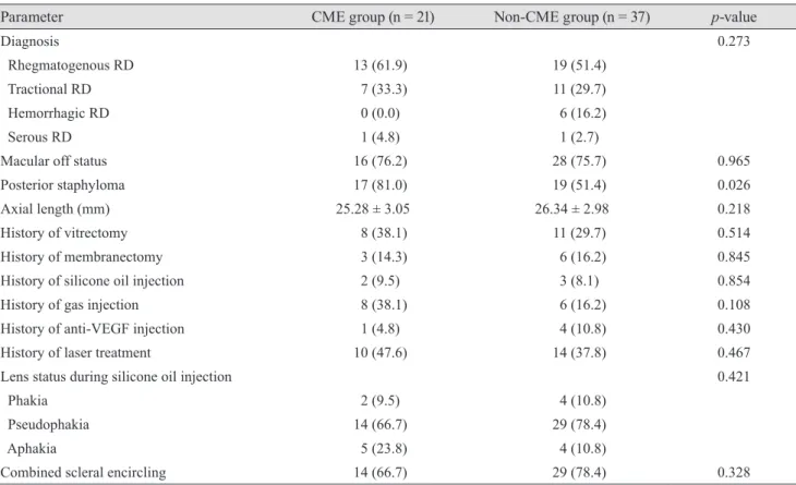
Table 2. Comparison of clinical characteristics between patients with and without cystoid macular edema
Parameter CME group (n = 21) Non-CME group (n = 37) p-value
Diagnosis 0.273
Rhegmatogenous RD 13 (61.9) 19 (51.4)
Tractional RD 7 (33.3) 11 (29.7)
Hemorrhagic RD 0 (0.0) 6 (16.2)
Serous RD 1 (4.8) 1 (2.7)
Macular off status 16 (76.2) 28 (75.7) 0.965
Posterior staphyloma 17 (81.0) 19 (51.4) 0.026
Axial length (mm) 25.28 ± 3.05 26.34 ± 2.98 0.218
History of vitrectomy 8 (38.1) 11 (29.7) 0.514
History of membranectomy 3 (14.3) 6 (16.2) 0.845
History of silicone oil injection 2 (9.5) 3 (8.1) 0.854
History of gas injection 8 (38.1) 6 (16.2) 0.108
History of anti-VEGF injection 1 (4.8) 4 (10.8) 0.430
History of laser treatment 10 (47.6) 14 (37.8) 0.467
Lens status during silicone oil injection 0.421
Phakia 2 (9.5) 4 (10.8)
Pseudophakia 14 (66.7) 29 (78.4)
Aphakia 5 (23.8) 4 (10.8)
Combined scleral encircling 14 (66.7) 29 (78.4) 0.328
Values are presented as mean ± deviation or number (%). Student’s t-test was used for all continuous variables. Chi-square test was used for all categorical variables.
CME = cystoid macular edema; RD = retinal detachment; VEGF = vascular endothelial growth factor.
OCT data acquired during SO tamponade (n = 2), uncer- tain medical history (n = 2), and diagnosis of CME by OCT before surgery for RD (n = 1). Thus, 58 eyes of 58 patients who had undergone vitrectomy with SO tampon- ade were finally enrolled.
Of the 58 eyes, 21 were diagnosed with CME by SD- OCT and assigned to the CME group. The incidence of CME after vitrectomy with SO tamponade was 36.2%.
Median patient age in the CME and non-CME groups was 51.48 and 50.27 years, respectively (p = 0.826). Median du- ration of SO tamponade in the CME and non-CME groups was 276 and 273 days, respectively (p = 0.966). There were no significant differences between the two groups in terms of sex, laterality, medical history of hypertension or diabe-
tes mellitus, preoperative best-corrected visual acuity, or IOP. Table 1 summarizes the demographic and clinical characteristics of the two groups.
Table 2 summarizes the clinical characteristics of RD and the treatment history of the two groups. The presence of posterior staphyloma in the CME group was significant- ly higher than in the non-CME group (p = 0.026). There were no significant differences between the two groups in the cause of RD, axial length, macular on/off status, surgi- cal history, history of combined surgery, or lens status during SO tamponade.
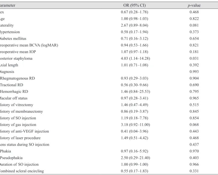
Table 3 summarizes the results of univariate logistic re- gression analyses of risk factors for CME after vitrectomy with SO tamponade. The presence of posterior staphyloma Table 3. Results of univariate logistic regression analyses of risk factors for cystoid macular edema after SO injection
Parameter OR (95% CI) p-value
Sex 0.67 (0.28–1.78) 0.468
Age 1.00 (0.98–1.03) 0.822
Laterality 2.67 (0.89–8.04) 0.081
Hypertension 0.58 (0.17–1.94) 0.373
Diabetes mellitus 0.71 (0.16–3.12) 0.654
Preoperative mean BCVA (logMAR) 0.94 (0.53–1.66) 0.821
Preoperative mean IOP 1.07 (0.97–1.18) 0.181
Posterior staphyloma 4.03 (1.14–14.28) 0.031
Axial length 1.01 (0.71–1.08) 0.392
Diagnosis 0.993
Rhegmatogenous RD 0.93 (0.29–3.03) 0.904
Tractional RD 0.56 (0.30–9.66) 0.690
Hemorrhagic RD 1.46 (0.84–25.53) 0.795
Macular off status 0.97 (0.28–3.41) 0.965
History of vitrectomy 1.46 (0.47–4.49) 0.515
History of membranectomy 0.86 (0.19–3.87) 0.845
History of SO injection 1.19 (0.18–7.78) 0.854
History of gas injection 3.18 (0.92–11.00) 0.068
History of anti-VEGF injection 0.41 (0.04–3.96) 0.443
History of laser procedure 1.49 (0.51–4.42) 0.468
Lens status during SO injection 0.437
Phakia 0.97 (0.16–5.92) 0.970
Pseudophakia 2.50 (0.29–21.40) 0.403
Duration of SO injection 1.00 (0.99–1.00) 0.966
Combined scleral encircling 0.55 (0.17–1.83) 0.331
SO = silicone oil; OR = odds ratio; CI = confidence interval; BCVA = best-corrected visual acuity; logMAR = logarithm of the minimum angle resolution; IOP = intraocular pressure; RD = retinal detachment; VEGF= vascular endothelial growth factor.
(odds ratio, 4.03 [1.14 to 14.28]; p = 0.031) was significantly associated with CME.
Of the 21 eyes in the CME group, 8 did not receive SO removal or exchange immediately after SO tamponade.
The remaining 13 eyes received SO removal, of which 11 experienced resolution of CME after the procedure (Fig. 1), while 2 did not exhibit any recovery (Fig. 2). Of the 11 eyes that recovered, 1 exhibited complete resolution of CME af- ter posterior subtenon triamcinolone injection (PSTI) and 1 after intravitreal Avastin injection; CME in the remain- ing 9 eyes subsided spontaneously without further inter- vention. The average recovery time was 70 days (range, 6 to 287 days).
Discussion
In the present study, the incidence of CME after vitrec- tomy with SO tamponade was found to be 36.2%, which is higher than that reported by previous studies (13.6% to 27.5%) [11-14]. Upon comparative evaluation of preopera- tive and postoperative SD-OCT images of 46 eyes that re- ceived vitrectomy with SO tamponade, Bae et al. [12] re- ported CME in 9 (19.6%) eyes before SO removal. Kiss et al. [13] reported the development of CME after SO tam- ponade only in eyes with RD caused by PVR (7 / 39 eyes, 17.1%). Rashad et al. [11] reported a 27.5% incidence of CME (14 of 51 eyes) using SD-OCT–based diagnosis. Kar- ahan et al. [14] reported a 13.6% incidence of CME (3 of 22 eyes) among patients with rhegmatogenous RD who re- ceived SO tamponade. In these previous studies, SO was removed at earlier time points (3.6 to 9 months postopera- tively) than in our study. It is likely that the incidence of CME was higher in this study than in other studies be- cause of a longer presence of SO. On average, CME oc- curred on postoperative day 171 in our study (not shown), while SO was removed before this time point in most pre- vious studies. In order to reduce the incidence of CME, it would be better to remove the SO as soon as possible.
In the present study, of the 13 eyes that received SO re- moval, 11 experienced resolution of CME; 2 eyes exhibited recovery after anti-vascular endothelial growth factor ther- apy or PSTI, while 9 exhibited spontaneous recovery. In the study by Bae et al. [12], 8 of 9 eyes recovered from CME within 6 months after surgery, regardless of the pro- cedure for peeling of the internal limiting membrane
Fig. 1. Rhegmatogenous retinal detachment due to retinal hole in the left eye of a 75-year-old female patient. The patient had no history of hypertension or diabetes mellitus and had received cat- aract surgery in both eyes. Preoperative visual acuity and intraoc- ular pressure were 0.02 Snellen units and 13 mmHg, respectively.
The patient received combined vitrectomy with scleral encircling and additional laser treatment. Cystoid macular edema was de- tected on spectral-domain optical coherence tomography images acquired 237 days after surgery. (A) Oil removal and posterior subtenon triamcinolone injection were performed 251 days after silicone oil injection. (B) Remission of cystoid macular edema was observed on spectral-domain optical coherence tomography images acquired 1 month later.
A
B
Fig. 2. Rhegmatogenous retinal detachment due to retinal hole in the left eye of a 23-year-old female patient. The patient had no history of hypertension or diabetes mellitus. Preoperative visual acuity and intraocular pressure were 0.05 Snellen units and 23 mmHg, respectively. The patient received combined vitrectomy with scleral encircling and additional laser treatment. Cystoid macular edema was detected on spectral-domain optical coher- ence tomography images acquired 218 days after surgery. (A) Oil removal was performed 343 days after silicone oil injection. (B) However, remaining cystoid macular edema was observed on spectral-domain optical coherence tomography images acquired 293 days after oil removal.
A
B
during SO removal. In the study by Karahan et al. [14], CME subsided in all 3 eyes within 1 month after SO re- moval. Upon evaluation of 12 eyes, Lo et al. [15] reported spontaneous resolution of CME within 9 to 12 months post-surgery in all 4 eyes with increased retinal thickness.
In these previous studies, the time of recovery from CME ranged from 1 to 12 months after SO removal without any treatment. In the present study, the majority of eyes that did not receive any treatment exhibited spontaneous reso- lution of CME within approximately 2 months. In two pa- tients with persistent CME, the final OCT evaluation was performed only 286 and 293 days after SO removal. Since complete resolution of CME requires a maximum of 12 months, these two patients are expected to recover through anti-vascular endothelial growth factor therapy or PSTI.
In this study, the presence of posterior staphyloma (odds ratio, 4.03) was determined to be a risk factor for CME.
SO tends to form a sphere due to its surface tension.
Therefore, the greater is the sphericity of the vitreous cavi- ty, the easier it would be for SO to fill the cavity complete- ly. It is likely that a retro-oil space is formed at the site of the posterior staphyloma. The development of CME has been attributed to elevated levels of inflammatory factors, such as interleukin 6, as well as growth factors in retro-SO fluid between the SO and the retina [16,17]. In our study, CME subsided spontaneously in 9 eyes; as in the previous studies, CME resolved only after SO removal without any intervention. These results support the hypothesis that SO removal helps resolve CME by redistribution of inflamma- tory factors into the vitreous cavity of the eye [18,19].
The presence of posterior staphyloma and axial length are known to be risk factors for the development of myo- pia [20]. However, in our study, the presence of posterior staphyloma was found to be a risk factor for CME devel- opment, whereas axial length was not. Staphyloma can oc- cur in eyes without long axial lengths. Curtin [20] showed that, in eyes with type I staphyloma—which is the most common type—axial length ranged from 25 to 38 mm.
Therefore, Curtin [20] pointed out that axial length is not a reliable marker to define pathologic myopia and concluded that pathologic myopia should be defined by the presence of staphyloma. Wang et al. [21] recently reported clinical features of staphylomas in eyes with axial lengths less than 26.5 mm. Thus, eye wall outpouching without long axial length should also be considered as posterior staphyloma.
Therefore, it seems reasonable to conclude that the shape
of the eyeball relative to the area of the retro-SO space is more important in the development of CME than the de- gree of myopia.
There have not been many studies regarding the risk factors of the development of macular edema related to SO tamponade status. Azzolini et al. [22] reported that macu- lar off status and longer SO permanence affect the inci- dence of ME associated with long-term SO tamponade.
Scheerlinck et al. [23] also reported that the duration of SO tamponade was a statistically significant factor related to the incidence of unexplained visual loss. In contrast, in the present study, macular on/off status and duration of SO tamponade were determined to have an insignificant effect on the incidence of ME. Further studies are needed on this subject.
This study has a few limitations, mostly because of its retrospective nature. The actual development of CME and its resolution might be faster than those determined in the present study, because OCT images were not acquired at every outpatient visit. Further prospective studies involv- ing larger cohorts are required to confirm our findings.
In conclusion, the present study indicates that the inci- dence of CME after vitrectomy with SO tamponade is as high as 36.2% in patients with RD. Additionally, the pres- ence of posterior staphyloma might be a risk factor for CME. Most eyes that underwent SO removal experienced spontaneous resolution of CME. In patients with RD with posterior staphyloma, the potential risk of CME due to SO injection should be evaluated carefully by SD-OCT during SO tamponade.
Conflict of Interest
No potential conflict of interest relevant to this article was reported.
References
1. Vitrectomy with silicone oil or sulfur hexafluoride gas in eyes with severe proliferative vitreoretinopathy: results of a randomized clinical trial. Silicone Study Report 1. Arch Ophthalmol 1992;110:770-9.
2. Vitrectomy with silicone oil or perfluoropropane gas in eyes with severe proliferative vitreoretinopathy: results of a
randomized clinical trial. Silicone Study Report 2. Arch Ophthalmol 1992;110:780-92.
3. Laqua H, Lucke K, Foerster MH. Development and current status of silicone oil surgery. Klin Monbl Augenheilkd 1988;192:277-83.
4. Riedel KG, Gabel VP, Neubauer L, et al. Intravitreal sili- cone oil injection: complications and treatment of 415 con- secutive patients. Graefes Arch Clin Exp Ophthalmol 1990;228:19-23.
5. Casswell AG, Gregor ZJ. Silicone oil removal. I. The effect on the complications of silicone oil. Br J Ophthalmol 1987;71:893-7.
6. Casswell AG, Gregor ZJ. Silicone oil removal. II. Operative and postoperative complications. Br J Ophthalmol 1987;71:898-902.
7. Franks WA, Leaver PK. Removal of silicone oil: rewards and penalties. Eye (Lond) 1991;5(Pt 3):333-7.
8. Hutton WL, Azen SP, Blumenkranz MS, et al. The effects of silicone oil removal. Silicone Study Report 6. Arch Oph- thalmol 1994;112:778-85.
9. La Heij EC, Hendrikse F, Kessels AG. Results and compli- cations of temporary silicone oil tamponade in patients with complicated retinal detachments. Retina 2001;21:107-14.
10. Colin J. The role of NSAIDs in the management of postop- erative ophthalmic inflammation. Drugs 2007;67:1291-308.
11. Rashad MA, Mohamed AA, Ahmed AI. Value of optical coherence tomography in the detection of macular patholo- gy before the removal of silicone oil. Clin Ophthalmol 2016;10:121-35.
12. Bae SH, Hwang JS, Yu HG. Comparative analysis of macu- lar microstructure by spectral-domain optical coherence tomography before and after silicone oil removal. Retina 2012;32:1874-83.
13. Kiss CG, Richter-Muksch S, Sacu S, et al. Anatomy and function of the macula after surgery for retinal detachment
complicated by proliferative vitreoretinopathy. Am J Oph- thalmol 2007;144:872-7.
14. Karahan E, Tuncer I, Zengin MO, et al. Spontaneous reso- lution of macular edema after silicone oil removal. Int J Ophthalmol 2014;7:1005-9.
15. Lo DM, Flaxel CJ, Fawzi AA. Macular effects of silicone oil tamponade: optical coherence tomography findings during and after silicone oil removal. Curr Eye Res 2017;
42:98-103.
16. Christensen UC, la Cour M. Visual loss after use of intra- ocular silicone oil associated with thinning of inner retinal layers. Acta Ophthalmol 2012;90:733-7.
17. Wolf S, Schon V, Meier P, Wiedemann P. Silicone oil- RMN3 mixture ("heavy silicone oil") as internal tampon- ade for complicated retinal detachment. Retina 2003;23:
335-42.
18. Asaria RH, Kon CH, Bunce C, et al. Silicone oil concen- trates fibrogenic growth factors in the retro-oil fluid. Br J Ophthalmol 2004;88:1439-42.
19. Funatsu H, Noma H, Mimura T, et al. Association of vitre- ous inflammatory factors with diabetic macular edema.
Ophthalmology 2009;116:73-9.
20. Curtin BJ. The posterior staphyloma of pathologic myopia.
Trans Am Ophthalmol Soc 1977;75:67-86.
21. Wang NK, Wu YM, Wang JP, et al. Clinical characteristics of posterior staphylomas in myopic eyes with axial length shorter than 26.5 millimeters. Am J Ophthalmol 2016; 162:
180-90.
22. Azzolini C, Donati S, Caprani SM, et al. Macular edema and silicone oil tamponade. J Clin Exp Ophthalmol 2014;
5:366.
23. Scheerlinck LM, Schellekens PA, Liem AT, et al. Incidence, risk factors, and clinical characteristics of unexplained vi- sual loss after intraocular silicone oil for macula-on retinal detachment. Retina 2016;36:342-50.