Endobronchial Metastases from Extrathoracic Malignancy
Sang Hoon Lee,1 Ji Ye Jung,1 Do Hoon Kim,2 Sang Kook Lee,1 Song Yee Kim,1 Eun Young Kim,1 Young Ae Kang,1 Moo Suk Park,1 Young Sam Kim,1 Joon Chang,1 and Se Kyu Kim1
1Division of Pulmonology, Department of Internal Medicine, Yonsei University College of Medicine, Seoul;
2Department of Internal Medicine, Bundang Jesaeng General Hospital, Seongnam, Korea.
Received: February 20, 2012 Revised: June 7, 2012 Accepted: June 15, 2012
Corresponding author: Dr. Se Kyu Kim, Division of Pulmonology,
Department of Internal Medicine, Yonsei University College of Medicine, 50 Yonsei-ro, Seodaemun-gu, Seoul 120-752, Korea.
Tel: 82-2-2228-1954, Fax: 82-2-393-6884 E-mail: [email protected]
∙ The authors have no financial conflicts of interest.
© Copyright:
Yonsei University College of Medicine 2013 This is an Open Access article distributed under the terms of the Creative Commons Attribution Non- Commercial License (http://creativecommons.org/
licenses/by-nc/3.0) which permits unrestricted non- commercial use, distribution, and reproduction in any medium, provided the original work is properly cited.
Purpose: Endobronchial metastasis is defined as documented extrathoracic malig- nancies metastatic to the endobronchus within a bronchoscopically visible range.
Although the clinical and radiologic findings of endobronchial metastasis are simi- lar to primary lung cancer, treatment and prognosis may be different. We hereby investigated the clinical, radiologic and bronchoscopic aspects of endobronchial metastases (EBM) in Korean patients. Materials and Methods: A total of 43 pa- tients with EBM who underwent bronchoscopic biopsies from June 1991 to De- cember 2009 at Severance Hospital, Yonsei University College of Medicine in Seoul, Korea, were analyzed retrospectively. We evaluated clinical, radiologic and bronchoscopic characteristics of EBM. Results: The patients consisted of 27 males and 16 females and their ages ranged from 18 to 77 years. The common pri- mary cancers related to EBM were rectal (16.3%), colon (11.6%), breast (9.3%) and uterine (9.3%) cancers. The mean interval from diagnosis of primary cancer to EBM was 36 months, and the mean survival duration from diagnosis of EBM was 16.1 months in 33 deceased patients. Conclusion: EBM develop in various types of malignancies at various times with unremarkable manifestations. Therefore, physicians should consider the possibility of EBM, especially if a patient has a his- tory of any malignancy, regardless of respiratory symptoms. Respiratory symp- toms related with EBM can be treated by various safe procedures.
Key Words: Endobronchial, metastasis, bronchoscopy
INTRODUCTION
Endobronchial lesions have many different histological causes, the most frequent being bronchogenic small cell carcinoma, well-differentiated neuroendocrine car- cinoma, bronchogenic squamous cell carcinoma and non-small cell carcinoma.1 Only 1.1% of endobronchial tumors are metastatic.2 However, the incidence of en- dobronchial metastases (EBM) differs according to the status of disease, patient group, and study program used.3-5 According to the study by Oshikawa, et al.,4 the incidence of EBM was reported as 15 cases (23%) out of 65 patients with meta- static pulmonary disease. In another study by Braman and Whitcomb,3 EBM were readily visible bronchoscopically in the main bronchus and lobar bronchus, and
We retrospectively reviewed the medical records of all 43 patients and collected data on baseline characteristics, his- topathological results, the interval of time from the primary tumor diagnosis to EBM diagnosis, clinical symptoms, ra- diologic findings, including chest X-ray and computed to- mography, developmental modes, treatment modalities, and survival time.
A flexible bronchoscopy was performed by a pulmonolo- gist under sedation for each patient. Histopathologic diag- nosis was confirmed in all cases by direct biopsy with bron- choscopy. EBM were defined as bronchoscopically visible involvement of the subsegmental or more proximal central bronchus with histologically verified extrathoracic primary malignancy. Primary lung cancer, esophageal cancer and lymphoma were excluded in this study because these can- cers can invade the bronchus directly without metastasis.
The developmental mode of EBM was classified into four groups, according to Kiryu, et al.,18 using chest images, bronchoscopic findings, and histology, as follows: type I, direct metastasis to the bronchus; type II, endobronchial in- vasion of parenchymal mass; type III, endobronchial inva- sion of mediastinal or hilar lymphadenopathy; and type IV, extension of peripheral tumor along the proximal bronchus.
This study protocol was approved by the Institutional Re- view Board of Severance Hospital.
RESULTS
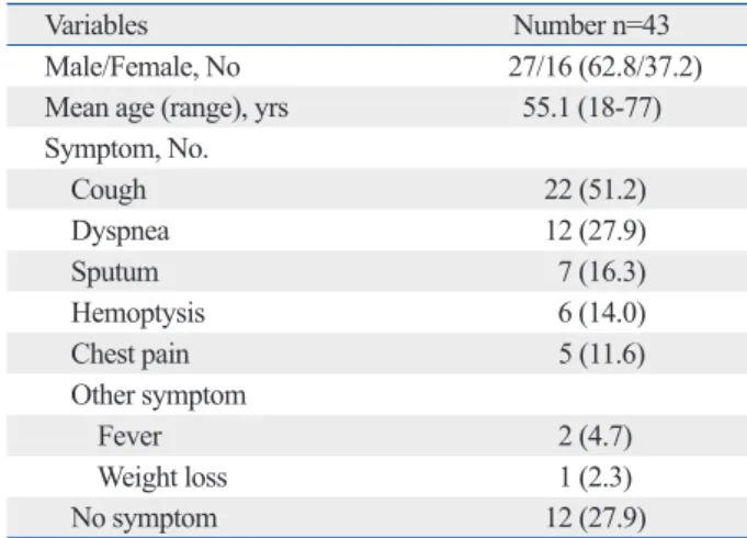
Patient characteristics and presenting symptoms Patient characteristics and presenting symptoms are sum- marized in Table 1. The 43 patients consisted of 27 males and 16 females. Their ages ranged from 18 to 77, with a mean age of 55.1 years. The most common presenting symp- toms were cough in 22 (51.2%) patients and dyspnea in 12 (27.9%) patients. Twelve patients (27.9%) with EBM were asymptomatic. Nine patients were detected by routine fol- low-up examination, two patients while evaluating other symptoms and one patient by medical check-up.
Clinical findings of patients with endobronchial metastases
Clinical findings of patients with endobronchial metastases are shown in Table 2. Common primary cancers related with EMB were rectal (7 patients, 16.3%), colon (5 pa- tients, 11.6%), breast (4 patients, 9.3%) and uterine (4 pa- tients, 9.3%) cancers. The others included stomach, thyroid, the prevalence of EBM was 2%.
From a histopathologic standpoint, EBM are observed in various types of malignancies including colorectal, breast, kidney, stomach, ovarian, thyroid, uterine, testicular, naso- pharynx, prostate, adrenal carcinomas, sarcomas, histocyto- ma and plasmacytomas.6-10 In addition, many other benign etiologic tumors have been reported, such as fungal disease, inflammatory pseudopolyp, lipoma and broncholith.11 The most common EBM are colorectal, breast and kidney carci- nomas.12,13 Although there are many underlying causes, most have similar clinical presentations such as cough, dys- pnea and sputum.
Metastases from non-pulmonary malignancies to the lungs are very common, but EBM from extrathoracic ma- lignancy are rare. Moreover, it is difficult to distinguish bronchogenic carcinoma from metastasis of extrathoracic malignancies. However, the treatment modality of EBM is determined by the histologic features of the primary tumor, biologic behavior, anatomic location, evidence of other metastatic sites, present symptoms, patient performance status and life expectancy. Therefore, the accurate diagnosis of EMB is very important for decisions regarding treatment modality.14 Only a small number of cases have been report- ed in Korea.15-17 The objective of this study was to investi- gate the clinical, radiologic and bronchoscopic aspects of EMB in Korean patients.
MATERIALS AND METHODS
We retrospectively reviewed all fibrobronchoscopic reports from June 1991 to December 2009 at Severance Hospital, Yonsei University College of Medicine, in Seoul, Korea and identified 43 patients who were diagnosed with EMB.
Table 1. Patient Characteristics
Variables Number n=43
Male/Female, No 27/16 (62.8/37.2) Mean age (range), yrs 55.1 (18-77) Symptom, No.
Cough 22 (51.2)
Dyspnea 12 (27.9)
Sputum 7 (16.3)
Hemoptysis 6 (14.0)
Chest pain 5 (11.6)
Other symptom
Fever 2 (4.7)
Weight loss 1 (2.3)
No symptom 12 (27.9)
deceased patients.
Location of endobronchial metastases
There were 50 bronchoscopically visible endobronchial le- sions, and their locations are listed in Table 3. EMB were observed at the left bronchus in 21 patients (48.8%) and at the right bronchus in 28 patients (65.1%). One colon cancer patient had three bronchoscopically visible endobronchial hepatoma, melanoma, nasopharynx, osteosarcoma, pros-
tate, kidney, urothelial, fibrosarcoma originating from left calf, salivary gland, parotid gland, cervix, and malignant fi- brous histocytoma originating from the left thigh. The pri- mary tumor site could not be detected in one of the patients with melanoma. Thirty-six patients (83.7%) had a previous history of extrathoracic malignancy. In seven patients, EBM were diagnosed at the same time as diagnosis of the following primary tumors: melanoma (2), prostate (2), pa- rotid gland (1), salivary gland (1) and stomach (1) cancers.
The interval time from diagnosis of the primary tumor to the diagnosis of EBM was between 0 to 160 months, with a median period of 36 months. The time interval between detection of the primary tumors and EMB was relatively long for thyroid (156 months), breast (111 months) and rectal (96 months) cancers. The treatment modalities were surgery in nine, chemotherapy in 17, radiotherapy in 10, chemotherapy combined with radiotherapy in one, and supportive care in 11 patients. Two patients received che- motherapy and surgery. The mean survival time from diag- nosis of endobronchial metastasis was 16.1 months in 33
Table 2. Clinical Findings in 43 Patients with Endobronchial Metastases Diagnosed by Bronchoscopy with Biopsy Primary site No. of
patients Time*
(median), months Treatment Survival†,
(median), months
Surgery CTx RTx Others SC
Rectum 7 17-156 (96.0) 3 3 1 1‡ - 3-24 (9.5)
Colon 5 1-67 (24.0) 2 1 - - 2 3-59 (14.0)
Breast 4 34-132 (111.0) - 1 1 - 2 13-57 (16.5)
Uterine 4 17-40 (31.0) - - 2 - 2 5-10 (8.3)
Stomach 3 0-60 (41.0) - 3 1 - - 4-18 (8)
Thyroid 3 18-160 (156.0) - - 2 1§ - 49
Hepatoma 2 69-95 (82.0) 1 2 1 - - 14-22 (18)
Melanoma 2 0 - 1 - - 1 2-34 (18)
Nasopharynx 2 3-38 (20.5) - 1 1 - 1 3-6 (4.5)
Osteosarcoma 2 22-26 (24.0) 1 1 - - 1 31
Prostate 2 0 - 2 - - - 5
Kidney 1 65 - 1 - - - -||
Urothelial 1 19 - - 1 - - 4
Fibrosarcoma 1 84 1 - - - - 14
Salivary 1 0 - - - - 1 13
Parotid 1 0 - 1 - - - 33
Cervix 1 23 - - - - 1 0
Histocytoma 1 28 1 - - - - 15
Total 43 2187 (36.0) 9 17 10 2 11 0-59 (16.1)
RTx, radiotherapy; CTx, chemotherapy; SC, supportive care.
*Interval between diagnosis of primary tumor and endobronchial metastasis.
†Survival time from diagnosis of endobronchial metastasis.
‡Photodynamic therapy.
§Radioactive iodine therapy.
||Survival is unknown.
Table 3. Location of Endobronchial Metastases Diagnosed by Bronchoscopy with Biopsy
Location
No. of patients Bronchus
Left Right
Trachea 1 - -
Multiple sites 6 - -
Main bronchus - 9 6
Upper lobe bronchus - 4 8
Middle lobe bronchus - - 6
Lingular - 1 -
Lower lobe bronchus - 7 8
Total 7 21 28
lesions. One melanoma, two prostate cancer, one rectal can- cer, and one stomach cancer patient had two visible endo- bronchial lesions.
Radiologic findings of endobronchial metastases The radiologic findings of 50 EMB are summarized in Ta- ble 4. Six patients had multiple lesions. Hilar mass (15 pa- tients, 34.9%) was the most frequent finding on chest radi- ography, followed by visible tumors (11 patients, 25.6%), atelectasis (10 patients, 23.3%) and multiple nodules (10 Table 4. Radiologic Findings in 43 Patients with Endobron-
chial Metastasis Diagnosed by Bronchoscopy with Biopsy Radiologic findings No. of patients (%)
Hilar mass 15 (34.9)
Visible tumors 11 (25.6)
Atelectasis 10 (23.3)
Multiple nodules 10 (23.3)
Normal 4 (9.3)
Pleural fluid 3 (7.0)
Mediastinal mass 2 (4.7)
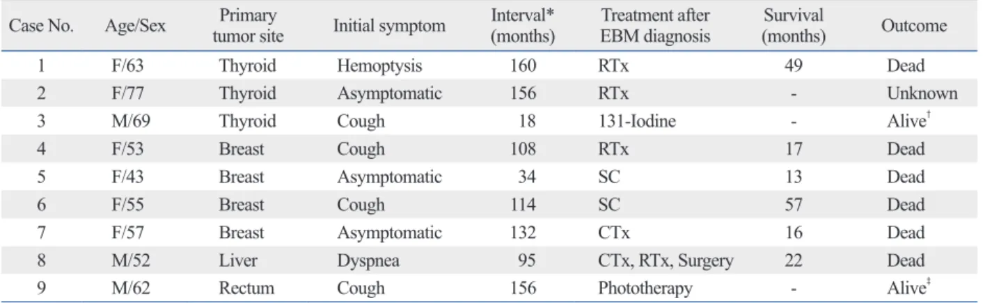
Table 5. Treatment and Outcome after EBM Diagnosis Case No. Age/Sex Primary
tumor site Initial symptom Interval*
(months) Treatment after
EBM diagnosis Survival
(months) Outcome
1 F/63 Thyroid Hemoptysis 160 RTx 49 Dead
2 F/77 Thyroid Asymptomatic 156 RTx - Unknown
3 M/69 Thyroid Cough 18 131-Iodine - Alive†
4 F/53 Breast Cough 108 RTx 17 Dead
5 F/43 Breast Asymptomatic 34 SC 13 Dead
6 F/55 Breast Cough 114 SC 57 Dead
7 F/57 Breast Asymptomatic 132 CTx 16 Dead
8 M/52 Liver Dyspnea 95 CTx, RTx, Surgery 22 Dead
9 M/62 Rectum Cough 156 Phototherapy - Alive‡
RTx, radiotherapy; CTx, chemotherapy; SC, supportive care; EBM, endobronchial metastases.
*Interval between diagnosis of primary tumor and endobronchial metastasis.
†The patient was still alive at 2 months.
‡The patient was still alive at 6 months.
Table 6. Classification of Developmental Modes of Endobronchial Metastases and Metastases to Other Organs Primary site No. of
patients
Developmental mode of EBM (type) Metastases to other organ
I II III IV Paren-
chyme Pleura Brain Liver Bone Other
Rectum 7 4 3 - - 2 - - - - -
Colon 5 2 - - 3 3 - 1 1 1 -
Breast 4 - 2 1 1 2 - - - 2 -
Uterus 4 1 1 1 1 1 1 - 1 3 -
Stomach 3 - - 3 - - - - - 2 Ovary
Thyroid 3 2 1 - - 2 1 - - - -
Hepatoma 2 1 1 - - 1 - - - 1 -
Melanoma 2 1 - - 1 - - - 1 - -
Nasopharynx 2 1 - - 1 1 - - 1 - -
Osteosarcoma 2 1 1 - - - - - - - Kidney
Prostate 2 - 1 - 1 1 - - - - -
Kidney 1 - - 1 - 1 - - - - -
Urothelial 1 - 1 - - 1 - - - - -
Fibrosarcoma 1 1 - - - 1 - - - - -
Salivary 1 1 - - - - - - - - -
Parotid 1 1 - - - 1 - - - - Elbow
Cervix 1 - 1 - - 1 - - - - -
Histocytoma 1 - - - 1 - 1 - - - -
Total 43 16 12 6 9 18 3 1 4 9 3
EBM, endobronchial metastases.
important to select treatment modalities for individual pa- tients. However, EBM are probably underestimated, be- cause bronchoscopy is not used routinely in patients with malignancy history. Staining bronchoscopy, macroscopic bronchoscopy or auto-fluorescence bronchoscopy can be used to detect less invasive endobronchial tumors early, thus allowing earlier treatment and a better prognosis. Moreover, electronic noses are also available in early detection of bronchogenic carcinoma.19 In many studies, EBM are clini- cally, radiologically and bronchoscopically indistinguish- able from bronchogenic carcinoma in most cases. They should be confirmed by histological analysis and the patho- logic comparison of EBM and the primary site is important.
Rosenblatt, et al.20 showed that the distinct features of meta- static bronchial lesions in the early stage included an intact epithelium covering the tumor mass with subepithelial lym- phatics. Clear cell renal cancer is easily diagnosed by im- munohistochemistry using markers such as CK7, CK20, thyroid transcription factor-1 and apoprotein-A1.21-23 These molecular biologic tools are very useful to distinguish ex- trathoracic lung metastasis from primary lung cancer.
The interval between diagnosis of primary cancer and de- tection of EBM is reported to be about 50 months and our study revealed an average interval of 36 months.13 This peri- od was long compared to the average intervals for metasta- sis to other organs in these cancers.24 This might mean that EBM frequently occur in “well-controlled” primary cancer.
However, there is no obvious explanation for this phenome- non. As our study demonstrated, EBM may not indicate a poor prognosis and should not be thought a bad prognostic factor in choosing treatment modalities. Akoglu, et al. showed that mean survival time was longer in patients with chemo- therapy or radiotherapy than in patients with supportive care.14 After diagnosis of EBM, the median survival dura- tion was 16.1 months in our study, similar to the 15.5 months found by Kiryu, et al.18 Larger studies may be required to evaluate molecular markers for genes that would explain the characteristics of these time intervals.
EBM are known to manifest late in the course of cancer progression. However, there are cases of lesions being diag- nosed at the same time as primary tumors.25 In our study, EBM and primary cancer were diagnosed simultaneously in seven patients with melanoma, prostate, parotid, salivary and stomach cancers. The most common metastatic sites of melanoma are lung and liver, and EBM were found at the same time as diagnosis of all melanomas in this study.26 Moreover, prostate cancer is commonly diagnosed simulta- patients, 23.3%). Four patients (9.3%) showed normal ra-
diologic findings and three of them had rectal cancer.
Treatment and outcome after EBM diagnosis in long- term survivors
Mean survival time after EBM diagnosis in long-term sur- vivors who were treated was 17.4 months, whereas that of patients given supportive care was 12.4 months. Treatment and outcome after EBM diagnosis in some long-term sur- viors are shown in Table 5. EBM related with thyroid, breast or rectal cancer appear less aggressive than in other malig- nancies. One of the thyroid cancer patients who received a total thyroidectomy due to recurrence six years after initial partial thyroidectomy showed re-recurrence as EBM after an additional seven-year period. One patient with hepato- cellular carcinoma received chemotherapy, radiotherapy and surgery, and he lived for more than ten years after these multidisciplinary treatments. One patient with rectal cancer received photodynamic therapy and one patient with thy- roid cancer received radioactive iodine therapy.
Classification of developmental modes of endobronchial metastases and metastases to other organs
Classifications of the developmental modes of endobron- chial metastases and the involvement of other organs are shown in Table 6. Patients were divided into four groups ac- cording to the classification of developmental modes: type I, 16 patients (37.2%); type II, 12 patients (27.9%); type III, 6 patients (14.0%); and type IV, 9 patients (20.9%). In 13 pa- tients, extrathoracic metastases were present at the same time as EBM diagnosis. The most common extrathoracic metastatic site was bone.
DISCUSSION
In this study, we analyzed the clinical, radiologic and bron- choscopic aspects of EBM in Korean patients. EBM were most frequently detected in colorectal, breast and uterine cancers and the main bronchus was the most common site of the primary tumor. Cough and a hilar mass on radiogra- phy were the most frequent clinical manifestations.
EBM from an extrathoracic primary tumor are very rare.13 When an endobronchial mass is detected, it is impor- tant to distinguish primary lung cancer from lung metasta- sis of extrathoracic primary tumors. Primary lung cancer and EBM have different prognoses, so diagnosis is very
astatic spread is needed.
The present study had several limitations. First, the types of primary tumors with EBM varied in this study, so overall prognosis would not represent the characteristics of each malignancy. Second, there were many improvements in the diagnosis and treatment of malignancies during this study period between 1991 and 2009. These improvements would influence the disease progression even in the same type of malignancy.
In conclusion, this study suggests that EBM develop in various types of malignancies at various times. However, the incidence of EBM has been underestimated because of its unremarkable manifestations. Some patients with EBM show no clinical symptoms or normal image findings. How- ever, EBM should be distinguished from primary lung can- cer through histological confirmation, and bronchoscopy in these asymptomatic or radiologically free patients with ma- lignancy is useful. Moreover, respiratory symptoms of EBM may be treated by various safe procedures, and in some cas- es, intrabronchial therapy may prolong patient survival.7 Therefore, physicians should consider the possibility of EBM when they encounter patients with colorectal or breast cancer, especially when respiratory symptoms are present.
REFERENCES
1. Salud A, Porcel JM, Rovirosa A, Bellmunt J. Endobronchial meta- static disease: analysis of 32 cases. J Surg Oncol 1996;62:249-52.
2. Kreisman H, Wolkove N, Finkelstein HS, Cohen C, Margolese R, Frank H. Breast cancer and thoracic metastases: review of 119 pa- tients. Thorax 1983;38:175-9.
3. Braman SS, Whitcomb ME. Endobronchial metastasis. Arch In- tern Med 1975;135:543-7.
4. Oshikawa K, Ohno S, Ishii Y, Kitamura S. Evaluation of broncho- scopic findings in patients with metastatic pulmonary tumor. In- tern Med 1998;37:349-53.
5. Shepherd MP. Endobronchial metastatic disease. Thorax 1982;
37:362-5.
6. Amer E, Guy J, Vaze B. Endobronchial metastasis from renal ade- nocarcinoma simulating a foreign body. Thorax 1981;36:183-4.
7. Fournel C, Bertoletti L, Nguyen B, Vergnon JM. Endobronchial metastases from colorectal cancers: natural history and role of in- terventional bronchoscopy. Respiration 2009;77:63-9.
8. Gallivan GJ, Emery RW. Endobronchial metastasis from cancer of the breast. Chest 1978;74:320.
9. Hanyu T, Kanda T, Matsuki A, Hasegawa G, Yajima K, Tsuchida M, et al. Endobronchial metastasis from adenocarcinoma of gas- tric cardia 7 years after potentially curable resection. World J Gas- trointest Surg 2010;2:270-4.
10. Shen Q, Yao Y, Teng X, Zhou J. Endobronchial metastasis from prostate cancer mimicking primary lung cancer. Intern Med 2010;
49:1613-5.
neously with other organ metastasis including EBM be- cause it is relatively indolent and slow-growing compared to other malignancies. Breast cancer and colorectal cancer are the most frequently reported endobronchial metastatic cancers, probably because they have high incidences and good prognoses, compared to other malignancies. Head and neck cancers such as parotid and salivary gland cancers were also found to frequently cause EBM in this study.
Therefore, although physicians should suspect the possibili- ty of EBM in all oncologic patients, more attention should be paid to patients with breast cancer, colorectal cancer and kidney cancer. It remains unclear why EBM are more fre- quently detected in these tumors.
Several studies reported that asymptomatic patients range from 20 to 62.5%.13,18,27 In our study, there were 12 patients (27.9%) who were asymptomatic. This result suggests that physicians should consider the possibility of EBM when they encounter patients previously diagnosed with cancer and with manifested respiratory symptoms. Moreover, reg- ular follow-up is important, even in patients without any re- spiratory symptoms.
Atelectasis and visible tumors with hilar masses are com- mon manifestations observed in the chest radiographs of patients with EBM.13,28 In our study, hilar mass, visible tu- mor and atelectasis were common findings, but 9.3% of pa- tients had normal chest X-rays. Therefore, physicians need to pay attention to the overall symptoms of patients with a history of malignancy and keep the possibility of endobron- chial metastasis in mind.
Most of our treatments of EBM were limited to conven- tional anti-cancer therapy such as chemotherapy, radiothera- py, and surgery; however, there are various treatment meth- ods to relieve dyspnea, hemoptysis or obstructive pneumonia caused by endobronchial metastasis. Intrabronchial therapy such as stent insertion, brachytherapy, and photodynamic therapy, or laser evaporation have been performed.13 These procedures are safe and effective as palliative treatment and can prolong survival in selected patients.29 One patient with rectal cancer and EBM was treated with phototherapy and showed good prognosis in the aspect of survival duration and symptom control.
Kiryu, et al.18 divided EBM patients into mode types of metastasis according to four developmental conditions and type IV was the most common condition. However, in our study, type I was most common, probably because it is dif- ficult to differentiate type II and IV.14 Our experience sug- gests that a more reliable classification of the mode of met-
nosis of bronchogenic carcinoma. Dis Chest 1967;51:587-95.
21. Kummar S, Fogarasi M, Canova A, Mota A, Ciesielski T. Cyto- keratin 7 and 20 staining for the diagnosis of lung and colorectal adenocarcinoma. Br J Cancer 2002;86:1884-7.
22. Srodon M, Westra WH. Immunohistochemical staining for thyroid transcription factor-1: a helpful aid in discerning primary site of tumor origin in patients with brain metastases. Hum Pathol 2002;
33:642-5.
23. Wilson RW, Moran CA. Primary melanoma of the lung: a clinico- pathologic and immunohistochemical study of eight cases. Am J Surg Pathol 1997;21:1196-202.
24. Coriat R, Diaz O, de la Fouchardière C, Desseigne F, Négrier S.
Endobronchial metastases from colorectal adenocarcinomas: clini- cal and endoscopic characteristics and patient prognosis. Oncolo- gy 2007;73:395-400.
25. Dursun AB, Demirag F, Bayiz H, Sertkaya D. Endobronchial me- tastases: a clinicopathological analysis. Respirology 2005;10:510-4.
26. Chen JT, Dahmash NS, Ravin CE, Heaston DK, Putman CE, Sei- gler HF, et al. Metastatic melanoma in the thorax: report of 130 patients. AJR Am J Roentgenol 1981;137:293-8.
27. Heitmiller RF, Marasco WJ, Hruban RH, Marsh BR. Endobron- chial metastasis. J Thorac Cardiovasc Surg 1993;106:537-42.
28. Ikezoe J, Johkoh T, Takeuchi N, Ishida T, Morimoto S, Kitamura I, et al. CT findings of endobronchial metastasis. Acta Radiol 1991;
32:455-60.
29. Carlin BW, Harrell JH 2nd, Olson LK, Moser KM. Endobronchial metastases due to colorectal carcinoma. Chest 1989;96:1110-4.
11. Magro CM, Ross P Jr. Endobronchial mimics of primary endo- bronchial carcinoma: a clinical study of 25 cases. Can Respir J 2005;12:123-7.
12. Berg HK, Petrelli NJ, Herrera L, Lopez C, Mittelman A. Endo- bronchial metastasis from colorectal carcinoma. Dis Colon Rec- tum 1984;27:745-8.
13. Sørensen JB. Endobronchial metastases from extrapulmonary sol- id tumors. Acta Oncol 2004;43:73-9.
14. Akoglu S, Uçan ES, Celik G, Sener G, Sevinç C, Kilinç O, et al.
Endobronchial metastases from extrathoracic malignancies. Clin Exp Metastasis 2005;22:587-91.
15. Kim YS, Chang J, Kim YS, Shin DH, Kim HS, Kim SK, et al.
Endobronchial metastasis of uterine cervix cancer: a two case re- ports and a review of the literature. Yonsei Med J 2002;43:547-52.
16. Lee KY, Ryu SJ, Joo M. Endobronchial metastasis of hepatocellu- lar carcinoma. Yonsei Med J 2003;44:544-7.
17. Park YB, Byun YS, Kim SK, Yang DG, Chang J, Kim JH, et al.
Endobronchial metastasis from stomach cancer. Respirology 1999;4:89-92.
18. Kiryu T, Hoshi H, Matsui E, Iwata H, Kokubo M, Shimokawa K, et al. Endotracheal/endobronchial metastases: clinicopathologic study with special reference to developmental modes. Chest 2001;119:768-75.
19. Machado RF, Laskowski D, Deffenderfer O, Burch T, Zheng S, Mazzone PJ, et al. Detection of lung cancer by sensor array analyses of exhaled breath. Am J Respir Crit Care Med 2005;171:1286-91.
20. Rosenblatt MB, Lisa JR, Collier F. Criteria for the histologic diag-