Mouse Single Oral Dose Toxicity Test of Lactobacillus-fermented Araliae Continentalis Radix Aqueous Extracts (fACR)
Young-Mi Jung1†, Sae-Kwang Ku2†, Dong Sub Lee3 and Kisang Kwon4*
1Department of Genetic Engineering, College of Natural Sciences, Kyungpook National University, Daegu 41566, Korea
2College of Oriental Medicine, Daegu Haany University, Gyeongsan 38610, Korea
3Department of Health Care & Biotechnology, Kyungwoon University, Gumi 39160, Korea
4Department of Biomedical Laboratory Science, Kyungwoon University, Gumi 39160, Korea
Received December 24, 2015 /Revised January 19, 2016 /Accepted January 29, 2016
The objective of this study was to obtain acute (single) oral dose toxicity information on Lactobacillus- fermented Araliae Continentalis Radix aqueous extracts (fACR) in female and male ICR mice, as com- pared with Araliae Continentalis Radix aqueous extracts (ACR). After administering a single oral dose of fACR, no treatment-related mortalities were observed within 14 days after the end of treatment up to 2,000 mg/kg, the maximum dosage for rodents of both sexes; moreover, no fACR treatment-related changes in the body and organ weights, clinical signs, necropsy, and histopathological findings were detected in this experiment. In addition, no ACR 2,000 mg/kg treatment-related mortalities, clinical signs, body and organ weights, or gross and histopathological findings were observed, as compared with equal genders of vehicle control. The results obtained in this study suggest that fACR is non-tox- ic in mice and is, therefore, likely to be safe for clinical use. The LD50 and approximate LD in female mice and male mice, respectively, were considered after a single oral dose of fACR over 2,000 mg/kg, the maximum dosage for rodents. In addition, no specific targets or clinical signs were detected in the present study. ACR 2,000 mg/kg-treated mice also did not show any treatment-related mortalities, clinical signs, changes to body and organ weights, or gross and histopathological findings, as com- pared with equal genders of vehicle control.
Key words : Histopathology, Lactobacillus fermented Araliae Continentalis Radix aqueous extracts (fACR), mouse, single oral dose toxicity
†Authors contributed equally.
*Corresponding author
*Tel : +82-54-479-1284, Fax : +82-54-479-1282
*E-mail : [email protected]
This is an Open-Access article distributed under the terms of the Creative Commons Attribution Non-Commercial License (http://creativecommons.org/licenses/by-nc/3.0) which permits unrestricted non-commercial use, distribution, and reproduction in any medium, provided the original work is properly cited.
Journal of Life Science 2016 Vol. 26. No. 2. 204~211 DOI : http://dx.doi.org/10.5352/JLS.2016.26.2.204
Introduction
Natural products are gaining space and importance in the pharmaceutical industry as well as inspiring the search for new potential sources of bioactive molecules [4, 29]. Herbs, medicinal plants and crude drug substances are considered to be a potential source of antioxidants to combat various diseases [22]. As increase of the concern in the functional food and well being in life, the demands and consumption of functional food originated from natural sources are in- creased [17]. However, the toxicological aspects about these natural origin-functional foods have been neglected because of the reasons that they have been used as various purposes
for long times [28]. Therefore, it is considered that more de- tail and systemic toxicological studies should be tested for control the abuse and potential toxicities even if they have been used as traditional folk medicine [26].
Araliae Continentalis Radix is a dried root parts of Aralia continentalis, a perennial plant and a member of the Araliaceae family. It is cultivated extensively in Asia, Siberia, China, and Korea [11] and has a long tradition of use in Korean traditional medicine to reduce pain and inflamma- tion [25, 20]. In addition, various pharmacological effects of Araliae Orientalis Radix have been reported through in vivo experiments as inhibiting effects against the cariogenic prop- erties of Streptococcus mutans [11], anticancer properties [23], hepatoprotectiv [8] and antibacterial [10] activities.
In recent years, fermented herbs have highlighted as a new source of medicinal ingredient or pharmaceutics, be- cause bioactivity of natural herbs increased by various fer- mentation through biotransformation or probiotic effects [1, 14, 15, 32]. Moreover, fermentation of herbs using a variety of edible microorganisms seems to further enhance the phar-
macological efficacy of parent herbs [12, 15, 32]. However, these are also indicated that systemic toxicological evalua- tions of fermented herbs should be tested, regardless of the safety of parent herb [30].
The objective of this study was to obtain acute (single) oral dose toxicity information of Lactobacillus fermented Araliae Continentalis Radix aqueous extracts (fACR) in fe- male and male ICR mice as compared with those ACR.
Because there are no available toxicological data after oral treatment in female and male mice of ACR or fACR, the highest dosage used in the present study were selected as 2,000 mg/kg in a volume of 20 ml of distilled water - the limited dosages of rodents [6], and 1,000 and 500 mg/kg were selected as middle and lower dosage groups according to KFDA Guidelines [16]. In addition each female and male vehicle controls were added with ACR 2,000 mg/kg. In or- der to investigate the toxicity and identify target organs, fACR were once orally administered to female and male ICR mice at dose levels of 2,000, 1,000, 500 and 0 (vehicle control) mg/kg (body weights) in a volume of 20 ml/kg, dissolved in distilled water, and the mortality and changes on the body weight and clinical signs were monitored during 14 days after treatment with gross observation, changes on the organ weights and histopathology of principle organs based on the recommendation of KFDA Guidelines [16], as compared with those of vehicle control and ACR 2,000 mg/kg treated mice.
Material and Methods
Animals and husbandry
Each of twenty-seven female and male specific patho- gen-free VAF Outbred CrljOri:CD1 [ICR] mice (6-wk old upon receipt, OrientBio, Sungnam, Korea; ANNEX I and II) were used after acclimatization for 9 days. Animals were allocated five per polycarbonate cage in a temperature (20-25
℃) and humidity (40-45%) controlled room. Light : dark cy- cle was 12 hr : 12 hr and feed (Samyang, Korea) and water were supplied free to access. All animals were overnight fasted (about 18 hrs) before treatment and terminal necropsy.
Animals were marked by picric acid. This animal experiment was conducted according to the international regulations of the usage and welfare of laboratory animals, and approved by the Institutional Animal Care and Use Committee in Daegu Haany University (Gyeongsan, Korea, DHU 2014- 013).
Groupings and administration
The animals were distributed into 10 groups, five mice per group after 9 days of acclimatization based on the body weights (Male: 34.47±1.12 g, ranged in 31.0~37.7 g; Female:
26.09±1.56 g, ranged in 23.4~29.3 g), respectively. The high- est dosage used in the present study was selected as 2,000 mg/kg in a volume of 20 ml, the limited dosages of rodents and the recommended oral dose volume in mouse[6, 16, 24]
using distilled water as vehicle, and 1,000 and 500 mg/kg were selected as middle and lower dosage groups recom- mended by KFDA Guidelines [16]. In addition, each female and male vehicle controls were added with ACR 2,000 mg/kg treated female and male groups. The test article was single orally administered using a zonde attached to 1ml syringe, and only 20 ml/kg of distilled water was once ad- ministered by gastric gavage, instead of test solutions in ve- hicle control mice.
Changes of body weights
Body weights were measured at the day of dosing (Day 0) immediately before treatment, 1, 2, 7, 13 and 14 days after treatment using automatic electronic balance (Precisa Instrument, Zuerich, Switzerland). In addition, to reduce in- dividual body weight differences of animals at treatment, body weight gains during Day 0 ~ Day 7, Day 7 ~ Day 13 and Day 0 ~ Day 14 was also calculated based on measured body weight at each point.
Necropsy
All unscheduled died animals were grossly observed im- mediately after finding them and all survived animals were subjected to terminal necropsy. Animals were asphyxiated by carbon dioxide, and gross necropsy was performed in all animals at 14 days after overnight fasting (about 18 hr, water was not restricted).
Specific organs grossly observed
Lung, Heart, Thymus, Kidney, Adrenal Glands, Spleen, Liver, Testis, Pancreas, Brain, Epididymis, Urinary Bladder, Gastrointestinal Tracts, Skins, Prostate, Urinary bladder, Submandibular Lymph Nodes and Injection Sites.
Organ Weight
The absolute organ weight was measured using automatic electronic balance (Precisa Instrument, Zuerich, Switzer- land), and then relative organ weight (% of body weight) was calculated for the following organs of all experimental
Table 1. Body weight gains in mice after single oral treatment of fACR or ACR
Groups Interval
Day 0*~7 Day 7~13 Day 0~13 Vehicle control
Male Female
6.14±0.55 3.88±0.92
1.20m.69 1.26±2.01
7.34±0.71 5.14±2.16 ACR 2,000 mg/kg treated group
Male Female
5.50±0.75 3.92±1.01
1.38±0.38 0.80±0.97
6.88±0.84 4.72±1.25 fACR-treated male groups
2,000 mg/kg 1,000 mg/kg 500 mg/kg
6.00±0.99 5.34±1.40 5.14±1.66
0.98±0.26 1.16±0.59 1.46±0.70
6.98±1.11 6.50±1.21 6.60±1.22 fACR-treated female groups
2,000 mg/kg 1,000 mg/kg 500 mg/kg
3.72±1.85 3.38±1.03 2.88±1.18
1.84±2.09 0.92±1.95 0.88±0.33
5.56±1.53 4.30±1.43 3.76±1.17 Values are expressed as mean ± SD of five mice, g
*Day of treatment after overnight fasted.
ACR: Araliae Continentalis Radix aqueous extracts
fACR: Lactobacillus fermented Araliae Continentalis Radix aqueous extracts
animals when they were sacrificed.
Measured organs: Lung, Heart, Thymus, Liver, Left Kidney, Left Adrenal Gland, Spleen, Left Testis/Ovary, Splenic lobe of Pancreas, Brain, Left Epididymis/total Uterus and Left Submandibular Lymph node (Total 14 organs).
Histopathology
Principle organs listed below were sampled at terminal necropsy, and fixed in 10% NBF (neutral buffered formalin).
After 18 hrs of fixation, paraffin embedding was conducted and 3-4 μm sections were prepared by routine histological methods. Representative sections of each specified organs were stained with Hematoxylin & eosin for light micro- scopical examination, and any histological findings were re- corded with representative images using a computer based image analysis program (iSolution FL ver 9.1, IMT i-solution Inc., Quebec, Canada).
Specific organs sampled
Lung-left lateral lobes, Heart, Liver-left lateral lobe, Thymus, Kidney-left side, Adrenal Gland- left side, Spleen, Testis/Ovary-left sides, Splenic lobe of Pancreas, Brain, Epididymis-head of left side, total Uterus and Submandibu- lar Lymph node-left side (Total 14 organs).
Statistical analysises
Multiple comparison tests for different dose groups were conducted. Variance homogeneity was examined using the Levene test [19]. If the Levene test indicated no significant deviations from variance homogeneity, the obtain data were analyzed by one way ANOVA test followed by Scheffe test to determine which pairs of group comparison were sig- nificantly different. In case of significant deviations from variance homogeneity were observed at Levene test, a non-parametric comparison test, Kruskal-Wallis H test was conducted. When a significant difference is observed in the Kruskal-Wallis H test, the Mann-Whitney U (MW) test was conducted to determine the specific pairs of group compar- ison, which are significantly different. Statistical analyses were conducted using SPSS for Windows (Release 14.0K, SPSS Inc., Chicago, IL, USA) [21]. In addition, degree of clin- ical signs, gross and histopathological findings were sub- divided into 3 degrees: 3+ Severe, 2+ moderate, 1+ slight [3, 18, 28]
Results
Mortalities
No fACR treatment related mortalities were recorded up to 2,000 mg/kg, the limited dosage in rodents, treated groups of the both female and male mice, and also ACR 2,000 mg/kg treatment related mortalities were not recorded in this experiment; All of animals (5/5; 100%) in all test groups including both female and male mice were survived during 14 days of experimental periods, and all of them sub- jected to the terminal necropsy.
Clinical signs
No fACR 2,000, 1,000 and 500 mg/kg or ACR 2,000 mg/kg treatment related clinical signs were recorded in this experiment.
Changes on body weights and gains
No meaningful changes on the body weight and gains were detected in all fACR or ACR treated mice as compared with equal genders of vehicle control, respectively (Table 1).
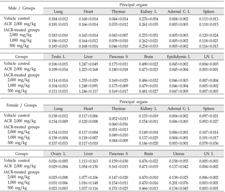
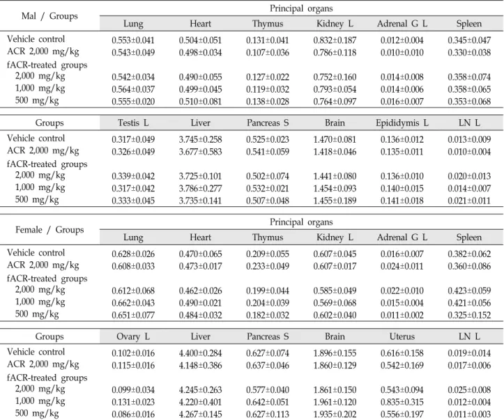
Changes on the organ weight
No significant changes on the organ weights were de-
Table 2. Changes on the absolute organ weights observed in mice after single oral treatment of fACR or ACR
Male / Groups Principal organs
Lung Heart Thymus Kidney L Adrenal G L Spleen
Vehicle control ACR 2,000 mg/kg fACR-treated groups 2,000 mg/kg 1,000 mg/kg 500 mg/kg
0.184±0.012 0.181±0.015 0.183±0.010 0.186±0.012 0.185±0.015
0.168±0.014 0.166±0.014 0.165±0.014 0.164±0.012 0.168±0.014
0.044±0.014 0.035±0.012 0.043±0.007 0.039±0.010 0.046±0.010
0.276±0.054 0.261±0.035 0.253±0.051 0.262±0.021 0.254±0.033
0.004±0.002 0.003±0.003 0.005±0.003 0.005±0.002 0.005±0.002
0.115±0.013 0.110±0.015 0.120±0.024 0.118±0.021 0.116±0.015
Groups Testis L Liver Pancreas S Brain Epididymis L LN L
Vehicle control ACR 2,000 mg/kg fACR-treated groups 2,000 mg/kg 1,000 mg/kg 500 mg/kg
0.106±0.015 0.109±0.014 0.114±0.014 0.104±0.013 0.111±0.015
1.247±0.049 1.223±0.168 1.255±0.029 1.248±0.091 1.246±0.117
0.175±0.011 0.180±0.018 0.169±0.025 0.175±0.009 0.169±0.017
0.490±0.022 0.473±0.023 0.486±0.032 0.479±0.031 0.481±0.027
0.045±0.002 0.045±0.004 0.046±0.003 0.046±0.004 0.047±0.005
0.004±0.003 0.003±0.001 0.007±0.004 0.005±0.002 0.007±0.003
Female / Groups Principal organs
Lung Heart Thymus Kidney L Adrenal G L Spleen
Vehicle control ACR 2,000 mg/kg fACR-treated groups 2,000 mg/kg 1,000 mg/kg 500 mg/kg
0.158±0.012 0.154±0.009 0.154±0.010 0.158±0.004 0.157±0.015
0.117±0.006 0.120±0.008 0.117±0.004 0.118±0.007 0.117±0.010
0.052±0.013 0.060±0.016 0.051±0.013 0.049±0.010 0.044±0.009
0.153±0.019 0.154±0.011 0.149±0.018 0.137±0.025 0.146±0.020
0.004±0.002 0.006±0.003 0.006±0.003 0.004±0.001 0.003±0.001
0.097±0.021 0.092±0.027 0.107±0.014 0.101±0.017 0.078±0.036
Groups Ovary L Liver Pancreas S Brain Uterus LN L
Vehicle control ACR 2,000 mg/kg fACR-treated groups 2,000 mg/kg 1,000 mg/kg 500 mg/kg
0.026±0.005 0.029±0.004 0.025±0.008 0.031±0.006 0.021±0.003
1.113±0.163 1.054±0.150 1.077±0.106 1.016±0.148 1.037±0.116
0.159±0.030 0.161±0.015 0.147±0.020 0.154±0.011 0.151±0.025
0.476±0.022 0.471±0.035 0.470±0.010 0.470±0.016 0.466±0.013
0.158±0.055 0.137±0.042 0.138±0.025 0.201±0.076 0.134±0.045
0.005±0.003 0.004±0.002 0.006±0.002 0.003±0.001 0.003±0.001 Values are expressed as mean ± SD of five mice, g
tected in all fACR or ACR treated mice as compared with equal genders of vehicle control, respectively (Table 2, 3).
Necropsy findings
Slight [1+] to moderate [2+] congestion of lung, thymic atrophy, splenic hypertrophy, edematous changes of uterus or hypertrophy of submandibular lymph nodes were spor- adically detected throughout all experimental groups tested in the present including both gender of vehicle controls.
Histopathological findings
Slight [1+] lung congestional spots – thickening of alveo- lar lung inflammatory cell infiltration with focal hemor- rhages, focal decreases of thymic cortex lymphoid cells, hy-
perplasia of splenic red pulp lymphoid cells, focal in- flammatory cell infiltrations in the liver parenchyma and dif- fused hyperplasia of lymphoid cells in the submandibular lymph nodes were sporadically detected throughout all ex- perimental groups tested in the present study including both gender vehicle controls. In addition, slight kidney cyst for- mation was detected in 1 female (1/5; 20%) mouse of fACR 500 mg/kg treated group (Table 4, 5).
LD50, approximate LD and target organs
The LD50 and the approximate LD of fACR after single oral treatment is calculated as over 2,000 mg/kg–the lim- ited dosages in rodents, in the both female and male ICR mice, because no fACR treatment related mortalities were
Table 3. Changes on the relative organ weights observed in mice after single oral treatment of fACR or ACR
Mal / Groups Principal organs
Lung Heart Thymus Kidney L Adrenal G L Spleen
Vehicle control ACR 2,000 mg/kg fACR-treated groups 2,000 mg/kg 1,000 mg/kg 500 mg/kg
0.553±0.041 0.543±0.049 0.542±0.034 0.564±0.037 0.555±0.020
0.504±0.051 0.498±0.034 0.490±0.055 0.499±0.045 0.510±0.081
0.131±0.041 0.107±0.036 0.127±0.022 0.119±0.032 0.138±0.028
0.832±0.187 0.786±0.118 0.752±0.160 0.793±0.054 0.764±0.097
0.012±0.004 0.010±0.010 0.014±0.008 0.014±0.006 0.016±0.007
0.345±0.047 0.330±0.038 0.358±0.074 0.358±0.065 0.353±0.068
Groups Testis L Liver Pancreas S Brain Epididymis L LN L
Vehicle control ACR 2,000 mg/kg fACR-treated groups 2,000 mg/kg 1,000 mg/kg 500 mg/kg
0.317±0.049 0.326±0.049 0.339±0.042 0.317±0.042 0.333±0.045
3.745±0.258 3.677±0.583 3.725±0.101 3.786±0.277 3.735±0.141
0.525±0.023 0.541±0.059 0.502±0.074 0.532±0.021 0.507±0.048
1.470±0.081 1.418±0.046 1.441±0.080 1.454±0.093 1.455±0.189
0.136±0.012 0.135±0.011 0.136±0.010 0.140±0.015 0.141±0.018
0.013±0.009 0.010±0.004 0.020±0.013 0.014±0.007 0.021±0.011
Female / Groups Principal organs
Lung Heart Thymus Kidney L Adrenal G L Spleen
Vehicle control ACR 2,000 mg/kg fACR-treated groups 2,000 mg/kg 1,000 mg/kg 500 mg/kg
0.628±0.026 0.608±0.033 0.612±0.068 0.662±0.043 0.651±0.077
0.470±0.065 0.473±0.017 0.462±0.026 0.490±0.021 0.484±0.032
0.209±0.055 0.233±0.049 0.199±0.044 0.204±0.039 0.182±0.032
0.607±0.045 0.607±0.017 0.585±0.049 0.569±0.068 0.602±0.040
0.016±0.007 0.024±0.011 0.022±0.010 0.015±0.004 0.011±0.002
0.382±0.062 0.360±0.086 0.423±0.059 0.421±0.056 0.325±0.152
Groups Ovary L Liver Pancreas S Brain Uterus LN L
Vehicle control ACR 2,000 mg/kg fACR-treated groups 2,000 mg/kg 1,000 mg/kg 500 mg/kg
0.102±0.016 0.115±0.016 0.099±0.034 0.131±0.023 0.086±0.016
4.400±0.284 4.148±0.386 4.245±0.263 4.220±0.401 4.267±0.145
0.627±0.074 0.637±0.046 0.577±0.040 0.642±0.051 0.627±0.113
1.896±0.155 1.860±0.129 1.861±0.150 1.961±0.120 1.935±0.202
0.616±0.158 0.542±0.169 0.543±0.094 0.835±0.315 0.556±0.197
0.019±0.014 0.017±0.006 0.025±0.008 0.012±0.004 0.011±0.003 Values are expressed as mean ± SD of five mice, % of body weights at sacrifice. L, left sides; S, splenic lobes; G, gland; LN, submandibular lymph node.
recorded up to 2,000 mg/kg treated female and male mice in the present study. In addition, no specific targets or clin- ical signs were also observed in the present study. ACR 2,000 mg/kg treated mice also did not showed any treatment re- lated mortalities, clinical signs, body and organ weights, gross and histopathological findings were also observed as compared with equal genders of vehicle control, in this ex- periment (Table 6).
Discussion
We reported that functional properties of ACR were en- hanced by fermentation, using Lactobacillus. Continentalic acid, an index component, was increased. The total poly-
phenol and the amount of amino acids was increased. In addition, the inhibitory effects of arthritis-related in- flammatory factors in cells were confirmed [13]. In the pres- ent study, we investigated the single oral dose toxicity of fACR in female and male ICR mice. In order to investigate the toxicity and identify target organs, fACR were once or- ally administered to female and male ICR mice at dose levels of 2,000, 1,000, 500 and 0(vehicle control) mg/kg (body weights) in a volume of 20 ml/kg, dissolved in distilled wa- ter, and the mortality and changes on the body weight and clinical signs were monitored during 14 days after treatment with gross observation, changes on the organ weights and histopathology of principle organs based on the recom- mendation of KFDA Guidelines [16], as compared with those
Table 4. Histopathological findings Observedin male mice after single oral treatment of fACR or ACR
Necropsy Findings at Sacrifice (Day 14) [Group summary]
Groups Vehicle control
ACR fACR treated as (mg/kg) 2,000 2,000 1,000 500 Lung
Normal Congestion 1+
Thymus Normal cDE 1+
Spleen Normal rHP 1+
Lymph nodea) Normal HP 1+
Others Normal
4/5 1/5 4/5 1/5 4/5 1/5 4/5 1/5 5/5
5/5 0/5 4/5 1/5 5/5 0/5 4/5 1/5 5/5
4/5 1/5 5/5 0/5 3/5 2/5 4/5 1/5 5/5
4/5 1/5
5/5 0/5 4/5 1/5 4/5 1/5 5/5
4/5 1/5
5/5 0/5 5/5 0/5 3/5 2/5 5/5 Values are expressed as observed animals/total observed animals (five mice per group).
a)Bilateral submandibular lymph node.
cDE = decreases of thymic cortex lymphoid cells rHP = hyperplasia of splenic red pulp lymphoid cells IF = focal inflammatory cell infiltration
Degrees = 1+, Slight; 2+, Moderate; 3+, Severe.
Table 5. Histopathological findings Observed in female mice after single oral treatment of fACR or ACR Necropsy Findings at Sacrifice (Day 14) [Group summary]
Groups Vehicle control
ACR fACR treated as (mg/kg) 2,000 2,000 1,000 500 Lung
Normal Congestion 1+
Kidney Normal Cyst 1+
Spleen Normal rHP 1+
Lymph nodea) Normal HP 1+
Others Normal
4/5 1/5 5/5 0/5 3/5 2/5 4/5 1/5 5/5
5/5 0/5 5/5 0/5 4/5 1/5 4/5 1/5 5/5
4/5 1/5 5/5 0/5 3/5 2/5 4/5 1/5 5/5
4/5 0/5
5/5 0/5 3/5 2/5 4/5 1/5 5/5
4/5 1/5
4/5 1/5 3/5 2/5 4/5 1/5 5/5
Values are expressed as observed animals/total observed animals (five mice per group).
a) Bilateral submandibular lymph node.
Degrees = 1+, Slight; 2+, Moderate; 3+, Severe.
Table 6. LD50, ALD, MTD and target organs detected in mice after single oral treatment of fACR or ACR
Animal Index
LD50 Approximated LD Targets and clinical signs
Genders Male Female
> 2,000 mg/kg
> 2,000 mg/kg
> 2,000 mg/kg
> 2,000 mg/kg
Not detected up to 2,000 mg/kg
95% confidence limits of could not calculated because of no regularities of mortalities detected in the present study. LD50, 50%
lethal dose; LD, lethal dose
of vehicle control and ACR 2,000 mg/kg treated mice.
In KFDA Guidelines [16] and OECD Guidelines [24], the recommended highest dose of test materials were 2,000 mg/kg or the maximum solubility, and they also recom- mended that in case of acute toxicity in mouse, the dosage volume were below 20 ml/kg [6]. Because there are no avail- able toxicological data after oral treatment in female and male mice of ACR or fACR, the highest dosage was selected as 2,000 mg/kg in a volume of 20 ml, the limited dosages of rodents and the recommended oral dose volume in mouse [6, 16, 24], and 1,000 and 500 mg/kg were selected as middle and lower dosage groups recommended by KFDA Guide- lines [16], in the present study. Each female and male vehicle control groups were added with ACR 2,000 mg/kg treated
female and male mice. Test material was once administered by gastric gavages using distilled water as vehicle in the present study.
As the results of single oral treatment of fACR, no treat- ment related mortalities were observed within 14 days after end of treatment up to 2,000 mg/kg, the limited dosage of rodents in the both genders, and also no fACR treatment related changes on the body and organ weights, clinical signs, necropsy and histopathological findings were detected in this experiment. In addition, no ACR 2,000 mg/kg treat- ment related mortalities, clinical signs, body and organ weights, gross and histopathological findings were also ob- served as compared with equal genders of vehicle control, in the present study. These findings are suggested that the fACR is non-toxic in mice and is therefore, likely to be safe for clinical use as similar to ACR itself.
All mice used in this study, showed normal body weight and organ weight changes ranged in normal age-matched mice regardless of treatment in the present study [7, 31].
Slight to moderate congestion of lung, thymic atrophy, splenic hypertrophy, edematous changes of uterus or hyper- trophy of submandibular lymph nodes detected as gross findings, and slight lung congestional spots–thickening of alveolar lung inflammatory cell infiltration with focal hem- orrhages, focal decreases of thymic cortex lymphoid cells, hyperplasia of splenic red pulp lymphoid cells, focal in- flammatory cell infiltrations in the liver parenchyma and dif- fused hyperplasia of lymphoid cells in the submandibular lymph nodes detected as histopathological findings were considered as accidental findings not toxicological signs re- lated to the fACR or ACR treatment, because they were spor- adically detected throughout the whole experimental groups tested in the present study including both genders of vehicle control. Especially, the edematous changes in uterus were considered as secondary changes from different physio- logical estrus cycles [2, 27]. In addition, most of them were also observed in normal mice as sporadic accidental findings [3, 18, 28]. In addition, slight kidney cyst formation was de- tected in 1 female (1/5; 20%) mouse treated with fACR 500 mg/kg were also difficult to considered as fACR treatment related toxicological signs, because no obvious dos- age-dependencies were detected in this study.
References
1. Bae, E. A., Hyun, Y. J., Choo, M. K., Oh, J. K., Ryu, J. H.
and Kim, D. H. 2004. Protective effect of fermented red gin- seng on a transient focal ischemic rats. Arch. Pharm. Res.
27, 1136-1140.
2. Banks, W. J. 1986. Female reproductive system. In: Banks WJ (Ed). Applied veterinary histology, 2nd ed., Williams &
Wilkins: Baltimore. 506-526.
3. Choi, J. S., Kim, J. W., Kim, K. Y., Ku, S. K. and Sohn, J.
H. 2014. Single-dose oral toxicity of fermented rice extracts (FREs): a 14-day observation. Pak. J. Pharm. Sci. 27, 129-137.
4. Devipriya, N., Srinivasan, M., Sudheer, A. R. and Menon, V. P. 2007. Effect of ellagic acid, a natural polyphenol, on alcohol induced prooxidant and antioxidant imbalance: a drug dose dependent study. Singapore Med. J. 48, 311-318.
5. Dourish, C. T. 1987. Effects of drugs on spontaneous motor activity. In Experimental Psychopharmacology (Greenshaw AJ, Dourish CT, Eds), pp.325-334, Humana Press, Clifton, 6. Flecknell, P. 1996. Laboratory Animal Anesthesia, 2nd Ed,
p 269, Harcourt Brace & Company.
7. Fox, J. G., Cohen, B. J. and Loew, F. M. 1984. Laboratory animal medicine. Academic Press. Inc., Orlando.
8. Hwang, Y. P., Choi, J. H. and Jeong, H. G. 2009. Protective effect of the Aralia continentalis root extract against carbon tetrachloride-induced hepatotoxicity in mice. Food Chem.
Toxicol. 47, 75-81.
9. Irwin, S. 1968. Comprehensive observational assessment: Ia.
A systemic, quantitative procedure for assessing the behavioral and physiological state of the mouse.
Psychopharmacologia 13, 222-257.
10. Jeong, S. I., Han, W. S., Yun, Y. H. and Kim, K. J. 2006.
Continentalic acid from Aralia continentalis shows activity against methicillin-resistant Staphylococcus aureus. Phytother.
Res. 20,511-514.
11. Jeong, S. I., Kim, B. S., Keum, K. S., Lee, K. H., Kang, S.
Y., Park, B. I., Lee, Y. R. and You, Y. O. 2013. Kaurenoic acid from aralia continentalis inhibits biofilm formation of Streptococcus mutans. Evid. Based Complement Alternat Med.
2013, 160592.
12. Jung, H. J., Choi, H., Lim, H. W., Shin, D., Kim, H., Kwon, B., Lee, J. E., Park, E. H. and Lim, C. J. 2012. Enhancement of anti-inflammatory and antinociceptive actions of red gin- seng extract by fermentation. J. Pharm. Pharmacol. 64, 756- 762.
13. Jung, Y. M., Lee, D. S. and Kwon, K. S. 2016. Anti- Inflammatory Effects of Fusion-Fermented Aralia continen- talis Radix (fACR) on THP-1 cells. J. Digital Convergence 14, 353-361.
14. Jung, Y. M., Lee, S. H., Lee, D. S., You, M. J., Chung, I.
K., Cheon, W. H., Kwon, Y. S., Lee, Y. J. and Ku, S. K. 2011.
Fermented garlic protects diabetic, obese mice when fed a high-fat diet by antioxidant effects. Nutr. Res. 31, 387-396.
15. Kim, C. M., Yi, S. J., Cho, I. J. and Ku, S. K. 2013. Red-koji fermented red ginseng ameliorates high fat diet-induced metabolic disorders in mice. Nutrients 5, 4316-4332.
16. Korea Food and Drug Administration. Testing Guidelines for Safety Evaluation of Drugs (Notification No. 2013-121, issued by the Korea Food and Drug Administration on April 05, 2013), 2013.
17. Lee, J. E., Kim, H. J., Lee, C. H., Lee, K. C., Choi, E. K., Chai, H. Y., Yun, Y. W., Kim, D. J., Nam, S. Y., Lee, B. J.
and Ahn, B. W. 2003. Four-week repeated-dose toxicity study on pinellia extract. Kor. J. Lab. Anim. Sci. 19, 127-141.
18. Lee, W. H., Gam, C. O., Ku, S. K. and Choi, S. H. 2011.
Single oral dose toxicity test of platycodin D, a saponin from platycodin radix in mice. Toxicol. Res. 27, 217-224.
19. Levene, A. 1981. Pathological factors influencing excision of tumours in the head and neck. Part I. Clin. Otalary. 6, 145-151.
20. Lim, H., Jung, H. A., Choi, J. S., Kim, Y. S., Kang, S. S.
and Kim, H. P. 2009. Anti-inflammatory activity of the con- stituents of the roots of Aralia continentalis. Arch. Pharm. Res.
32, 1237-1243.
21. Ludbrook, J. 1997. Update: microcomputer statistics pack- ages. A personal view. Clin. Exp. Pharmacol. Physiol. 24, 294- 296.
22. Noh, J. R., Kim, Y. H., Gang, G. T., Hwang, J. H., Kim, S. K., Ryu, S. Y., Kim, Y. S., Lee, H. S. and Lee, C. H. 2011.
초록:독활의 복합 유산균 발효 추출액의 마우스에 대한 단회경구투여 독성시험
정영미1․구세광2․이동섭3․권기상4*
(1경북대학교 생명과학부, 2대구한의대학교 한의예과, 3경운대학교 보건바이오학과, 4경운대학교 임상병리학과)
본 실험에서는 유산균발효 독활의 마우스 단회 경구 투여 독성 자료를 얻기 위해 식품의약품안전청 고시 제 2013-121 “의약품 등의 독성시험 기준”에 의거하여, 설치류 투여 한계 용량인 2,000 mg/kg을 최고 투여군을 설정 하고 공비 2로 1,000 및 500 mg/kg 투여군을 중간 및 저용량 투여군으로 설정하여 실험을 실시하였으며, 그 결과 는 독활 열수 추출물 2,000 mg/kg 암수 투여군 및 암수 매체 대조군과 비교 평가 하였다. 본 실험의 결과, 설치류 투여한계 용량인 2,000 mg/kg 투여군까지, 유산균발효 독활 열수 추출물 투여와 관련된 사망례, 임상증상, 체중, 장기중량, 육안부검 및 조직병리학적 소견이 인정되지 않았다. 따라서 유산균발효 독활 열수 추출물의 마우스에 대한 단회 경구 투여 반수 치사량 및 개략적 치사량은 암수 각각 2,000 mg/kg이상으로 산출되었으며, 특정 임상 증상 및 표적 장기 역시 없는 것으로 판단되어, 유산균발효 독활은 매우 안전한 물질로 판단된다. 또한 독활 열수 추출물 2,000 mg/kg 투여와 관련된 사망례, 임상 증상, 체중, 장기중량, 육안 및 조직병리학적 변화 역시 인정되지 않았다. 이러한 결과는 독활의 활용도를 증대시키는 기초 자료가 될 것으로 사료된다.
Hepatoprotective effect of Platycodon grandiflorum against chronic ethanol-induced oxidative stress in C57BL/6 mice.
Ann. Nutr. Metab. 58, 224-231.
23. Oh, H. L., Lee, D. K., Lim, H. and Lee, C. H. 2010. HY253, a novel decahydrofluorene analog, from Aralia continentalis, induces cell cycle arrest at the G1 phase and cytochrome c-mediated apoptosis in human lung cancer A549 cells. J.
Ethnopharmacol. 129, 135-139.
24. Oh, H. L., Lim, H., Cho, Y. H., Koh, H. C., Kim, H., Lim, Y. and Lee, C. H. 2009. HY251, a novel cell cycle inhibitor isolated from Aralia continentalis, induces G1 phase arrest via p53-dependent pathway in HeLa cells. Bioorg. Med.
Chem. Lett. 19, 959-961.
25. Organization for Economic Co-Operation and Development (Ed.). OECD guideline (423) for testing of chemicals – acute oral toxicity-acute toxic class method. 2001.
26. Park, H. J., Hong, M. S., Lee, J. S., Leem, K. H., Kim, C.
J., Kim, J. W. and Lim, S. 2005. Effects of Aralia continentalis on hyperalgesia with peripheral inflammation. Phytother.
Res. 19, 511-513.
27. Park, J. H., Seo, B. I., Cho, S. Y., Park, K. R., Choi, S. H., Han, C. K., Song, C. H., Park, S. J. and Ku, S. K. 2013. Single
oral dose toxicity study of prebrewed armeniacae semen in rats. Toxicol. Res. 29, 91-98.
28. Pineda, M. H. 1989. Female reproductive system. In:
McDonald LE and Pineda MH (Eds). Veterinary endocrinol- ogy and reproduction, Lea & Febiger: Philadelphia, 303-354.
29. Roh, S. S. and Ku, S. K. 2010. Mouse single oral dose toxicity study of DHU001, a polyherbal formula. Toxicol. Res. 26, 53-59.
30. Saravanan, N., Rajasankar, S. and Nalini, N. 2007. Antiox- idant effect of 2-hydroxy-4-methoxy benzoic acid on ethanol induced hepatotoxicity in rats. J. Pharm. Pharmacol. 59, 445- 453.
31. Seo, S. H., Hwang, Y. H., Lee, J. H., Oh, S. Y., Kim, T. S.
and Ma, J. Y. 2012. Single dose oral toxicity study of fer- mented Soshiho-tang extract in mice. Kor. J. Orient. Physiol.
Pathol. 26, 47-52.
32. Tajima, Y. (Ed). 1989. Biological reference data book on ex- perimental animals. Soft Science Inc., Tokyo
33. Trinh, H. T., Han, S. J., Kim, S. W., Lee, Y. C. and Kim, D. H. 2007. Bifidus fermentation increases hypolipidemic and hypoglycemic effects of red ginseng. J. Microbiol.
Biotechnol. 17, 1127-1133.

![Table 5. Histopathological findings Observed in female mice after single oral treatment of fACR or ACR Necropsy Findings at Sacrifice (Day 14) [Group summary]](https://thumb-ap.123doks.com/thumbv2/123dokinfo/5008756.549020/6.892.462.807.214.518/histopathological-findings-observed-treatment-necropsy-findings-sacrifice-summary.webp)
