․교신저자: 최해윤 경상북도 포항시 남구 대잠동 907-8 대구한의대학교 부속 포항한방병원 TEL: 054-281-0055
E-mail: [email protected]
金銀花 추출물의 마우스 단회 경구투여 독성실험
유효정, 박미연, 최해윤, 김종대 대구한의대학교 한의과대학 내과학 교실
Mouse Single Oral Dose Toxicity Test of Lonicerae Flos Aqueous Extracts
Hyo-jeong Yoo, Mee-yeon Park, Hae-yun Choi, Jong-dae Kim
Dept. of Internal Medicine, College of Oriental Medicine, Dae-Gu Haany University ABSTRACT
Objectives :The object of this study was to obtain accurate information (single oral dose toxicity) of Lonicerae Flos (LF; Dried flower bud parts of Lonicera japonica Thunb (Caprifoliaceae)), which has traditionally been used in Korean medicine for treating various inflammatory diseases.
Methods :In order to observe the 50% lethal dose (LD 50), approximate lethal dosage (ALD) and target organs, test articles were once orally administered to female and male ICR mice at dose levels of 2,000, 1,000, 500 and 0 (control) ㎎/㎏ (body weight.). The mortality and changes on body weight, clinical signs and gross observation were monitored for 14 days after single oral treatment ofLF aqueous extracts with organ weights and histopathological observations of 12 types of principle organs.
Results :
1. After single oral treatment of LF aqueous extracts, we could not find any mortality and toxicological evidences up to 2,000 ㎎/㎏ treated group, the limited dosages in rodents at body and organ weights, clinical signs, gross and histopathological observations.
2. Slight diarrhea was detected in most mice treated with 2,000 ㎎/㎏ ofLF aqueous extracts and male mice of LF aqueous extracts 1,000 ㎎/㎏ within 2 days after end of treatment, respectively.
Conclusion :The results obtained in this study suggest that the LD 50 and ALD ofLF aqueous extracts in both female and male mice after single oral treatment were considered as over 2,000 ㎎/㎏ because no mortalities were detected up to 2000
㎎/㎏, the highest dose recommended by KFDA and OECD. However, we also observed the possibility of digestive disorders like diarrhea when over 1,000 ㎎/㎏ ofLF aqueous extracts were administered in the present study.
Key words : Lonicerae Flos, Toxicity
Ⅰ. 서 론
金銀花 (Lonicerae Flos;LF)는 인동과 (Caprifoliaceae) 에 속한 인동 (Lonicera japonica Thunb)의 花蕾를 건조한 것으로 여름철 꽃이 피기 전에 채취한 것
을 이용하는 약재로 性은 寒하고, 味는 甘하며, 淸 熱解毒, 凉散風熱 등의 효능이 있어 消炎, 淸血, 利 尿, 殺菌 작용을 나타내어 熱性病, 身熱無汗, 化膿 性疾患, 急慢性淋疾, 梅毒, 痢疾, 膿瘍, 癰疽, 疥癬腫 毒惡瘡, 咽喉腫痛을 치료하는데 사용되어 왔다1.
근래에 들어 한약 자체의 독성 평가가 진행되어 단미제에 대한 독성연구로는 半夏2, 竹瀝3, 麻黃4, 大茴香5, 漢防己6, 人蔘7-8, 玉竹9, 桑葉 추출물10등에 서 수행되어져 왔으며, 복합 처방에 대한 연구로는
Ninomiya 등11은 八味地黃丸의 독성을, Ryu 등12은 補中治濕湯의 독성을, 황 등13은 加味歸脾湯의 독성 을, 배 등14은 補中益氣湯合大七氣湯의 독성실험을 각각 수행하였다. 그러나 한약이 장기복용 약물이 며 또한 생약 복합물이기 때문에 실험의 진행이 매우 어렵고 동물의 생체 내에서 약물의 동태를 파악하기 어려워 잔류 가능성이 있는 잠재적인 독 성 평가는 거의 이루어 지지 않고 있다. 이에 따라 의약품의 독성평가에 준하여 안전성 평가가 이루 어져야 할 것으로 사료된다.
이에 본 연구에서는 대표적인 항염증 약물로 다 양한 질환에 널리 사용되어 온 金銀花의 일반적인 독성시험 중 현재 의약품 등의 독성시험기준15에 명시되어 있는 마우스 경구 단회투여독성 시험을 수행하여 유의한 결과를 얻어 보고하고자 한다.
Ⅱ. 실 험
1. 재 료 1) 약 재
본 실험에 사용된 金銀花는 약업사 (옴니허브, Korea)에서 매입한 것을 현미경하에서 관능검사를 통하여 선정하여 사용하였다.
2) 金銀花 추출
선정된 金銀花 100g을 취하여 정제수 2,000㎖로 80℃에서 3시간 동안 3번 가열 추출한 후, 흡인 여 과한 여과액을 rotary vacuum evaporator (Buchi Rotavapor R144, Buchi Labortechnik AG, Switzland) 로 감압․농축하여 점조성의 추출물을 얻은 다음 programmable freeze dryer (Labconco Freezone1, Labconco Corp., MO, USA)를 사용하여 동결 건조 시켜 총 23.80g (수율 약 23.80%)의 연갈색의 물 추출물을 얻어 실험에 사용하였다. 물 추출한 金銀 花 동결 건조물은 -20℃의 냉장고에 보관 후 실험 에 사용하였으며, 본 실험에서 사용한 용매인 증류 수에 100㎎/㎖의 농도까지 비교적 잘 용해되었다.
2. 마우스 경구 단회투여독성 실험 1) 실험동물 및 사양관리
이 독성 실험은 대구한의대 자체 동물 윤리 위 원회를 통과하여 시행되었으며, 암수 각 20마리의 ICR 마우스 (6-wk old upon receipt, SLC, Japan)를 7일간의 순화과정을 거쳐 실험에 사용하였으며, 순화 과정 및 실험 전 기간 동안 온도 (20-25℃)와 습도 (30-35%)가 조절된 사육실에서 마우스용 polycarbonate 사육 상자에 5마리씩 수용하여 사육하였고, 명암 주기 (light: dark cycle)는 12시간 주기로 조절하 였으며, 사료 (Samyang, Korea)와 음수는 자유롭 게 공급하였다. 모든 실험동물은 투여일 및 최종 부검일 18시간 전 절식을 실시하였으며, picric acid 로 개체를 식별하였다.
2) 군분리 및 약물의 투여
실험동물은 군당 5마리씩 Table 1에 기록한 8그 룹으로 구분하였다. 金銀花는 한의학에서 오랫동안 사용되어온 대표적인 약재로, 현재까지 金銀花의 독성에 대한 보고를 찾아볼 수 없어 한국식품의약 품 안전청 기준15과 OECD 실험기준16에 의거하여, 설치류 최고 한계투여용량인 2,000㎎/㎏을 최고 용 량으로 설정하였으며, 공비 2로 1,000 및 500㎎/㎏
을 중간 및 저용량 투여군으로 설정하였다. 또한 암수 각각에 대한 매체 대조군을 추가 하였다. 모 든 투여군에서는 金銀花 추출물을 멸균 증류수에 용해시켜 20㎖/㎏의 용량으로 overnight 절식 후 존데가 부착된 1㎖ 주사기를 이용하여 경구 투여 하였다. 식이와 음수에 따른 약물의 흡수 변화를 최소화하기 위해, 투여 후 대략 3시간 동안 사료와 음수 공급을 제한하였다.
3) 임상증상의 관찰
모든 실험동물의 임상증상을 투여 전후에 각각 functional observational battery test17-18를 기초하 여 동물의 행동, 자극에 대한 반응성, 각성도 및 경 계성, 자세 및 보행 이상 등에 관한 일반증상을 관 찰, 기록하였으며, 투여일 이후에도 하루에 최소한 2 번씩 모든 실험동물의 임상증상을 관찰, 기록하였다.
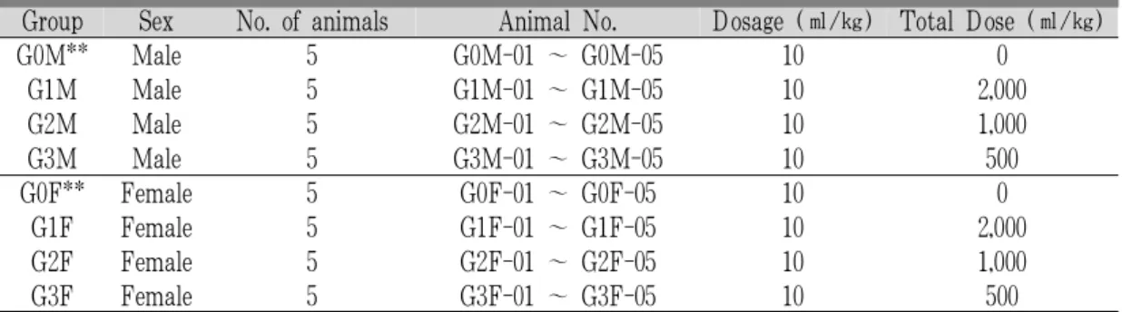
Group Sex No. of animals Animal No. Dosage (㎖/㎏) Total Dose (㎖/㎏)
G0M** Male 5 G0M-01 ~ G0M-05 10 0
G1M Male 5 G1M-01 ~ G1M-05 10 2,000
G2M Male 5 G2M-01 ~ G2M-05 10 1,000
G3M Male 5 G3M-01 ~ G3M-05 10 500
G0F** Female 5 G0F-01 ~ G0F-05 10 0
G1F Female 5 G1F-01 ~ G1F-05 10 2,000
G2F Female 5 G2F-01 ~ G2F-05 10 1,000
G3F Female 5 G3F-01 ~ G3F-05 10 500
** ; Vehicle control; distilled water 20㎖/㎏ as vehicle in this study; All test articles in vehicle were once orally dosed in a volume of 20㎖/㎏, dissolved in distilled water.
Table 1. Experimental Design Used in Single Dose Toxicity Test
4) 체중의 측정
모든 실험동물의 체중을 투여 전 1일, 투여직전, 투여 후 1, 2, 7, 13 및 14일에 각각 측정하였으며, 실험 시작 시 개체 차이에 따른 체중의 변화를 최 소화하기 위하여, 투여일 부터 투여 후 7일, 투여 후 7일부터 13일 및 투여일 부터 투여 후 13일간의 체중 증가량인 증체량을 각각의 체중을 이용하여 산출하였다.
5) 육안부검
투여 14일 후 모든 실험동물은 overnight 절식을 실시하였으며, ethyl ether (덕산공업주식회사, Korea) 마취하에 부검을 실시하고, 폐, 심장, 신장, 비장, 고환, 간, 췌장, 부고환 (Epididymis), 슬와 임파절 (Popliteal lymph node), 난소, 뇌, 및 자궁 등 12개 의 주요 장기를 위주로 이상 육안소견을 각각 관 찰, 기록하였다.
6) 장기중량의 측정
모든 실험동물은 육안부검 소견을 관찰 기록한 후 폐, 심장, 좌측 신장, 비장, 좌측 고환, 간, 췌장 비장엽 (Pancreas-splenic lobes), 좌측 부고환 (Epididymis -left), 좌측 슬와 임파절 (Popliteal lymph node-left), 좌측 난소, 뇌 및 자궁에 대한 절대 중량을 각각 측정하였으며, 체중의 변화에 수반된 이차적 변화 를 최소화하기 위해 체중에 대한 각각의 장기 절 대중량의 비율인 상대 중량을 산출하였다.
7) 조직병리학적 관찰
폐, 심장, 좌측 신장, 비장, 좌측 고환, 간, 췌장 비장 엽 (Pancreas-splenic lobes), 좌측 부고환 (Epididymis -left), 좌측 슬와 임파절 (Popliteal lymph node-left), 좌측 난소, 뇌 및 자궁 등의 12개 주요 장기의 일 부 조직을 10% 중성포르말린에 18시간 이상 고정 시킨 다음, 탈수를 거쳐 파라핀 포매 후 4㎛의 절 편을 제작하였다. 이후 hematoxylin & eosin 염색 을 실시하고, 광학현미경 하에서 이상 유무를 관찰, 기록하였다.
3. 통계 처리
모든 수치는 평균 ± 표준편차로 표시하였으며, 다중비교검증을 이용하여 통계처리를 실시하였고, 분산동질성을 Levene test를 실시하여 검증하였다.
등분산일 경우, one way ANOVA test를 실시한 다 음 Scheffe test로 사후 검증을 실시하여 군간의 유 의성을 측정하였다. 비등분산일 경우에는 비모수 검증인 Kruskal-Wallis H test를 실시하여 유의성 이 인정된 경우에는, Mann-Whitney U-Wilcoxon Rank Sum W를 실시하여 군간의 유의성을 검증하 였다. 단회 투여독성시험의 경우, 반수치사량 및 95% 신뢰한계 (confidence limits)를 Probit 방법으 로 측정하였으며, 임상 증상 및 육안부검 및 조직 병리학적 소견은 각각 그 정도에 따라 0 (normal),
1+ (slight), 2+ (moderate) 및 3+ (severe)로 구 분하였다. 통계처리 및 Probit 방법은 SPSS for Windows (Release 14.0K, SPSS Inc., USA)를 이용 하여 평가하였으며, p-value가 0.05 이하인 경우 통 계적 유의성을 인정하였다.
Ⅲ. 결 과
1. 마우스 경구 단회 투여독성 실험 1) 사망률
金銀花 추출물 투여와 관련 있는 사망례는 실험 전 기간 동안 관찰되지 않아, 모든 실험동물(5/5;
100%)을 최종부검을 실시하였다(Table 2).
Group IDb 0c 1 2 3 4 Day after dosing5 6 7 8 9 10 11 12 13 Total**
MALE
G0M 0 0 0 0 0 0 0 0 0 0 0 0 0 0 0/5(0%)
G1M 0 0 0 0 0 0 0 0 0 0 0 0 0 0 0/5(0%)
G2M 0 0 0 0 0 0 0 0 0 0 0 0 0 0 0/5(0%)
G3M 0 0 0 0 0 0 0 0 0 0 0 0 0 0 0/5(0%)
FEMALE
G0F 0 0 0 0 0 0 0 0 0 0 0 0 0 0 0/5(0%)
G1F 0 0 0 0 0 0 0 0 0 0 0 0 0 0 0/5(0%)
G2F 0 0 0 0 0 0 0 0 0 0 0 0 0 0 0/5(0%)
G3F 0 0 0 0 0 0 0 0 0 0 0 0 0 0 0/5(0%)
a ; number of died animals, b ; Group ID was listed in Table 1, c ; Dosing day, **; total mortalities during 14 days of observation periods - died animals/total observed animals (n = 5) (percentages)
Table 2. Mortalities Observed in Female and Male Mice of Single Dose Toxicity Testa
2) 임상증상
경미한 설사 소견이 암컷 金銀花 추출물 2,000㎎
/㎏ 투여군과 수컷 金銀花 추출물 2,000 및 1,000㎎
/㎏ 투여군에 국한되어 관찰되었으며 이들 설사 소견은 투여 3일 후부터 정상으로 회복되었다. 탈
모 소견이 수컷 金銀花 추출물 1,000㎎/㎏ 투여군 에 국한되어 투여 9일 후부터 최종부검 시까지 2 례(2/5; 40%) 인정되었다. 이 외 金銀花 추출물 투여와 관련된 임상 증상은 실험 전 기간 동안 인 정되지 않았다(Table 3).
Group IDb Clinical signs
Normal Diarrhea Hair losses
MALE
G0M 5/5 (100%) 0/5 (0%) 0/5 (0%)
G1M 0/5 (0%) 5/5 (100%) 0/5 (0%)
G2M 0/5 (0%) 4/5 (80%) 2/5 (40%)
G3M 5/5 (100%) 0/5 (0%) 0/5 (0%)
FEMALE
G0F 5/5 (100%) 0/5 (0%) 0/5 (0%)
G1F 2/5 (40%) 3/5 (60%) 0/5 (0%)
G2F 5/5 (100%) 0/5 (0%) 0/5 (0%)
G3F 5/5 (100%) 0/5 (0%) 0/5 (0%)
a ; Observed animals/total observed animals of five mice (percentages),b ; Group ID was listed in Table 1.
Table 3. Clinical Signs Observed in Female and Male Mice of Single Dose Toxicity Testa
3) 체중의 변화
金銀花 추출물 투여와 관련된 체중 및 체중 증
가량의 변화는 인정되지 않았다(Table 4).
Group IDb Interval
Day 0c - 7 Day 7 - 13 Day 0 -13
MALE
G0M 9.38±1.76 1.66±1.30 5.36±2.03
G1M 8.12±1.20 2.10±0.31 4.84±1.09
G2M 8.70±1.32 0.44±1.71 4.98±0.51
G3M 9.66±1.72 2.94±0.73 6.82±1.50
FEMALE
G0F 6.74±0.86 2.72±0.76 5.64±0.52
G1F 6.06±1.04 3.04±1.50 4.90±1.11
G2F 6.72±1.07 1.18±1.63 2.74±2.36
G3F 6.68±0.70 3.42±1.93 5.26±1.41
a ; Values are expressed as mean ± S. D. of five mice, g, b ; Group ID was listed in Table 1,c ; Day of dosing after overnight fasted.
Table 4. Body Weight Gains in Female and Male Mice of Single Dose Toxicity Testa
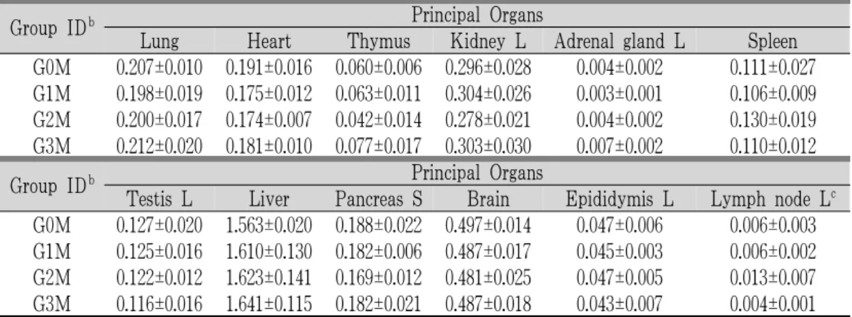
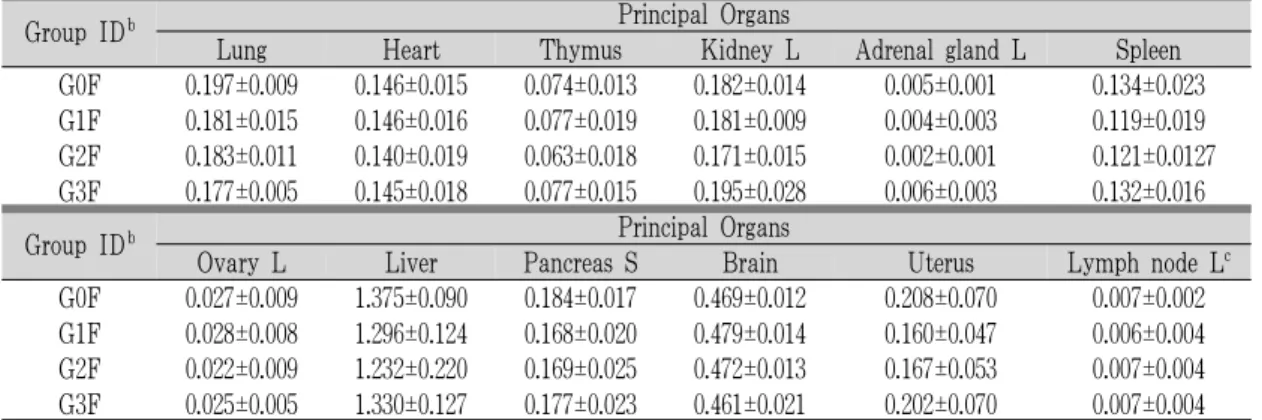
4) 장기중량의 변화
金銀花 추출물 1,000㎎/㎏ 암컷 투여군에서 동일 성별의 매체 대조군에 비해 뇌 상대 중량의 유의 성 있는(p<0.05) 감소가 인정된 이외에, 각각의 동
일 성별 매체 대조군에 비해 의미 있는 장기중량 의 변화는 모든 金銀花 추출물 투여군에서 인정되 지 않았다(Table 5-8).
Group IDb Principal Organs
Lung Heart Thymus Kidney L Adrenal gland L Spleen G0M 0.207±0.010 0.191±0.016 0.060±0.006 0.296±0.028 0.004±0.002 0.111±0.027 G1M 0.198±0.019 0.175±0.012 0.063±0.011 0.304±0.026 0.003±0.001 0.106±0.009 G2M 0.200±0.017 0.174±0.007 0.042±0.014 0.278±0.021 0.004±0.002 0.130±0.019 G3M 0.212±0.020 0.181±0.010 0.077±0.017 0.303±0.030 0.007±0.002 0.110±0.012
Group IDb Principal Organs
Testis L Liver Pancreas S Brain Epididymis L Lymph node Lc G0M 0.127±0.020 1.563±0.020 0.188±0.022 0.497±0.014 0.047±0.006 0.006±0.003 G1M 0.125±0.016 1.610±0.130 0.182±0.006 0.487±0.017 0.045±0.003 0.006±0.002 G2M 0.122±0.012 1.623±0.141 0.169±0.012 0.481±0.025 0.047±0.005 0.013±0.007 G3M 0.116±0.016 1.641±0.115 0.182±0.021 0.487±0.018 0.043±0.007 0.004±0.001
a ; Values are expressed as mean ± S. D. of five mice, g, b ; Group ID was listed in Table 1, L ; left side, S ; splenic lobe,c ; Popliteal lymph node.
Table 5. Changes on the Absolute Organ Weights Observed in Male Mice of Single Dose Toxicity Testa
Group IDb Principal Organs
Lung Heart Thymus Kidney L Adrenal gland L Spleen
G0F 0.197±0.009 0.146±0.015 0.074±0.013 0.182±0.014 0.005±0.001 0.134±0.023 G1F 0.181±0.015 0.146±0.016 0.077±0.019 0.181±0.009 0.004±0.003 0.119±0.019 G2F 0.183±0.011 0.140±0.019 0.063±0.018 0.171±0.015 0.002±0.001 0.121±0.0127 G3F 0.177±0.005 0.145±0.018 0.077±0.015 0.195±0.028 0.006±0.003 0.132±0.016
Group IDb Principal Organs
Ovary L Liver Pancreas S Brain Uterus Lymph node Lc
G0F 0.027±0.009 1.375±0.090 0.184±0.017 0.469±0.012 0.208±0.070 0.007±0.002 G1F 0.028±0.008 1.296±0.124 0.168±0.020 0.479±0.014 0.160±0.047 0.006±0.004 G2F 0.022±0.009 1.232±0.220 0.169±0.025 0.472±0.013 0.167±0.053 0.007±0.004 G3F 0.025±0.005 1.330±0.127 0.177±0.023 0.461±0.021 0.202±0.070 0.007±0.004
a ; Values are expressed as mean ± S. D. of five mice, g, b ; Group ID was listed in Table 1, L ; left side, S ; splenic lobe,c ; Popliteal lymph node.
Table 6. Changes on the Absolute Organ Weights Observed in Female Mice of Single Dose Toxicity Testa
Group IDb Principal Organs
Lung Heart Thymus Kidney L Adrenal gland L Spleen
G0M 0.600±0.047 0.554±0.043 0.174±0.018 0.859±0.095 0.012±0.006 0.321±0.068 G1M 0.579±0.046 0.511±0.038 0.183±0.027 0.886±0.042 0.010±0.002 0.311±0.031 G2M 0.581±0.027 0.508±0.038 0.122±0.037 0.811±0.060 0.011±0.004 0.381±0.066 G3M 0.585±0.0047 0.501±0.048 0.211±0.043 0.834±0.066 0.019±0.005 0.304±0.027
Group IDb Principal Organs
Testis L Liver Pancreas S Brain Epididymis L Lymph node Lc
G0M 0.369±0.052 4.517±0.182 0.546±0.063 1.449±0.165 0.134±0.007 0.018±0.010 G1M 0.364±0.044 4.702±0.296 0.533±0.027 1.424±0.080 0.133±0.005 0.018±0.005 G2M 0.356±0.035 4.724±0.278 0.494±0.039 1.403±0.106 0.138±0.013 0.038±0.020 G3M 0.321±0.047 4.524±0.199 0.501±0.044 1.364±0.064 0.120±0.017 0.012±0.003
a; Values are expressed as mean ± S. D. of five mice, g,b; Group ID was listed in Table 1, L ; left side, S, splenic lobe, c; Popliteal lymph node.
Table 7. Changes on the Relative Organ Weights Observed in Male Mice of Single Dose Toxicity Testa
Group IDb Principal Organs
Lung Heart Thymus Kidney L Adrenal gland L Spleen
G0F 0.658±0.040 0.488±0.038 0.245±0.032 0.608±0.038 0.016±0.004 0.449±0.077 G1F 0.621±0.049 0.499±0.057 0.261±0.055 0.621±0.027 0.015±0.011 0.406±0.056 G2F 0.685±0.051 0.527±0.073 0.235±0.055 0.641±0.033 0.009±0.004 0.450±0.077 G3F 0.606±0.014 0.494±0.056 0.263±0.059 0.665±0.071 0.019±0.010 0.450±0.041
Group IDb Principal Organs
Ovary L Liver Pancreas S Brain Uterus xymph node Lc
G0F 0.089±0.031 4.596±0.215 0.615±0.073 1.570±0.048 0.694±0.219 0.023±0.008 G1F 0.096±0.026 4.428±0.171 0.574±0.062 1.643±0.076 0.574±0.154 0.020±0.012 G2F 0.082±0.029 4.586±0.567 0.637±0.133 1.775±0.140* 0.622±0.195 0.025±0.015 G3F 0.085±0.017 4.529±0.249 0.605±0.078 1.575±0.087 0.689±0.233 0.024±0.015
a ; Values are expressed as mean ± S. D. of five mice, g, b ; Group ID was listed in Table 1, L ; left side, S ; splenic lobe,c ; Popliteal lymph node, * ; p<0.05 as compared with vehicle control by Scheffe test.
Table 8. Changes on the Relative Organ Weights Observed in Female Mice of Single Dose Toxicity Testa
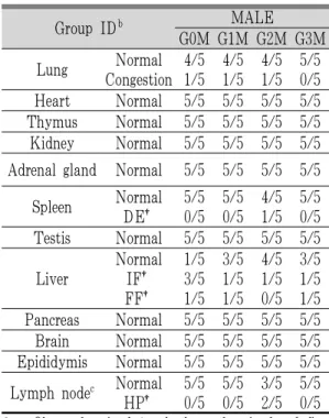
5) 부검소견
경미하거나, 중등도의 폐 충혈, 가슴샘 위축, 비 장 위축 및 자궁 부종 소견이 암수 매체 대조군을 포함한 모든 실험군에 걸쳐 산발적으로 관찰되었 으며, 金銀花 추출물 1,000㎎/㎏ 수컷 투여군에 국 한되어 슬와 임파절 종대 및 biting wound가 각각 2례(2/5; 40%) 인정된 이외에, 金銀花 추출물 투 여와 관련된 의미 있는 육안 부검소견은 인정되지 않았다(Table 9-10).
Group IDb MALE
G0M G1M G2M G3M Lung Normal
Congestion 4/5 1/5 4/5
1/5 4/5 1/5 5/5
0/5 Heart Normal 5/5 5/5 5/5 5/5 Thymus Normal
Atrophy 3/5 2/5 4/5
1/5 4/5 1/5 5/5
0/5 Kidney Normal 5/5 5/5 5/5 5/5 Adrenal gland Normal 5/5 5/5 5/5 5/5
Spleen Normal Atrophy 3/5
2/5 4/5 1/5 4/5
1/5 5/5 0/5 Testis Normal 5/5 5/5 5/5 5/5 Liver Normal 5/5 5/5 5/5 5/5 Pancreas Normal 5/5 5/5 5/5 5/5 Brain Normal 5/5 5/5 5/5 5/5 Epididymis Normal 5/5 5/5 5/5 5/5 Lymph nodec Normal
Hypertrophy 4/5 0/5 0/5
0/5 3/5 2/5 4/5
0/5 Others Normal
Biting wound 5/5 0/5 5/5
0/5 5/5 2/5 5/5
0/5
a; Observed animals/total observed animals of five mice
b; Group ID was listed in Table 1,c; Bilateral popliteal lymph node.
Table 9. Necropsy Findings Observed Male Mice of Single Dose Toxicity Testa
Group IDb FEMALE G0F G1F G2F G3F Lung Normal
Congestion 4/5 1/5 4/5
1/5 3/5 2/5 5/5
0/5 Heart Normal 5/5 5/5 5/5 5/5 Thymus Normal
Atrophy 4/5 1/5 5/5
0/5 4/5 1/5 4/5
0/5 Kidney Normal 5/5 5/5 5/5 5/5 Adrenal gland Normal 5/5 5/5 5/5 5/5
Spleen Normal Atrophy 5/5
0/5 4/5 1/5 5/5
0/5 4/5 1/5 Ovary Normal 5/5 5/5 5/5 5/5 Liver Normal 5/5 5/5 5/5 5/5 Pancreas Normal 5/5 5/5 5/5 5/5 Brain Normal 5/5 5/5 5/5 5/5 Uterus Normal
Edematous 3/5 2/5 3/5
2/5 3/5 2/5 3/5
2/5 Lymph nodec Normal 5/5 5/5 5/5 5/5 Others Normal 5/5 5/5 5/5 5/5
a; Observed animals/total observed animals of five mice,
b; Group ID was listed in Table 1,c ; Bilateral popliteal lymph node.
Table 10. Necropsy Findings Observed Female Mice of Single Dose Toxicity Testa
6) 조직병리학적 소견
경미한 폐 충혈 - 폐포 벽의 비후와 염증세포 및 적혈구 축적(Fig. 1), 비장 적색 수질내 임파구 감소(Fig. 2), 간의 국소 지방변화(Fig. 3) 및 염증 세포 침윤(Fig. 4) 소견이 암수 매체 대조군을 포 함한 모든 실험군에 걸쳐 산발적으로 관찰되었으 며, 수컷 1,000㎎/㎏ 투여군에 국한되어 슬와 임파 절의 임파구 증생(Fig. 5) 소견이 2례(2/5; 40%) 인정된 이외에 金銀花 추출물 투여와 관련된 조직 병리학적 소견은 인정되지 않았다(Table 11-12).
Vehicle control male 02
(G0M-02) Congestion 1+ Vehicle control female 03 (G0F-03) Congestion 1+
LFe 2,000㎎/㎏ male 05
(G1M-05) Congestion 1+ LFe 2,000㎎/㎏ female 02 (G1F-02) Congestion 1+
LFe 1,000㎎/㎏ male 05
(G2M-05) Congestion 1+ LFe 1,000㎎/㎏ female 01 (G2M-01) Congestion 1+
Fig. 1. Histopathological changes detected on the lung after single oral treatment ofLonicerae Flosaqueous extracts(LFe)
Note that slight(1+) lung focal congestional spots - thickening of alveolar lung inflammatory cell infiltration with/without focal hemorrhages were randomly detected throughout the all experimental groups tested regardless of genders including both genders of vehicle controls as sporadic finings not LFe treatment related toxicological signs; A, alveolar sac-respiratorybronchiole; B, bronchiole. All Hematoxylin & Eosin stain;
Scale bars = 160µm.
Vehicle control female 03 (G0F-03) Decreases of lymphoid cells 1+
LFe 1,000㎎/㎏ male 01 (G2M-01) Decreases of lymphoid cells 1+
Fig. 2. Histopathological changes detected on the
spleen after single oral treatment ofLonicerae Flosaqueous extracts(LFe)
Note that slight(1+) decreases of lymphoid cells in the red pulp(as seen empty vacuoles) were restrictly detected one(1/5; 20%) rat of female vehicle control and male 1,000㎎/㎏ treated groups, respectively as sporadic finings not LFe treatment related toxicological signs. All Hematoxylin & Eosin stain;
Scale bars = 160µm
Vehicle control male 05 (G0M-05) Focal fatty changes 1+
LFe 2,000㎎/㎏ male 04 (G1M-04) Focal fatty changes 1+
LFe 2,000㎎/㎏ female 04 (G1F-04) Focal fatty changes 1+
LFe 1,000㎎/㎏ male 04 (G2F-04) Focal fatty changes 1+
LFe 500㎎/㎏ male 01 (G3F-01) Focal fatty changes 1+
Fig. 3. Histopathological changes detected as focal fatty changes in the liver after single oral treatment of Lonicerae Flos aqueous extracts (LFe)
Note that slight (1+) focal fatty changes in the hepatic parenchyma - hypertrophy of hepatocytes or deposition of lipid droplets were randomly detected throughout the all experimental groups tested regardless of genders including both genders of vehicle controls as sporadic finings notLFe treatment related toxicological signs; C, central vein. All Hematoxylin & Eosin stain; Scale bars = 160µm.
Vehicle control male 04
(G0M-04) IF 1+ Vehicle control female 04 (G0F-04) IF 1+
LFe 2,000㎎/㎏ male 03
(G1M-03) IF 1+ LFe 2,000㎎/㎏ female 04 (G1F-04) IF 1+
LFe 500㎎/㎏ male 02
(G3M-02) IF 1+ LFe 500㎎/㎏ female 02 (G3F-02) IF 1+
Fig. 4. Histopathological changes detected as focal inflammatory cell infiltrations(IF) in the Liver after Single Oral Treatment of Lonicerae Flos aqueous extracts(LFe)
Note that slight(1+) IF were randomly detected in the hepatic parenchyma throughout the all experimental groups tested regardless of genders including both genders of vehicle controls as sporadic finings not LFe treatment related toxicological signs; C, central vein. All Hematoxylin & Eosin stain; Scale bars
= 160µm.
LFe 1,000㎎/㎏ male 02 (G2M-02) Decreases of lymphoid cells 1+
Enlarged of right F areas
Fig. 5. Histopathological changes detected on the popliteal lymph node after single oral treatment
of Lonicerae Flosaqueous extracts (LFe) Note that slight(1+) hyperplasia of lymphoid cells were restrictly detected two(2/5; 40%) rats of male treated with 1,000㎎/㎏ treated groups, respectively as sporadic finings not LFe treatment related toxicological signs. They did not showed any dose - dependencies; M, medullary sinus; F, cortex -follicle. All Hematoxylin & Eosin stain; Scale bars = 160µm.
Group IDb MALE
G0M G1M G2M G3M Lung Normal
Congestion 4/5 1/5 4/5
1/5 4/5 1/5 5/5
0/5 Heart Normal 5/5 5/5 5/5 5/5 Thymus Normal 5/5 5/5 5/5 5/5 Kidney Normal 5/5 5/5 5/5 5/5 Adrenal gland Normal 5/5 5/5 5/5 5/5
Spleen Normal DE† 5/5
0/5 5/5 0/5 4/5
1/5 5/5 0/5 Testis Normal 5/5 5/5 5/5 5/5
Liver Normal IF† FF†
1/53/5 1/5
3/51/5 1/5
4/51/5 0/5
3/51/5 1/5 Pancreas Normal 5/5 5/5 5/5 5/5 Brain Normal 5/5 5/5 5/5 5/5 Epididymis Normal 5/5 5/5 5/5 5/5 Lymph nodec Normal
HP† 5/5 0/5 5/5
0/5 3/5 2/5 5/5
a ; Observed animals/total observed animals of five0/5 mice, b; Group ID was listed in Table 1, c; Bilateral popliteal lymph node, †; DE ; decreases of lymphoid cells in the red pulp, IF ; focal inflammatory cell infiltration, FF; focal fatty change, HP; hyperplasia of lymphoid cells.
Table 11. Histopathological Findings Observed Male Mice of Single Dose Toxicity Testa
Group IDb FEMALE G0F G1F G2F G3F Lung Normal
Congestion 4/5 1/5 4/5
1/5 3/5 2/5 4/5
1/5 Heart Normal 5/5 5/5 5/5 5/5 Thymus Normal 5/5 5/5 5/5 5/5 Kidney Normal 5/5 5/5 5/5 5/5 Adrenal gland Normal 5/5 5/5 5/5 5/5
Spleen Normal DE† 4/5
1/5 5/5 0/5 5/5
0/5 5/5 0/5 Ovary Normal 5/5 5/5 5/5 5/5
Liver Normal IF† FF†
3/52/5 0/5
4/51/5 1/5
5/50/5 0/5
4/51/5 0/5 Pancreas Normal 5/5 5/5 5/5 5/5 Brain Normal 5/5 5/5 5/5 5/5 Uterus Normal 5/5 5/5 5/5 5/5 Lymph nodec Normal 5/5 5/5 5/5 5/5
a ; Observed animals/total observed animals of five mice, b ; Group ID was listed in Table 1,
c ; Bilateral popliteal lymph node, †; DE ; decreases of lymphoid cells in the red pulp, IF;
focal inflammatory cell infiltration, FF; focal fatty change.
Table 12. Histopathological Findings Observed Female Mice of Single Dose Toxicity Testa
Ⅳ. 고 찰
최근 중국산 한약재의 수입과 대량 생산에 따른 농약 등의 오염에 의한 독성문제가 심각한 사회문 제로 대두됨에 따라 오랫동안 사용되어 온 한약 역시 독성으로부터 완전히 벗어나지 못하게 되었 다. 따라서 최근 한약 자체에 대한 독성에 대한 문 제가 폭넓게 제기되어 왔으나19-20 한약이 장기복용 약물이며 또한 생약 복합물이기 때문에 실험의 진 행이 매우 어렵고 동물의 생체 내에서 약물의 동 태를 파악하기 어려워 잔류 가능성이 있는 잠재적 인 독성 평가는 거의 이루어 지지 않고 있다. 따라 서 서양의학에서 말하는 의약품의 독성평가에 준 하여 한의학의 처방을 적용하여 안전성 평가가 이
루어져야 할 것으로 사료된다21. 또한 최근에 들어 생약 성분을 함유한 기능성 식품에 대한 독성 검 시 기준 역시 강화되고 있는 실정이며, 곧 이들 역 시 의약품 등과 동일한 독성 평가의 수행이 요구 될 것으로 사료된다.
현재까지 金銀花의 추출물 및 金銀花 유래 성분, 특히 Flavone 유도체로서 luteolin(5, 7, 3’, 4 - tetrahydroxyflavone)의 약리효과에 대한 많은 연구 가 진행되어 왔으며, 이중 항암 효과22-25, 항바이러 스 효과26, 간 보호 효과27, 접촉성 피부염 억제 효 과28-29, 항균 효과30, 항염 효과31-34, 면역 증강35-37및 혈소판 활성 억제 효과38와 함께 비교적 강력한 free radical 형성 억제 효과, 즉 항산화 효과22,39-41 가 비교적 잘 알려져 있으나, 독성학적 측면에 대 한 보고는 가장 기본적인 설치류 단회 투여 독성 에 대한 보고조차 없다.
따라서 본 연구에서는 대표적인 항염증 약물로 다양한 질환에 널리 사용되어 온, 金銀花의 일반적 인 독성시험 중 현재 한국식품의약품 안전청의 의 약품 등의 독성시험기준15에 명시되어 있는 마우스 경구 단회투여독성 시험을 실시하여, 장기투여 독 성 시험과 생식․발생독성 시험을 위시한 특수 독 성시험에 대한 기초자료를 제공하고자 하였다.
金銀花(Lonicerae Flos;LF)는 인동과(Caprifoliaceae) 에 속한 인동(Lonicera japonica Thunb)의 花蕾를 건조한 것으로 여름철 꽃이 피기 전에 채취한 것 을 이용하는 약재로 性은 寒하고, 味는 甘하며, 淸 熱解毒, 養血止血, 散風熱의 효능이 있다. 즉, 性이 寒하여, 淸熱시키고 解毒시키는 효능이 있어, 熱毒 瘡癰을 치료하는 療藥으로 알려져 있는 대표적인 항염증 약물의 하나이다1,37.
경구 단회투여독성 시험에서는 반수치사율, 개 략적 치사량, 최대 내성 용량 및 표적장기를 산출 하기 위하여, 한국식품의약품안전청 고시15에 따라 金銀花 추출물 2,000, 1,000 및 500㎎/㎏을 단회 경 구 투여한 다음 14일간 체중 및 임상증상을 관찰하 였으며, 14일 후 최종 부검을 통하여 12개의 주요
장기에 대한 장기 중량의 측정, 육안 부검 및 조직 병리학적 검사를 각각 실시하였다.
본 실험의 결과, 단회 투여 독성 시험에서 설치 류 최대 한계 투여 용량인 2,000㎎/㎏까지 金銀花 추출물 투여와 관련된 사망례가 인정되지 않았으 며, 金銀花 추출물 2,000㎎/㎏ 암수 투여군 및 1,000
㎎/㎏ 수컷 투여군에서 투여 후 1∼2일 동안 인정 된 경미한 설사 소견을 제외하고, 金銀花 추출물 투여와 관련된 임상증상은 인정되지 않았다. 또한, 金銀花 추출물 투여와 관련된 체중 및 체중증가량 의 변화는 모든 투여군에서 인정되지 않았고, 金銀 花 추출물 1,000㎎/㎏ 암컷 투여군에서 동일 성별 의 매체 대조군에 비해 뇌 상대 중량의 유의성 있 는 (p<0.05) 감소가 인정된 이외에, 각각의 동일 성 별 매체 대조군에 비해 의미 있는 장기 중량의 변 화는 모든 金銀花 추출물 투여군에서 인정되지 않 았으며, 경미하거나, 중등도의 폐 충혈, 가슴샘 위 축, 비장 위축 및 자궁 부종 소견이 암수 매체 대 조군을 포함한 모든 실험군에 걸쳐 산발적으로 관 찰되었고, 金銀花 추출물 1,000㎎/㎏ 수컷 투여군에 국한되어 슬와 임파절 종대 및 biting wound가 각 각 2례(2/5; 40%) 인정된 이외에, 金銀花 추출물 투여와 관련된 의미 있는 육안 부검소견은 인정되 지 않았다. 최종 부검 후 조직병리학적 검사에서도, 경미한 폐 충혈, 비장 적색 수질내 임파구 감소, 간 의 국소 지방변화 및 염증세포 침윤 소견이 암수 매체 대조군을 포함한 모든 실험군에 걸쳐 산발적 으로 관찰되었으며, 수컷 1,000㎎/㎏ 투여군에 국한 되어 슬와 임파절의 임파구 증가 소견이 2례 (2/5;
40%) 인정된 이외에 金銀花 추출물 투여와 관련된 조직병리학적 소견은 인정되지 않았다. 본 실험에 사용한 실험동물의 체중 및 장기 중량은 동일한 주령의 마우스들의 정상 범주에 포함되어 관찰되 었다42-43.
한국식품의약품 안전청 기준15 및 OECD 기준16 에 따르면, 설치류에서 투여한계 농도를 2,000㎎/㎏
또는 최대 용해농도로 규정하고 있고, 투여 용량은
용액의 경우 20㎖/㎏, 현탁액인 경우 10㎖/㎏을 넘 지 못하게 규정하고 있다. 본 실험에서 사용한 金 銀花 추출물은 멸균 증류수에 100㎎/㎖까지 비교 적 잘 용해되어, 멸균 증류수에 100㎎/㎖의 농도까 지 용해시켜 20㎖/㎏의 용량 즉, 2,000㎎/㎏을 최고 농도로 투여하였다.
金銀花 추출물 2,000㎎/㎏ 암수 투여군 및 1,000
㎎/㎏ 수컷 투여군에서 투여 후 1∼2일 동안 인정 된 경미한 설사 소견은 金銀花 추출물 투여와 관 련된 독성증상으로 판단되며, 1,000㎎/㎏ 이상의 金 銀花 추출물 투여는 설사와 같은 위장관 장애를 초래할 수 있을 것으로 판단된다. 또한, 수컷 1000
㎎/㎏ 투여군에 국한되어 인정된 탈모 소견은 투 여 용량 의존성이 전혀 인정되지 않았으며, 최종 부검시 biting wound 소견이 동일 동물에서 인정되 었고, 조직병리학적 검사에서 단순 외상성 임파구 증생 소견만 인정되어, 金銀花 추출물 투여와 관련 된 병변이 아니라, biting wound에 기인한 이차적 병소로 판단된다. 동일 성별의 매체 대조군에 비해, 金銀花 추출물 1,000㎎/㎏ 암컷 투여군에서 인정된 뇌 상대 중량의 감소는 투여 용량 의존성이 전혀 인정되지 않았으며, 육안부검 및 조직병리학적 검 사에서 뇌조직의 의미 있는 변화가 관찰되지 않은 점으로 미루어 보아, 金銀花 추출물 투여와 관련된 독성 증상으로 간주하기는 어렵다. 육안 부검시 인 정된 경미 하거나, 중등도의 폐 충혈, 가슴샘 위축, 비장 위축 및 자궁 부종 소견들과 조직병리학적 검사시 인정된 경미한 폐 충혈, 비장 적색 수질내 임파구 감소, 간의 국소 지방변화 및 염증세포 침 윤 소견은 암수 매체 대조군을 포함한 모든 실험군 에 걸쳐 산발적으로 관찰되었으며, 용량 의존성이 전혀 인정되지 않아, 金銀花 추출물 투여와 관련 없는 우발적 병소로 판단된다. 이들 병소들은 정상 마우스에서 드물게 인정되는 소견으로 알려져 있
고44-45, 특히 자궁은 estrus cycle에 따라 쉽게 변화
할 수 있어46-47, 자궁 부종은 estrus cycle에 따른 이 차적 변화로 판단된다. US Environmental Protection
Agency OPPTS 870.10048 에 따르면 일반적으로 반 수 치사량이 5000∼15000㎎/㎏인 물질을 무독성 물 질로, 500∼5,000㎎/㎏을 비교적 저독성(Class III) 물질로 규정하고 있으나, 한국식품의약품 안전청 고시 제 2005-60호15 및 OECD 기준16에 따르면, 설 치류에서 투여한계 농도를 2,000㎎/㎏으로 제한하 고 있다. 따라서 본 실험의 마우스 단회 경구투여 독성실험에서 金銀花 추출물의 반수치사량 및 개 략적 치사량은 각각 2,000㎎/㎏이상으로 관찰되었 으며, 설치류에서 투여한계 농도인 2,000㎎/㎏까지 金銀花 추출물 투여와 관련된 독성증상은 인정되 지 않았다.
Ⅴ. 결 론
金銀花 추출물의 마우스 경구단회투여 독성 실 험을 수행한 결과 다음과 같은 결과를 얻었다.
1. 경구단회투여 사망률; 金銀花 추출물 투여와 관 련 있는 사망례는 실험 전 기간동안 관찰되지 않았다.
2. 경구단회투여 임상증상; 수컷 金銀花 추출물 2,000 및 1,000㎎/㎏투여군과 암컷 金銀花 추출 물 2,000㎎/㎏ 투여군에 국한되어 경미한 설사 소견과 수컷 金銀花 추출물 1,000㎎/㎏ 투여군 에 국한되어 탈모 소견이 관찰된 이외에 金銀 花 추출물 투여와 관련된 임상증상은 실험은 실험 전 기간동안 인정되지 않았다.
3. 경구단회투여 체중 및 체중 증가량의 변화; 金 銀花 추출물 투여와 관련된 체중 및 증체량의 변화는 인정되지 않았다
4. 경구단회투여 장기중량의 변화; 金銀花 추출물 1,000㎎/㎏ 암컷 투여군에서 동일 성별의 매체 대조군에 비해 뇌 상대 중량의 유의성 있는 (p<0.05) 감소가 인정된 이외에, 각각의 동일 성 별 매체 대조군에 비해 의미 있는 장기중량의 변화는 모든 金銀花 추출물 투여군에서 인정되
지 않았다.
5. 경구단회투여 부검소견; 경미하거나, 중등도의 폐 충혈, 가슴샘 위축, 비장 위축 및 자궁 부종 소견이 암수 매체 대조군을 포함한 모든 실험군 에 걸쳐 산발적으로 관찰되었으며, 金銀花 추출 물 1,000㎎/㎏ 수컷 투여군에 국한되어 슬와 임 파절 종대 및 biting wound가 각각 2례 인정된 이외에, 金銀花 추출물 투여와 관련된 의미 있 는 육안 부검소견은 인정되지 않았다.
6. 경구단회투여 조직병리학적 소견; 경미한 폐 충 혈, 비장 적색 수질내 임파구 감소, 간의 국소 지방변화 및 염증 세포 침윤 소견이 암수 매체 대조군을 포함한 모든 실험군에 걸쳐 산발적으 로 관찰되었으며, 수컷 1,000㎎/㎏ 투여군에 국 한되어 슬와 임파절의 임파구 증생 소견이 2례 인정된 이외에 金銀花 추출물 투여와 관련된 조 직병리학적 소견은 인정되지 않았다.
참고문헌
1. 김인락, 김호철, 국윤범, 박성주, 박용기, 박지하, 서부일, 서영배, 송호준, 신민교, 이영종, 이영철, 이제현, 임강현, 조수인, 정종길, 주영승, 최호영.
本草學. 서울: 圖書出版 永林社; 2007, p. 240-2.
2. Lee JE, Kim HJ, Choi EK, Chai HY. Yun YW, Kim DJ, Nam SY, Lee BJ, Ahn BW, Kang HG.
and Kim, YB. Four-week repeated-dose toxicity study on Pinellia Extract. Korean J. Lab. Anim.
Sci. 2003;19:127-41.
3. 장인규, 홍남두. 죽력 (竹瀝)의 독성시험 및 약 효학적 연구. 대한한방내과학회지. 1985;2:83-101.
4. Theoharides TC. Sudden death of a healthy college student related to ephedrine toxicity from a mahuang-containing drink. J. Clin Psychopharmacol.
1997;17:437-9.
5. Ali BH, Blunden G. Pharmacological and toxicological properties of Nigella sativa. Phytother Res.
2003;17:299-305.
6. Chen J, Tong Y, Zhang X, Tian H, Chang Z.
Acute toxicity of Stephania cepharantha. Zhong Yao Cai. 1999;22:468-9.
7. Kitts D, Hu C. Efficacy and safety of ginseng.
Public Health Nutr. 2000;3:473-85.
8. 이혜정. 종류별 인삼수침엑기스의 독성 연구. 침 구과학회지. 1993;10:167-73.
9. Chen H, Feng R, Guo Y, Sun L, Zhou Y, Jiang J. Toxicity studies of Rhizoma Polygonati Odorati.
J Ethnopharmacol. 2001;74:221-4.
10. 황석연 외 12인. 상엽의 4주 반복투여독성 평 가. The korean journal of laboratory animal science. 2004;20(3):274-82.
11. Ninomiya H, Kato S, Okuda H. Effects of Hachimi-jio-gan in aged rats. J Altern Complement Med. 2001;7:355-9.
12. Ryu JC, Kim KR, Kim HJ, Youn JY, Myung SW, Kim GH, Lee MJ, Chang IM. Genotoxicity study of bojungchisup-tang, an oriental herbal decoction-in vitro chromosome aberration assay in Chinese hamster lung cells and in vivo supravital -staining micronucleus assay with mouse peripheral reticulocytes. Arch Pharm Res. 1998;21:391-7.
13. 황석연 외 12인. 가미귀비탕의 4주 반복투여독 성 평가. The korean journal of laboraty animal science. 2004;20(3):267-73.
14. 배열철, 최빈혜, 김동우, 허진일, 변준석, 김대 준. 보중익기탕합대칠기탕 추출물의 ICR 마우 스에서 경구단회투여독성 평가. 대한한방내과 학회. 2005;26(2):369-78.
15. 한국식품의약품안전청. 의약품 등의 독성시험기 준. 서울: 한국식품의약품안전청 고시 제 2005- 60호. 2005.
16. Organization for Economic C0-Operation and Development(Ed.). OECD guideline(423) for the testing of chemicals-acute oral toxicity-acute
toxic class method. 2001.
17. Irwin S. Comprehensive observational assessment:
Ia. A systematic, quantitative procedure for assessing the behavioral and physiologic state of the mouse. Psychopharmacologia. 1968;13:
222-57.
18. Dourish CT. Effects of drugs on spontaneous motor activity. In: Experimental Psychopharmacology.
Greenshaw AJ. and Dourish, CT. (Eds). Clifton:
Humana Press. 1987. p. 325-34.
19. 장인수, 양창섭, 이선동, 한창호. 최근 독성 문 제가 제기된 한약에 대한 오해와 실제. 대한한 방내과학회지. 2007;spr:67-75.
20. 이은, 박병욱, 허금정, 고흥. 한약과 민간약물의 독성 및 부작용에 대한 고찰. 대한한방내과학 회지. 2002;23(2):222-7.
21. 경희대학교 약학대학. 건강기능식품의 기능성 평가 체계 구축에 대한 연구. 서울: 식품의약 품안전청; 2002.
22. Leung, H.W., Kuo, C.L., Yang, W.H., Lin, C.H., Lee, H.Z.. Antioxidant enzymes activity involvement in luteolin - induced human lung squamous carcinoma CH27 cell apoptosis. Eur. J. Pharmacol.
2006;534:12-8.
23. Leung, H.W., Wu, C.H., Lin, C.H., Lee, H.Z..
Luteolin induced DNA damage leading to human lung squamous carcinoma CH27 cell apoptosis.
Eur. J. Pharmacol. 2005;508:77-83.
24. Park, E., Kum, S., Wang, C., Park, S.Y., Kim, B.S., Schuller - Levis. Anti - inflammatory activity of herbal medicines: inhibition of nitric oxide production and tumor necrosis factor - alpha secretion in an activated macrophage - like cell line. Am. J. Chin. Med. 2005;33:415-24.
25. Yip, E.C., Chan, A.S., Pang, H., Tam, Y.K., Wong, Y.H.. Protocatechuic acid induces cell death in HepG2 hepatocellular carcinoma cells
through a c - Jun N - terminal kinase - dependent mechanism. Cell Biol. Toxicol. 2006;
22:293-302.
26. Ko, H.C., Wei, B.L., Chiou, W.F.. The effect of medicinal plants used in Chinese folk medicine on RANTES secretion by virus - infected human epithelial cells. J. Ethnopharmacol. 2006;107:
205-10.
27. 박선관, 이은방, 최병기. 사염화탄소 유발 간독 성에 대한 金銀花의 작용. 응용약물학회지. 2002;
10:32-6.
28. 김상찬, 이재령, 최경임, 박숙자, 권영규, 변성 희. 金銀花 화장수가 DNCB로 유발된 접촉성 피부염에 미치는 영향. 大韓本草學會誌. 2006;
21:9-15.
29. 이정노, 정승일, 장선일. 수용성 金銀花 추출물 이 Trimellitic Anhydride 유도 마우스 접촉성 과민반응에 미치는 영향. 大韓本草學會誌. 2008;
23:51-8.
30. 배지현, 김미순, 강은혜. 식중독 유발세균의 증 식에 미치는 金銀花 추출물의 항균효과. 한국 식품과학회지. 2005;37:642-7.
31. Lee, J.H., Ko, W.S., Kim, Y.H., Kang, H.S., Kim, H.D., Choi, B.T.. Anti - inflammatory effect of the aqueous extract from Lonicera japonica flower is related to inhibition of NF - kappaB activation through reducing I - kappaBalpha degradation in rat liver. Int. J. Mol. Med. 2001;7:79-83.
32. Tae, J., Han, S.W., Yoo, J.Y., Kim, J.A., Kang, O.H., Baek, O.S., Lim, J.P., Kim, D.K., Kim, Y.H., Bae, K.H., Lee, Y.M.. Anti - inflammatory effect of Lonicera japonica in proteinase - activated receptor 2 - mediated paw edema. Clin. Chim.
Acta.. 2003;330:165-71.
33. Kim, J.A., Kim, D.K., Kang, O.H., Choi, Y.A., Park, H.J., Choi, S.C., Kim, T.H., Yun, K.J., Nah, Y.H., Lee, Y.M.. Inhibitory effect of luteolin
on TNF - alpha - induced IL-8 production in human colon epithelial cells. Int. Immunopharmacol..
2005;5:209-17.
34. Suh, S.J., Chung, T.W., Son, M.J., Kim, S.H., Moon, T.C., Son, K.H., Kim, H.P., Chang, H.W., Kim, C.H.. The naturally occurring biflavonoid, ochnaflavone, inhibits LPS - induced iNOS expression, which is mediated by ERK1/2 via NF - kappaB regulation, in RAW264.7 cells.
Arch. Biochem. Biophys. 2006;447:136-46.
35. Luo, Z.H. The combined modulating effects of cerium nitrate with certain Chinese traditional drugs on altered cell-mediated immunities in scald mice. Zhonghua Wai Ke Za Zhi. 1990;
28:562-5, 574-5.
36. 조성구. 金銀花 添加가 肉鷄 生産性과 臟器 發 育에 미치는 影響. 韓國家禽學會誌. 1992;19:27-34.
37. 김동현, 김형민, 류종훈, 엄재영, 김상찬, 양재 하, 조민경, 임종필, 홍승헌. 한방약리학. 서울:
신일상사; 2006. p. 133-5.
38. Chang, W.C., Hsu, F.L. Inhibition of platelet activation and endothelial cell injury by polyphenolic compounds isolated from Lonicera japonica Thunb.
Prostaglandins Leukot Essent Fatty Acids. 1992;
45:307-12.
39. Choi, C.W., Jung, H.A., Kang, S.S., Choi, J.S., Antioxidant constituents and a new triterpenoid glycoside from Flos Lonicerae. Arch. Pharm.
Res. 2007;30:1-7.
40. Tang, D., Li, H.J., Chen, J., Guo, C.W., Li, P..
Rapid and simple method for screening of natural antioxidants from Chinese herb Flos Lonicerae Japonicae by DPPH-HPLC-DAD- TOF/MS. J. Sep. Sci.. 2008;31:3519-26.
41. Wang, T., Jiang, X., Yang, L., Wu, S.. pH-gradient counter-current chromatography isolation of natural antioxidant chlorogenic acid from Lonicera
japonica Thumb. using an upright coil planet centrifuge with three multi-layer coils connected in series. J. Chromatogr. A.. 2008;1180:53-58.
42. Plata, E.J., Murphy, W.H., Growth and haematologic properties of the BALB/wm strain of inbred mice. Lab. Anim. Sci., 1972;22:712-20.
43. Yamaguchi, C., Fujita, S., Obara, T., Ueda, T.
Effects of room temperature on reproduction, body weight and organ weights, food and water intakes, and hematology in mice. Exp. Anim., 1983;
32:1-11.
44. Lee, H.S., Lee I.G., Ku S.K.. Single oral dose toxicity study of water extracts of Picrorrhiza Rhizoma in mice. J. Toxicol. Pub. Health. 2006;
22:117-26.
45. Lee, J.H, Yang, K.J., Shin, H.D., Park, B.R.,
Son, C.W., Jang, H.J., Park, D.C., Lee, H.S., Ku, S.K.. Single subcutaneous dose toxicity of Polycan®, a β-glucan originated from Aureobasidium in mice. Lab. Anim. Res.. 2005;21:299-305.
46. Banks, W.J.,. Female reproductive system. In:
Banks WJ (Ed). Applied veterinary histology, 2nd ed., Baltimore: Williams & Wilkins; 1986.
p. 506-26.
47. Pineda, M.H. Female reproductive system. In:
McDonald LE and Pineda MH (Eds). Veterinary endocrinology and reproduction, Lea & Febiger:
Philadelphia. 1989. p. 303-54.
48. U.S. Environmental Protection Agency. Health Effects Test Guidelines OPPTS 870.10 0, Acute Toxicity Testing Background. US EPA August, Washington, USA, 1998.