ⓒ 2013 The Korean Medicine Society For The Herbal Formula Study
This is an open access journal which permits unresticted access via the internet (URL, http://www.ompak.okdanche.com.) non-commercial use, dist ribution, reproduction and providing the original work is properly cited.
Original Article / 원저
홍국 발효 황금의 마우스 단회 경구투여 독성시험
박재찬
·
최해윤·
김종대*대구한의대학교 한의과대학 내과학 교실
Mouse Single Oral Dose Toxicity Test of Red Koji Fermented Scutellariae Radix Aqueous Extracts
Jae-Chan Park
·
Hae-Yun Choi·
Jong-Dae Kim.*Dept. of Korean Medicine, College of Korean Medicine, Daegu Haany University
ABSTRACT
Objectives : The objectives of this study was to obtain acute information (single oral dose toxicity) of Red-Koji (Monascus purpureus 12002) Fermented Scutellariae Radix Aqueous Extracts (fSR), has been traditionally used in Korean medicine for treating various diseases including inflammatory diseases.
Methods : In order to observe the 50% lethal dose (LD50), approximate lethal dosage (ALD) and target organs, fSR powders were once orally administered to female and male ICR mice at dose levels of 2,000, 1,000, 500 and 0 (control) ㎎/㎏ (body weight.). The mortality and changes on body weight, clinical signs and gross observation were monitored during 14days after single oral treatment of fSR with organ weights and histopathological observations of 12 types of principle organs.
Results : After single oral treatment of fSR, we could not find any mortality and toxicological evidences up to
2,000 ㎎/㎏ treated group, the limited dosages in rodents, on the body and organ weights, clinical signs, gross
and histopathological observations, except for some accidental findings.
Park et al., Mouse Single Oral Dose Toxicity Test Of Red Koji Fermented Scutellariae Radix Aqueous Extracts
Conclusions : The results obtained in this study suggest that the LD50 and ALD of fSR in both female and male mice after single oral treatment were considered as over 2,000 ㎎/㎏ because no mortalities were detected up to 2,000 ㎎/㎏ and can be safety used in clinics.
Keyword : Red Koji Fermented Scutellariae Radix, Toxicity
Ⅰ. 서 론
*
黃芩 (
Scutellariae Radix
; SR)은 꿀풀과 (La- biatae
)에 속한 황금 (Scutellaria baicalensis Georgi
)의 뿌리를 절편하여 건조한 약재로 性은 寒하고, 味는 苦하며, 淸熱燥濕, 瀉火解毒, 淸熱 凉血, 安胎의 效能이 있다1).최근 들어 유산균, 효모, 고초균 등 미생물을 이용한 발효기술의 진보로 천연물의 생리활성효 능이 증가된 발효산물을 얻거나 또는 상호간의 상 승효과에 의해 생리활성 효능이 상승되는 제품들 이 개발되고 있다2). 특히
Bifidobacterium
속 유 산균으로 발효시킨 발효홍삼의 항고지혈 및 혈당 강하 효능이 증가되며3),Bacilluslichenifomis
균주로 발효시킨 발효 홍삼 청국장에서도 약리활 성의 증가가 인정된 바 있어4), 발효 한약재의 개 발이 주목을 받고 있는 실정이다5). 최근 들어Monascus
속의 홍국균이 생산하는 홍국색소 그 자체가 식품첨가물로 이용되고 있으며, 홍국균 자 체가 콜레스테롤 생합성 효소 (HMG-CoA redu- ctase) 활성 억제, 혈압상승 억제 및 항산화 활성 을 가지고 있는 것으로 알려져 있다6). 따라서 홍 국의 우수한 생리활성 효능 때문에 이를 새로운 건강기능식품 개발에 활용하려는 연구가 활발하 게 이루어지고 있는 가운데 홍국을 소재로 한 된 장, 고추장, 발효메밀, 발효 콩 등이 상품화 되고 있는 실정이며7), 홍국 발효에 의해 인삼의 항고 지혈증 및 항산화 활성의 증가가 이미 잘 알려져 있으나6), 독성에 대한 연구는 미비한 실정이다.*교신저자 : 김종대, 대구시 수성구 상동 대구한의대학교 한의과대학
․ Tel : 053-770-2122
․ Fax : 053-770-2189
․ Email : [email protected]
∙접수 2013/05/31 ∙수정 2013/06/07 ∙채택 2013/06/10
본 연구에서는 한의학에서 청열약으로 널리 사 용되어온 황금 추출물을 생리활성을 증가시키기 위해 홍국균 (Monascus purpureus 12002;한국 식품개발연구원)으로 발효시킨 홍국 발효 황금 추 출물의 일반 독성시험 중 마우스 경구 단회투여 독성 시험을 실시하여, 장기투여 독성 시험과 생 식·발생독성 시험을 위시한 특수 독성시험에 대 한 기초자료를 제공하고자 하였다.
본 실험에서는 반수치사율 (50% lethal dose, LD50), 개략적 치사량 (approximate lethal dosage;
ALD), 최대 내성 용량 (maximum tolerance do- sage; MTD) 및 표적장기 (target organ)를 산출 하기 위하여, 한국식품의약품안전처 고시8)에 따 라 홍국 발효 황금 추출물 2,000, 1,000 및 500
㎎/㎏을 단회 경구 투여한 다음 14일간 체중 및 임상증상을 관찰하였으며, 14일 후 최종 부검을 통하여 12개의 주요 장기에 대한 장기 중량의 측 정, 육안 부검 및 조직병리학적 검사를 각각 실시 하였다.
Ⅱ. 재료 및 방법
1. 실험동물 및 사양관리
암수 각 20마리의 ICR 마우스 (6-wk old upon receipt, SLC, Japan)를 9일간의 순화과정을 거 쳐 실험에 사용하였으며, 순화과정 및 실험 전 기 간 동안 온도 (20-24℃)와 습도 (48-52%)가 조 절된 사육실에서 마우스용 polycarbonate 사육상 자에 5마리씩 수용하여 사육하였고, 명암 주기 (light : dark cycle)는 12시간 주기로 조절하였으며, 사 료 (Samyang, Korea)와 음수는 자유롭게 공급 하였다. 모든 실험동물은 투여일 및 최종 부검일
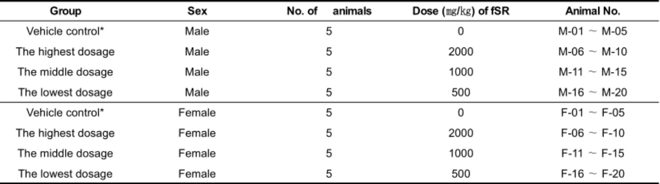
Group Sex No. of animals Dose (㎎/㎏) of fSR Animal No.
Vehicle control* Male 5 0 M-01 ∼ M-05
The highest dosage Male 5 2000 M-06 ∼ M-10
The middle dosage Male 5 1000 M-11 ∼ M-15
The lowest dosage Male 5 500 M-16 ∼ M-20
Vehicle control* Female 5 0 F-01 ∼ F-05
The highest dosage Female 5 2000 F-06 ∼ F-10
The middle dosage Female 5 1000 F-11 ∼ F-15
The lowest dosage Female 5 500 F-16 ∼ F-20
Table 1. Experimental Design Used in This Study
* Vehicle control; distilled water 20 ㎖/㎏ as vehicle in this study. All test articles in vehicle were once orally administered in a volume of 20 ㎖/㎏, dissolved in distilled water.
18시간 전 절식을 실시하였으며 (이 기간에도 음 수는 자유롭게 공급하였다), picric acid로 개체 를 식별하였다.
2. 홍국 발효 황금 추출물
본 실험에 사용된 황금 (
Scutellaria baicalensis Georgi
)은 지역 약업사 (Omniherb, Korea)에서 매입한 것을 대구한의대학교 한의과대학 본초학 교실에서 관능검사를 통하여 선정하여 사용하였 으며, 선정된 황금 100g을 취하여 정제수 1L로 80℃에서 3시간 동안 3번 가열 추출한 후, 흡인 여과한 여과액을 rotary vacuum evaporator (Buchi Rotavapor R144, Buchi Labortechnik AG, Switzland)로 감압·농축하여 점조성의 추 출물을 얻은 다음 programmable freeze dryer (Labconco Freezone1, Labconco Corp., MO, USA)를 사용하여 동결 건조시켜, 총 272.00g (수 율 약 27.20%)의 연갈색 물 추출물을 얻어 실험 에 사용하였다. 준비한 황금 물 추출물에 고체 배 양한 홍국균 1.15%를 첨가하여, 40℃에서 3일간 배양하여, 홍국 발효 황금 추출물을 준비하였다.홍국균의 고체 배양은 PDA (potato dextrose agar:
Difco Co., USA) 배지를 사용하여 24℃에서 4일 간 균체를 배양한 후, 배지 중량의 10%에 상당하 는 멸균수를 배지에 첨가하고, 분쇄하여 사용하였 다. 홍국 발효 황금 추출물은 고압 멸균기 (Hanback ST. Co., Korea)로 멸균한 다음, 전기 미세분쇄 기 (Sungchang Machine Co., Sungnam, Korea) 를 사용하여, 세말하여, 연갈색의 분말을 얻어 실
험에 사용하였다. 준비한 홍국 발효 황금 추출물 은 -20℃의 냉장고에 보관 후 실험에 사용하였 으며, 본 실험에서 사용한 용매인 증류수에 100
㎎/㎖의 농도까지 비교적 잘 용해되었다.
3. 군분리 및 약물의 투여
실험동물은 군당 5마리씩 Table 1에 기록한 8그 룹으로 구분하였다. 현재까지 홍국 발효 황금에 대 한 독성자료 역시 찾아 볼 수 없어, 한국식품의약 품안전처 고시8와 OECD 실험기준9)에 의거하여, 설치류 최고 한계투여용량인 2,000㎎/㎏을 최고 용량으로 설정하였으며, 공비 2로 1,000 및 500
㎎/㎏을 중간 및 저용량 투여군으로 설정하였다.
또한 암수 각각에 대한 매체 대조군(vehicle)을 추 가 하였다. 홍국 발효 황금은 멸균 증류수에 용해 시켜 20㎖/㎏의 용량으로 overnight 절식 후 존 데(sonde)가 부착된 1㎖ 주사기를 이용하여 경구 투여 하였으며, 매체 대조군에서는 동일한 용량의 멸균 증류수만 공급하였다. 식이와 음수에 따른 약물의 흡수 변화를 최소화하기 위해, 투여 후 대 략 3시간 동안 사료와 음수 공급을 제한하였다 (Table 1).
4. 임상증상의 관찰
모든 실험동물의 임상증상을 투여 전후에 각각 functional observational battery test10-11)를 기초하여 동물의 행동, 자극에 대한 반응성, 각성 도 및 경계성, 자세 및 보행 이상 등에 관한 일반 증상을 관찰, 기록하였으며, 투여일 이후에도 하
Park et al., Mouse Single Oral Dose Toxicity Test Of Red Koji Fermented Scutellariae Radix Aqueous Extracts
루에 최소한 2번씩 모든 실험동물의 임상증상을 관찰, 기록하였다.
5. 체중의 측정
모든 실험동물의 체중을 투여전 1일, 투여직전, 투여 후 1, 2, 7, 13 및 14일 (최종 희생일)에 각 각 측정하였으며, 실험 시작시 개체차이에 따른 체중의 변화를 최소화하기 위하여, 투여일 ∼ 투 여 후 7일, 투여 후 7일 ∼ 13일 및 투여일 ∼ 투 여 후 14일간의 체중 증가량인 증체량을 각각의 체중을 이용하여 산출하였다.
6. 육안 부검
투여 14일 후 모든 실험동물은 overnight 절식 을 실시하였으며, ethyl ether (Duksan Pure Chemical Co., Korea) 마취하에 부검을 실시하 고, 폐, 가슴샘, 부신, 심장, 신장, 비장, 고환, 전 립샘, 간, 췌장, 부고환, 악하임파절, 난소, 뇌, 피 부, 소화관, 기도, 난관 및 자궁 등 주요 장기를 위주로 이상 육안소견을 각각 관찰, 기록하였다.
7. 장기 중량의 측정
모든 실험동물은 육안부검 소견을 관찰 기록한 후 폐, 심장, 가슴샘, 좌측 신장, 좌측 부신, 비장, 좌측 고환, 간, 췌장 비장엽, 좌측 부고환, 좌측 악하임파절, 좌측 난소, 뇌 및 자궁에 대한 절대 중량을 각각 측정하였으며, 체중의 변화에 수반 된 이차적 변화를 최소화하기 위해 체중에 대한 각각의 장기 절대중량의 비율인 상대 중량을 산 출하였다.
8. 조직 병리학적 관찰
폐, 심장, 가슴샘, 좌측 신장, 좌측 부신, 비장, 좌측 고환, 간, 췌장 비장엽, 좌측 부고환, 좌측 악하임파절, 좌측 난소, 뇌 및 자궁 등의 일부 조 직을 10% 중성포르말린에 18시간 이상 고정시킨 다음, 탈수를 거쳐 파라핀 포매 후 4㎛의 절편을 제작하였다. 이후 hematoxylin & eosin (H&E) 염색을 실시하고, 광학현미경 하에서 이상 유무 를 관찰, 기록하였다.
9. 통계처리
모든 수치는 평균 ± 표준편차로 표시하였으 며, 다중비교검증을 이용하여 통계처리를 실시하 였고, 분산동질성을 Levene test를 실시하여 검 증하였다. 등분산일 경우, one way ANOVA test 를 실시한 다음 Scheffe test로 사후 검증을 실시 하여 군간의 유의성을 측정하였다. 비등분산일 경 우에는 비모수 검증인 Kruskal-Wallis H test 를 실시하여 유의성이 인정된 경우에는, Mann- Whitney U (MW) test를 실시하여 군간의 유의 성을 검증하였다. 단회 투여독성시험의 경우, 반 수치사량 및 95% 신뢰한계 (confidence limits) 를 Probit 방법으로 측정하였으며, 임상 증상, 육 안부검 및 조직병리학적 소견은 각각 그 정도에 따라 0 (normal), 1+ (slight), 2+ (moderate) 및 3+ (severe)로 구분하였다. 통계처리 및 Probit 방법은 SPSS for Windows (Release 14.0K, SPSS Inc., USA)를 이용하여 평가하였으며, p-value 가 0.05 이하인 경우 통계적 유의성을 인정하였다.
Ⅲ. 결 과
1. 사망률
홍국 발효 황금 추출물 투여와 관련 있는 사망 례는 실험 전 기간 동안 관찰되지 않았다 (Table 2).
2. 임상 증상
홍국 발효 황금 추출물 투여와 관련된 임상증 상은 실험 전 기간 동안 인정되지 않았다.
3. 체중의 변화
본 실험의 결과, 홍국 발효 황금 추출물 투여 와 관련된 체중 및 증체량의 변화는 인정되지 않 았다 (Table 3).
4. 장기 중량의 변화
각각의 동일 성별 매체 대조군에 비해 의미 있는 장기중량의 변화는 모든 홍국 발효 황금 추출물
Groups
Day after administration
Total*
0a 1 2 3 4 5 6 7 8 9 10 11 12 13
Vehicle control
Male 0 0 0 0 0 0 0 0 0 0 0 0 0 0 0/5 (0%)
Female 0 0 0 0 0 0 0 0 0 0 0 0 0 0 0/5 (0%)
fSR treated male groups
2000 ㎎/㎏ 0 0 0 0 0 0 0 0 0 0 0 0 0 0 0/5 (0%)
1000 ㎎/㎏ 0 0 0 0 0 0 0 0 0 0 0 0 0 0 0/5 (0%)
500 ㎎/㎏ 0 0 0 0 0 0 0 0 0 0 0 0 0 0 0/5 (0%)
fSR treated female groups
2000 ㎎/㎏ 0 0 0 0 0 0 0 0 0 0 0 0 0 0 0/5 (0%)
1000 ㎎/㎏ 0 0 0 0 0 0 0 0 0 0 0 0 0 0 0/5 (0%)
500 ㎎/㎏ 0 0 0 0 0 0 0 0 0 0 0 0 0 0 0/5 (0%)
Table 2. Mortalities Observed in Female and Male Mice after Single Oral Treatment of fSR
Values are expressed as number of died animals; a treatment day; fSR, Red-Koji (Monascus purpureus 12002) fermented Scutellariae Radix aqueous extracts (yield=27.20%); * total mortalities during 14 days of observation periods – died animals/total observed animals (percentages; 5 mice per group)
Groups
Intervals
Day 0a∼Day7 Day 7∼Day 13 Day 0∼Day 14b
Vehicle control
Male 8.14±1.19 1.96±0.29 4.40±1.02
Female 6.14±1.91 0.70±2.48 1.92±2.75
fSR treated male groups
2000 ㎎/㎏ 9.40±1.29 1.92±0.86 6.18±1.33
1000 ㎎/㎏ 8.42±1.23 1.46±0.93 4.29±1.34
500 ㎎/㎏ 7.94±1.42 0.80±1.57 3.84±2.36
fSR treated female groups
2000 ㎎/㎏ 5.54±2.48 0.90±1.37 1.44±2.69
1000 ㎎/㎏ 5.02±0.88 1.90±0.92 2.62±1.31
500 ㎎/㎏ 5.18±1.68 2.20±1.27 2.98±1.73
Table 3. Weight Gains in Female and Male Mice after Single Oral Treatment of fSR
Values are expressed as mean ± SD of five mice; fSR, Red-Koji (Monascus purpureus 12002) fermented Scutellariae Radix aqueous extracts (yield=27.20%); a Day of treatment after overnight fasted; b Day of sacrifice after overnight fasted
Park et al., Mouse Single Oral Dose Toxicity Test Of Red Koji Fermented Scutellariae Radix Aqueous Extracts
투여군에서 인정되지 않았다 (Table 4∼7).
5. 부검 소견
다양한 [1 ∼ 3+] 정도의 폐 충출혈(congestion), 가슴샘 위축, 비장 위축, 악하임파절 종대 및 자궁 부종 소견이 암수 매체 대조군을 포함한 모든 실 험군에 걸쳐 산발적으로 관찰된 이외에, 홍국 발 효 황금 추출물 투여와 관련된 의미 있는 육안 부 검소견은 인정되지 않았다 (Table 8∼9).
6. 조직병리학적 소견
다양한 정도의 폐 충출혈 – 폐포 벽의 비후와 염증세포 및 적혈구 축적 (Fig. 1), 가슴샘 피질 임 파구 감소 (Fig. 2), 비장 적색수질 임파구 증생 (Fig. 3), 간 국소 염증세포 침윤 (Fig. 4) 및 자 궁 점막 탈락 (Fig. 5) 소견이 암수 매체 대조군 을 포함한 모든 실험군에 걸쳐 산발적으로 관찰 된 이외에 홍국 발효 황금 추출물 투여와 관련된 조직병리학적 소견은 인정되지 않았다 (Table 10, 11).
Groups Principal Organs
Lung Heart Thymus Kidney L Adrenal gland L Spleen
Vehicle control 0.210±0.023 0.191±0.010 0.063±0.014 0.321±0.034 0.003±0.001 0.101±0.017 fSR treated groups
2000 ㎎/㎏ 0.207±0.012 0.191±0.008 0.058±0.013 0.325±0.048 0.003±0.002 0.119±0.014 1000 ㎎/㎏ 0.209±0.008 0.194±0.010 0.047±0.008 0.289±0.013 0.003±0.001 0.129±0.025 500 ㎎/㎏ 0.195±0.008 0.188±0.017 0.047±0.011 0.310±0.063 0.002±0.001 0.096±0.029
Table 4. Changes on the Absolute Organ Weights Observed in Male Mice after Single Oral Treatment of fSR
Testis L Liver Pancreas S Brain Epididymis L Lymph node La
Vehicle control 0.119±0.007 1.614±0.032 0.178±0.010 0.499±0.010 0.050±0.003 0.005±0.004 fSR treated groups
2000 ㎎/㎏ 0.128±0.017 1.607±0.133 0.194±0.013 0.488±0.012 0.048±0.004 0.004±0.002 1000 ㎎/㎏ 0.131±0.010 1.552±0.100 0.188±0.036 0.494±0.012 0.046±0.004 0.005±0.002 500 ㎎/㎏ 0.111±0.004 1.530±0.205 0.173±0.014 0.491±0.022 0.045±0.005 0.003±0.002 Values are expressed as mean ± SD of five mice; fSR, Red-Koji (Monascus purpureus 12002) fermented Scutellariae Radix aqueous extracts (yield=27.20%); L, left sides; S, splenic lobes; a Submandibular lymph node
Ovary L Liver Pancreas S Brain Uterus Lymph node La
Vehicle control 0.020±0.003 1.175±0.199 0.166±0.025 0.488±0.012 0.195±0.081 0.002±0.001 fSR treated groups
2000 ㎎/㎏ 0.018±0.009 1.180±0.202 0.150±0.016 0.486±0.030 0.177±0.083 0.003±0.002 1000 ㎎/㎏ 0.022±0.005 1.203±0.037 0.160±0.017 0.468±0.029 0.171±0.045 0.004±0.001 500 ㎎/㎏ 0.018±0.005 1.298±0.194 0.151±0.015 0.485±0.015 0.245±0.040 0.005±0.003
Table 5. Changes on the Absolute Organ Weights Observed in Female Mice after Single Oral Treatment of fSR
Groups Principal Organs
Lung Heart Thymus Kidney L Adrenal gland L Spleen
Vehicle control 0.179±0.021 0.149±0.021 0.058±0.021 0.197±0.020 0.004±0.001 0.139±0.047 fSR treated groups
2000 ㎎/㎏ 0.179±0.017 0.148±0.009 0.052±0.020 0.208±0.013 0.003±0.001 0.125±0.035 1000 ㎎/㎏ 0.172±0.015 0.140±0.007 0.069±0.014 0.196±0.010 0.005±0.002 0.117±0.030 500 ㎎/㎏ 0.180±0.008 0.149±0.008 0.048±0.020 0.215±0.020 0.005±0.003 0.107±0.018 Values are expressed as mean ± SD of five mice; fSR, Red-Koji (Monascus purpureus 12002) fermented Scutellariae Radix aqueous extracts (yield=27.20%); L, left sides; S, splenic lobes; a Submandibular lymph node
Groups
Principal Organs
Lung Heart Thymus Kidney L Adrenal gland L Spleen
Vehicle control 0.582±0.069 0.528±0.032 0.175±0.037 0.889±0.094 0.009±0.004 0.282±0.054 fSR treated groups
2000 ㎎/㎏ 0.545±0.045 0.505±0.030 0.153±0.033 0.853±0.096 0.009±0.005 0.314±0.045 1000 ㎎/㎏ 0.578±0.040 0.538±0.045 0.131±0.022 0.800±0.029 0.009±0.003 0.358±0.072 500 ㎎/㎏ 0.545±0.051 0.523±0.048 0.130±0.023 0.853±0.082 0.007±0.002 0.297±0.058
Table 6. Changes on the Relative Organ Weights Observed in Male Mice after Single Oral Treatment of fSR
Testis L Liver Pancreas S Brain Epididymis L Lymph node La
Vehicle control 0.331±0.020 4.470±0.165 0.492±0.023 1.383±0.047 0.138±0.009 0.014±0.012 fSR treated groups
2000 ㎎/㎏ 0.340±0.055 4.231±0.290 0.513±0.055 1.287±0.085 0.127±0.018 0.009±0.006 1000 ㎎/㎏ 0.364±0.040 4.292±0.283 0.520±0.094 1.368±0.074 0.129±0.016 0.013±0.005 500 ㎎/㎏ 0.309±0.024 4.234±0.160 0.484±0.059 1.372±0.143 0.129±0.020 0.007±0.003 Values are expressed as mean ± SD of five mice, % of body weight; fSR, Red-Koji (Monascus purpureus 12002) fermented Scutellariae Radix aqueous extracts (yield=27.20%); L, left sides; S, splenic lobes; a Submandibular lymph node
Groups
Principal Organs
Lung Heart Thymus Kidney L Adrenal gland L Spleen
Vehicle control 0.633±0.033 0.528±0.048 0.201±0.050 0.701±0.066 0.016±0.003 0.485±0.117 fSR treated groups
2000 ㎎/㎏ 0.649±0.072 0.536±0.054 0.185±0.053 0.752±0.059 0.011±0.004 0.446±0.088 1000 ㎎/㎏ 0.596±0.023 0.487±0.011 0.239±0.040 0.681±0.034 0.017±0.005 0.408±0.100 500 ㎎/㎏ 0.618±0.060 0.511±0.049 0.161±0.053 0.736±0.059 0.016±0.010 0.363±0.029
Table 7. Changes on the Relative Organ Weights Observed in Female Mice after Single Oral Treatment of fSR
Ovary L Liver Pancreas S Brain Uterus Lymph node La
Vehicle control 0.070±0.010 4.147±0.412 0.587±0.080 1.741±0.179 0.682±0.233 0.008±0.004 fSR treated groups
2000 ㎎/㎏ 0.064±0.024 4.221±0.313 0.541±0.046 1.766±0.250 0.624±0.241 0.009±0.004 1000 ㎎/㎏ 0.075±0.013 4.192±0.321 0.558±0.058 1.627±0.106 0.592±0.146 0.013±0.003 500 ㎎/㎏ 0.062±0.013 4.424±0.348 0.518±0.037 1.668±0.156 0.849±0.211 0.015±0.010 Values are expressed as mean ± SD of five mice, % of body weight; fSR, Red-Koji (Monascus purpureus 12002) fermented Scutellariae Radix aqueous extracts (yield=27.20%); L, left sides; S, splenic lobes; aSubmandibular lymph node
Park et al., Mouse Single Oral Dose Toxicity Test Of Red Koji Fermented Scutellariae Radix Aqueous Extracts
Groups Organs- Findings
Vehicle control
fSR treated as 2000
㎎/㎏
1000
㎎/㎏
500 ㎎/㎏
Lung Normal Congestion
4/5 1/5
4/5 1/5
4/5 1/5
4/5 1/5
Thymus Normal Atrophy
5/5 0/5
5/5 0/5
4/5 1/5
5/5 0/5
Spleen Normal Atrophy
3/5 2/5
4/5 1/5
5/5 0/5
3/5 2/5
Lymph nodea Normal Hypertrophy
3/5
2/5 4/5
1/5
4/5 1/5
4/5 1/5
Table 8. Necropsy Findings Observed Male Mice after
Single Oral Treatment of fSR
Observed animals/total observed animals of five mice; fSR, Red-Koji (Monascus purpureus 12002) fermented Scutellariae Radix aqueous extracts (yield=27.20%); a Bilateral submandibular lymph node.
Groups Organs- Findings
Vehicle control
fSR treated as 2000
㎎/㎏
1000 ㎎/㎏
500
㎎/㎏
Lung Normal Congestion
4/5 1/5
4/5 1/5
3/5 2/5
5/5 0/5 Thymus
Normal Atrophy
3/5 2/5
4/5 1/5
5/5 0/5
4/5 1/5 Spleen
Normal Atrophy
4/5 1/5
5/5 0/5
5/5 0/5
5/5 0/5 Uterus
Normal Edematous
2/5 3/5
3/5 2/5
2/5 3/5
0/5 5/5 Lymph nodea
Normal Hypertrophy
5/5 0/5
3/5 2/5
5/5 0/5
4/5 1/5
Table 9. Necropsy Findings Observed Female Mice
after Single Oral Treatment of fSR
Observed animals/total observed animals of five mice; fSR, Red-Koji (Monascus purpureus 12002) fermented Scutellariae Radix aqueous extracts (yield=27.20%); a Bilateral submandibular lymph node.
Groups Organs- Findings
Vehicle control
fSR treated as 2000
㎎/㎏
1000
㎎/㎏ 500 ㎎/㎏
Lung Normal Congestion
4/5 1/5
4/5 1/5
4/5 1/5
4/5 1/5 Thymus
Normal cDE†
5/5 0/5
5/5 0/5
4/5 1/5
5/5 0/5 Spleen
Normal rHP†
4/5 1/5
4/5 1/5
4/5 1/5
5/5 0/5 Liver
Normal IF†
3/5 2/5
5/5 0/5
4/5 1/5
5/5 0/5
Table 10. Histopathological Findings Observed Male
Mice
O bserved anim als/total observed anim als of five m ice; fSR , Red-Koji (M onascus purpureus 12002) ferm ented Scutellariae Radix aqueous extracts (yield=27.20% ); a Left subm andibular lym ph node; †Abbreviations: cDE, decreases of cortex lym phoid cells; rHP, hyperplasia of red pulp lym phoid cells; IF, focal inflam m atory cell infiltration
Groups Organs- Findings
Vehicle control
fSR treated as
2000 ㎎/㎏ 1000 ㎎/㎏ 500 ㎎/㎏
Lung Normal Congestion
4/5 1/5
4/5 1/5
3/5 2/5
5/5 0/5 Thymus
Normal cDE†
3/5 2/5
3/5 2/5
4/5 1/5
5/5 0/5 Spleen
Normal rHP†
3/5 2/5
4/5 1/5
3/5 2/5
4/5 1/5 Liver
Normal IF†
4/5 1/5
4/5 1/5
4/5 1/5
5/5 0/5 Uterus
Normal DM†
4/5 1/5
5/5 0/5
5/5 0/5
4/5 1/5
Table 11. Histopathological Findings Observed
Female Mice
O bserved anim als/total observed anim als of five m ice; fSR, Red-Koji (Monascus purpureus 12002) fermented Scutellariae Radix aqueous extracts (yield=27.20% ); a Left subm andibular lym ph node; †Abbreviations: cDE, decreases of cortex lym phoid cells; rHP, hyperplasia of red pulp lym phoid cells; IF, focal inflam m atory cell infiltration; DM , desquam ation of m ucosa layers
Fig. 1. Histopathological changes detected on the lung. Note that slight (1+) lung focal congestional spots-thickening of alveolar lung inflammatory cell infiltration with/without focal hemorrhages were randomly detected throughout the experimental groups tested regardless of genders including both genders of vehicle controls as sporadic findings not fSR treatment related toxicological signs. fSR, Red-Koji (Monascus purpureus 12002) fermented Scutellariae Radix aqueous extracts (yield=27.20%); A, alveolar sac-respiratory bronchiole; B, bronchiole; All hematoxylin & eosin stain; Scale bars = 200 ㎛.
Fig. 2. Histopathological changes detected in the thymus. Note that slight [1+] or moderate [2+] decreases of lymphoid cells in the thymic cortex (cDE; as seen vacuoles or loosening) were detected in the female vehicle control, female 2,000 ㎎ /㎏, male 1,000 ㎎/㎏ and female 500 ㎎/㎏ of fSR treated mice as sporadic findings; fSR, Red-Koji (Monascus purpureus 12002) fermented Scutellariae Radix aqueous extracts (yield=27.20%); C, cortex; M, medulla; All hematoxylin & eosin stain; Scale bars = 200 ㎛.
Park et al., Mouse Single Oral Dose Toxicity Test Of Red Koji Fermented Scutellariae Radix Aqueous Extracts
Fig. 3. Histopathological changes detected on the spleen. Note that slight [1+]
hyperplasia of lymphoid cells and megakar- yocytes in the red pulp (rHP) were randomly detected throughout the experimental groups including both genders of vehicle controls as sporadic findings not fSR treatment related toxicological signs.
fSR, Red-Koji (Monascus purpureus 12002) fermented Scutellariae Radix aqueous extracts (yield=27.20%); M, megakaryocytes, All hematoxylin & eosin stain; Scale bars = 200 ㎛.
Fig. 4. Histopathological changes detected in the liver. Note that slight [1+] focal inflammatory cell infiltrations (IF) were detected throughout the experimental groups including both genders of vehicle controls as sporadic findings not fSR treatment related toxicological signs. fSR, Red-Koji (Monascus purpureus 12002) fermented Scutellariae Radix aqueous extracts (yield=27.20%); C, central vein; All hematoxylin & eosin stain; Scale bars = 200 ㎛.
Fig. 5. Histopathological changes detected in the uterus. Note that slight [1+] or severe [3+] desquamation of uterus mucosa (DM) were observed in one (1/5; 20%) female mice in vehicle control, fSR 2,000 and 500 ㎎/㎏ treated group as a process of estrus cycle, respectively. fSR, Red-Koji (Monascus purpureus 12002) fermented Scutellariae Radix aqueous extracts (yield=27.20%); L, lumen; All hematoxylin & eosin stain; Scale bars = 200 ㎛.
Ⅳ. 고 찰
본 연구에서는 한의학에서 청열약으로 널리 사 용하는 황금은 氣味상 苦寒한데 苦味는 燥濕하고 寒性은 淸熱하여 淸熱燥濕하는 常用藥物이 되어, 濕熱諸症에 사용하는데, 특히 肺經의 熱을 瀉하 는데 더욱 우수하다. 그러므로 邪熱이 肺를 犯하 여 나타나는 肺熱咳嗽, 淡黃粘稠 등의 證에 반드 시 사용하는 대표적인 淸熱藥의 하나이다12). 황 금 추출물 및 황금 유래 성분, 특히 Flavonoids 로서 baicalein 및 wogonin의 약리효과에 대한 많 은 연구가 진행 되어 왔으며, 항바이러스 효과13), 항 당뇨 효과14), 신경계 보호 효과15), 조혈작용16), 항산화 효과17), 항암효과18), 면역활성19), 항염효 과20), ritonavir 독성완화 효과21), 항돌연변이 원 성22)이 비교적 잘 알려져 있다. 또한, 황금물 추 출물의 마우스 단회 경구투여 독성시험23), 황금 과 아카시아 복합 추출물에 대한 랫트 90일 반복 경구 투여 독성실험24), 황금 에탄올 추출물 및 황 금의 flavonoid 성분들에 대한 랫트 단회 투여 독 성실험25) 등 안전성과 관련된 연구도 다양하게 보 고되고 있다.
황금 추출물을 생리활성을 증가시키기 위해 홍 국균 (
Monascus purpureus
12002; 한국식품개 발연구원)으로 발효시킨 홍국 발효 황금 추출물 의 일반 독성시험 중 현재 한국식품의약품안전처고시 의약품 등의 독성시험기준8)에 명시되어 있 는 마우스 경구 단회투여독성 시험을 실시하여, 장기투여 독성 시험과 생식 발생독성 시험을 위 시한 특수 독성시험에 대한 기초자료를 제공하고 자 하였다.
현재까지 발효 황금 추출물에 대한 독성 자료 를 찾아 볼 수 없어, 본 실험에서는 한국식품의약 품안전처 고시8)와 OECD 실험기준9)에 의거하여, 설치류 최고 한계투여용량인 2,000㎎/㎏을 최고 용량으로 설정하였으며, 공비 2로 1000 및 500
㎎/㎏을 중간 및 저용량으로 설정하여, 단회 경구 투여하고, 14일간 체중 및 임상증상을 관찰하였 으며, 14일 후 최종 부검을 통하여 12개의 주요 장 기에 대한 장기 중량, 육안 부검 및 조직병리학적 관찰을 실시한 결과, 단회투여 독성 시험에서 설 치류 최대 한계투여 용량인 2,000㎎/㎏까지 홍국 발효 황금 추출물 투여와 관련된 사망례가 인정되 지 않았으며, 홍국 발효 황금 추출물의 투여와 관 련된 임상증상, 체중, 증체량 및 장기 중량의 변 화 역시 인정되지 않았다. 또한 육안부검시 다양 한 정도의 폐 충출혈, 가슴샘 위축, 비장 위축, 악 하임파절 종대 및 자궁 부종 소견이 매체 대조군 을 포함한 모든 실험군에서 산발적으로 인정되었 고, 조직병리학적 검사시 다양한 정도의 폐 충출 혈, 가슴샘 피질 임파구 감소, 비장 적색수질 임 파구 증생, 간 국소 염증세포 침윤 및 자궁 점막 탈락 소견이 암수 매체 대조군을 포함한 모든 실 험군에 걸쳐 산발적으로 관찰된 이외에 홍국 발
Park et al., Mouse Single Oral Dose Toxicity Test Of Red Koji Fermented Scutellariae Radix Aqueous Extracts
효 황금 추출물 투여와 관련된 육안 부검 및 조직 병리학적 소견은 인정되지 않았다.
본 실험에 사용한 실험동물의 체중 및 장기중 량은 동일한 주령의 마우스들의 정상 범주26-7)에 포함되어 관찰되었으며, 각각 동일 성별의 매체 대 조군과 비교하여, 홍국 발효 황금 추출물 투여와 관련된 체중 및 증체량의 변화는 인정되지 않았다.
한국식품의약품안전처 고시8) 및 OECD 기준9) 에 따르면, 설치류에서 투여한계 농도를 2000㎎/㎏
또는 최대 용해농도로 규정하고 있고, 투여 용량 은 20㎖/㎏을 넘지 못하게 규정하고 있다. 본 실 험에서 사용한 홍국 발효 황금 추출물은 멸균 증 류수에 100㎎/㎖까지 비교적 잘 용해되어, 20㎖
/㎏의 용량 즉, 2,000㎎/㎏을 최고 농도로 투여 하였다.
육안 부검시 인정된 다양한 정도의 폐 충출혈, 가슴샘 위축, 비장 위축, 악하임파절 종대 및 자 궁 부종 소견들과 조직병리학적 검사시 인정된 다 양한 정도의 폐 충출혈, 가슴샘 피질 임파구 감소, 비장 적색수질 임파구 증생, 간 국소 염증세포 침 윤 및 자궁 점막 탈락 소견은 암수 매체 대조군을 포함한 모든 실험군에 걸쳐 산발적으로 관찰되었 으며, 용량 의존성이 전혀 인정되지 않아, 홍국 발 효 황금 추출물 투여와 관련 없는 우발적 병소로 판단되고, 이들 병소들은 정상 마우스에서 드물게 인정되는 소견으로 알려져 있고28-9), 특히 자궁은 성주기(estrus cycle)에 따라 쉽게 변화할 수 있 어30) 자궁 부종은 성주기에 따른 이차적 변화로 판단된다.
US Environmental Protection Agency31)에 따 르면 일반적으로 반수 치사량이 5,000~15,000㎎
/㎏인 물질을 무독성 물질로, 500~5,000㎎/㎏을 비교적 저독성 (Class III) 물질로 규정하고 있으 나, 한국식품의약품 안전청 고시8) 및 OECD 기준9) 에 따르면, 설치류에서 투여한계 농도를 2,000
㎎/㎏으로 제한하고 있다. 따라서 본 실험의 마우 스 단회 경구투여 독성실험에서 홍국 발효 황금 추출물의 반수치사량 및 개략적 치사량은 각각 2,000㎎/㎏ 이상으로 관찰되었다.
Ⅴ. 결 론
설치류에서 투여한계 농도인 2,000㎎/㎏까지 홍국 발효 황금 추출물 투여와 관련된 사망례는 인정되지 않아, 마우스 단회 경구투여 독성실험 에서 홍국 발효 황금 추출물의 반수치사량 및 개 략적 치사량은 각각 2000㎎/㎏ 이상으로 관찰되 어, 비교적 안전한 약물로 판단된다.
참 고 문 헌
1. Kim DH, Kim HM, Ryu JH, UM JY, Kim SC, Y JH, Cho MK, Yim JP, Hong SH.
Herbal Pharmacology. Seoul:Shin-Il books.
2001:242-6.
2. Bae JH, Lee MJ, Lee SM. Antimicrobial Effect of Cutellaria baicalensis Geroge Ex- tracts on Food-Borne Pathogens. J Microbiol Biotechnol. 2005;33:35-40.
3. Trinh HT, Han SJ, Kim SW, Lee YC, Kim DH. Bifidus fermentation increases hypoli- pidemic and hypoglycemic effects of red ginseng. J Microbiol Biotechnol. 2007;17:
1127-33.
4. Lee SI, Shin JG, Kim SD. Effect of ginseng- chungkukjang effects on lipid profiles of serum in alcohol administered diabetic- induced rats. J Korean Soc Food Sci Nutr.
2005b;34:1362-6.
5. Cha JY, Park JC, Ahn HY, Eom KE, Park BK, Jun BS, Lee CH, Cho YS. Effect of Mo- nascus purpureus-Fermented Korean Red Ginseng Powder on the Serum Lipid Levels and Antioxidative Activity in Rats. J Korean Soc Food Sci Nutr. 2009;38:1153-60.
6. Tsukahara M, Shinzato N, Tamaki Y, Nami- hira T, Matsui T. Red yeast rice fermenta- tion by selected Monascus sp.with deep-red
color, lovastatin production but no citrinin, and effect of temperature-shift cultivation on lovastatin production. Appl Biochem Biotechnol. 2009;158:476-82.
7. Pyo YH, Lee TC. The potential antioxidant capacity and angiotensin I-converting enzyme inhibitory activity of Monascus-fermented soybean extracts:evaluation of Monascus- fermented soybean extracts as multifun- ctional food additives. J Food Sci. 2007;72:
S218-23.
8. KFDA. Toxicity Study Criteria of medicine and medical supplies. Seoul. KFDA. noti- fication No. 2009-116. 2009.
9. Organization for Economic Co-Operation and Development (Ed.). OECD guideline (423) for testing of chemicals - acute oral toxicity-acute toxic class method. 2001.
10. Irwin S. Comprehensive observational as- sessment: Ia. A systemic, quantitative procedure for assessing the behavioral and physiological state of the mouse. Psycho- pharmacology. 1968;13:222-57.
11. Dourish CT. Effects of drugs on spontaneous motor activity. In: Experimental Psychop- harmacology. Greenshaw AJ, Dourish CT, Ed. Clifton:Humana Press. 1987:325-34.
12. Kim IR, Kim HC, Kook YB, Park SJ, Park YG, Park JH, Seo BI, Seo YB, Song HJ, Shin MK, Lee YJ, Lee YC, Lee JH, Yim KH, Cho SI, Jeong JG, Joo YS, Choi HY.
Herbology. Seoul:Younglim.2007;216-18.
13. Błach-Olszewska Z, Jatczak B, Rak A, Lorenc M, Gulanowski B, Drobna A, Lamer- Zarawska E. Production of cytokines and stimulation of resistance to viral infection in human leukocytes by Scutellaria baicalensis flavones. J Interferon Cytokine Res. 2008;28:
571-81.
14. Waisundara VY, Hsu A, Huang D, Tan BK.
Scutellaria baicalensis enhances the anti- diabetic activity of metforminin strepto- zotocin-induced diabetic Wistar rats. Am J Chin Med. 2008;36:517-40.
15. Sun X, Chan LN, Gong X, Sucher NJ N- methyl-D-aspartate receptor antagonist activity in traditional Chinese stroke medi- cines. Neurosignals. 2003;12:31-8.
16. Udut EV, Zhdanov VV, Gur'iantseva LA, Minakova MIu, Dygai AM. Mechanisms of the erythropoiesis-stimulating effect of skullcap (Scutellaria baicalensis) extract.
Eksp Klin Farmakol. 2005;68:43-5.
17. Przybylska D, Długosz A. The research on Antoxid activity in free radicals processes.
Acta Pol Pharm. 2007;64:491-5.
18. Himeji M, Ohtsuki T, Fukazawa H, Tanaka M, Yazaki S, Ui S, Nishio K, Yamamoto H, Tasaka K, Mimura A. Difference of growth- inhibitory effectof Scutellaria baicalensis- producing flavonoid wogonin among human cancercells and normaldiploidcell. Cancer Lett. 2007;245:299-74.
19. Wang JY, Chuang HN, Chiu JH, Fu SL, Tsai TH, Tsou AP, Hu CP, Chi CW, Yeh SF, Lui WY, Wu CW, Chou CK. Effects of Scut- ellariabaicalensis Georgi on macrophage- hepatocyte interaction through cytokines related to growth control of murine hepa- tocytes. Exp Biol Med(Maywood). 2006;231:
444-55.
20. Huang ST, Lee Y, Gullen EA, Cheng YC.
Impacts of baicalein analogs with modification of the 6th position of A ring on the activity toward NF-kappaB-, AP-1-, or CREB- mediated transcription. Bioorg Med Chem Lett. 2008b;18:5046-9.
21. Mehendale S, Aung H, Wang CZ, Tong R, Foo A, Xie JT, Yuan CS. Scutellaria bai- calensis and a constituent flavonoid, bai -
Park et al., Mouse Single Oral Dose Toxicity Test Of Red Koji Fermented Scutellariae Radix Aqueous Extracts
calein, attenuate ritonavir-induced gastroin- testinal side-effects. J Pharm Pharmacol.
2007;59:1567-72.
22. Lee BH, Lee SJ, Kang TH, Kim DH, Sohn DH, Ko GI, Kim YC. Baicalein: an in vitro antigenotoxic compound from Scutellaria baicalensis. Planta Med. 2000;66:70-1.
23. Lee JW, Jung YS, Jung TY, Kim JD, Choi HY. Mouse Single Oral Dose Toxicity Test of Scutellariae Radix Aqueous Extracts.
Korean J Orient Int Med. 2013;34:46-59.
24. Yimam M, Zhao Y, Ma W, Jia Q, Do SG, Shin JH. 90-day oral toxicity study of UP446, a combination of defined extracts of Scutellaria baicalensis and Acacia catechu, in rats. Food Chem Toxicol. 2010;48:1202 -9.
25. Chang BY, Malla B, Sohn DH, Kim YC, Kim SY. Acute Toxicity of a Combined Preparation of the Standardized Extracts of Scutellaria baicalensis GEORGI and Salvia miltiorrhiza BUNGE. Kor J Pharmacogn.
2011;42: 295-70.
26. Plata EJ, Murphy WH. Growth and haema-
tologic properties of the BALB/wm strain of inbred mice. Lab Anim Sci. 1972;22:
712-20.
27. Yamaguchi C, Fujita S, Obara T, Ueda T.
Effects of room temperature on repro- duction, body weight and organ weights, food and water intakes, and hematology in mice. Exp Anim. 1983;32:1-11.
28. Lee JH, Yang KJ, Shin HD, Park BR, Son CW, Jang HJ, Park DC, Lee HS, Ku SK. Single subcutaneous dose toxicity of Polycan® ,a β-glucan originated from Aureobasidium in mice. Lab Anim Res. 2005a;21:299-305.
29. Lee HS, Lee IG, Ku SK. Single oral dose toxicity study of water extracts of Picrorrhiza Rhizoma in mice. J Toxicol Pub Health.
2006;22:117-29.
30. Banks WJ. Female reproductive system.
In: Banks WJ, ed. Applied veterinary histology, 2nd ed. Baltimore:Williams &
Wilkins. 1988:506-29.
31. US Enviromental Protection Agency Health Effects Test Guidelines OPPTS 870.100.
Acute Toxicity Testing Backround. US EPA August. Washington. USA. 1998.

![Fig. 2. Histopathological changes detected in the thymus. Note that slight [1+] or moderate [2+] decreases of lymphoid cells in the thymic cortex (cDE; as seen vacuoles or loosening) were detected in the female vehicle control, fe](https://thumb-ap.123doks.com/thumbv2/123dokinfo/5453811.435670/9.892.115.778.719.1005/histopathological-detected-moderate-decreases-lymphoid-vacuoles-loosening-detected.webp)
![Fig. 4. Histopathological changes detected in the liver. Note that slight [1+] focal inflammatory cell infiltrations (IF) were detected throughout the experimental groups including both genders of vehicle controls as sporadic findings not fS](https://thumb-ap.123doks.com/thumbv2/123dokinfo/5453811.435670/10.892.110.784.135.999/histopathological-detected-inflammatory-infiltrations-detected-experimental-including-controls.webp)
![Fig. 5. Histopathological changes detected in the uterus. Note that slight [1+] or severe [3+] desquamation of uterus mucosa (DM) were observed in one (1/5; 20%) female mice in vehicle control, fSR 2,000 and 500 ㎎/㎏ treated group as a process of estrus](https://thumb-ap.123doks.com/thumbv2/123dokinfo/5453811.435670/11.892.233.664.148.317/histopathological-changes-detected-desquamation-observed-vehicle-control-treated.webp)
