Abstract― Crystal structure of coumarin 6 has been solved by X-ray diffraction. The crystals are triclinic, space group P-1, with a=8.823(2) Å, b=8.898(2) Å, c=11.025(9) Å, α=86.41(3)°, β=85.39(3)°, γ=76.23(3)°, Mr=350.42, V=837.1(3) Å3, Z=2 and R=0.0516. The molecules are packed parallel to each other by weaker π···π and C–H···π interactions. The detailed geometry of C–H···π interactions were discussed. The hydrogen bonds and non-traditional C–H···O interactions join the no-parallel molecules together. All the molecules packed wall-like with the molecular brick.
Keywords: crystal structure, coumarin 6, π···π, C–H···π, C–H···O interactions, X-ray diffraction
C–H···π and C–H···O Interactions in Coumarin 6 : 3-(2-benzothiazolyl)-7-(diethylamino)-coumarin
Xiaochuan Li1 and Young-A Son†
BK 21 FTIT, Department of Organic Materials and Textile System Engineering, Chungnam National University, Daejeon, 305-764, South Korea
1College of Chemistry and Environmental Science, Henan Normal University, Xinxiang, Henan, 453-007, China (Received: January 25, 2010/Revised: March 19, 2010/Accepted: June 14, 2010)
1. Introduction
Coumarins are relatively old molecules, the discovery of which dates back to the end of the 19th century. Coumarins are of interest because they constitute an important class of compounds, occupying a special place in nature and they form sub-units of many natural products. Coumarin and its derivatives are also widely used as additives in food, perfumes, cosmetics, pharmaceuticals, and agrochemicals1,2).
Courmarins owe their commercial importance to their efficient light emission properties, their reasonable stability and their relative ease of synthesis. Coumarin fluorescent dyes are suitable for use in the coloration of synthetic fibers, in day light fluorescent pigments and in a range of applications which specifically exploit their light emission properties, including non-destructive flaw detection, tunable dye lasers and solar energy collectors. Coumarin 6, (3-(2-benzothiazolyl)-7- (diethylamino)-coumarin), is one of the most organic fluorescent materials with an electron- releasing group (N, N-diethylamino) in the 7- position and an electron-accepting heterocyclic group
†Corresponding author. Tel.: +82-42-821-6620; Fax.: +82-42-823-3736; e-mail: [email protected]
in the 3-position. There is some interest in the molecular design which could extend the available range of long-wavelength emitting fluorescent materials. The shift to longer wavelength has invariably been achieved by strengthening the electron-accepting character attached to the pyranone ring. The first single crystal of coumarin 6 was reported in 1995, in which the crystal data of it was investigated. In this paper, a single crystal of coumarin 6 with different packing model will be reported. Non-traditional C–H···O interactions were established in this crystal, which were not observed in the reported one. C–H···O interactions together with other interactions determined the packing model. It is helpful for us to understand and control the crystalline state and applied to the optical technologies.
2. Experimental
2.1 General Comments
Coumarin 6 is commercially available. And it can also be synthesized easily according to the reported methods3). Chemicals used for the synthesis were commercially available, were of AR grade,
and were used as received without further puri- fication. The yellow crystals of the title compound were obtained by slow evaporation of its solution in ethanol at room temperature. The crystalline solid product thus was separated by filtration, washed with ethanol and dried. Finally, single crystals suitable for X-ray diffraction were obtained.
2.2 Methodology
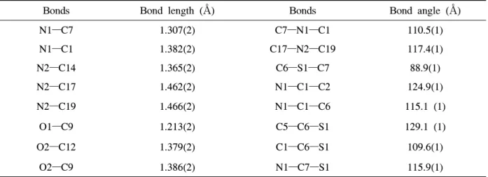
The crystal structure data collections was done on a ADSC Quantum 210 diffractometer using graphite synchrotron radiation (λ=0.76999 Å) at 293(2) K. A crystal with dimensions 0.20mm × 0.15mm × 0.10 mm was mounted on a glass fiber and detected. Cell constants and an orientation matrix for data collection were obtained from least-squares refinement. The structure was solved by direct methods with program SHELXS-97 and refined by full matrix least squares on F2 with SHELXL -974-6). Direct phase determination yielded the positions of all non-hydrogen atoms. All non- hydrogen atoms were subjected to anisotropic refine- ment. All hydrogen atoms were generated geo- metrically with C–H bond distances of 0.93–0.96 Å according to criteria described in the SHELXTL manual. The crystal used for the diffraction study showed no decomposition during data collection. A summary of the experimental and crystallographic data are presented in Table 1. Selected bond distances and angles can be seen in Table 2.
Bonds Bond length (Å) Bonds Bond angle (Å)
N1—C7 1.307(2) C7—N1—C1 110.5(1)
N1—C1 1.382(2) C17—N2—C19 117.4(1)
N2—C14 1.365(2) C6—S1—C7 88.9(1)
N2—C17 1.462(2) N1—C1—C2 124.9(1)
N2—C19 1.466(2) N1—C1—C6 115.1 (1)
O1—C9 1.213(2) C5—C6—S1 129.1 (1)
O2—C12 1.379(2) C1—C6—S1 109.6(1)
O2—C9 1.386(2) N1—C7—S1 115.9(1)
Table 1. Crystal data and structure refinement parameters
3. Results and Discussion
3.1 Geometry of the molecule
The molecular structure of coumarin 6 with atom numbering is given in Fig. 1. Crystal structure of coumarin 6 with triclinic(spacegroupP-1) has been reported3). In this paper, the space group of crystal coumarin 6 is same to the reported one with similar reference axes a, b, and c. However, the three angles between the axes are less than 90°, which is different from the reported crystal with two angle (α and β) larger than 90°. In this new crystal, the cormarin 6 crystallizes with two molecules per unit into triclinic crystal system with a space group of P-1. The bond lengths and bond angles of the two molecules in the crystal unit are all in the normal range. So the discussion can be limited to one of the molecules.
According to the crystal structure, the coumarin ring and the benzothiazoly ring systems in the molecule are planar. The pyrone ring is deviated 2.44°
compared to the thiazoly ring. Most of the basic data of this crystal is similar to the reported one.
However, the interactions generated in this crystal are completely different from the reported one.
3.2 C–H···O interactions
The different intermolecular interactions lead to the different assembles in the crystal lattice. Only one kind of intermolecular hydrogen bond is observed in the crystal(Fig. 2). The adjacent nonparallel molecules are connected by intermolecular hydrogen
Items Data Empirical formula C20H18N2O2S
Formula weight 350.42
Temperature (K) 293(2)
Wavelength (Å) 0.76999
Crystal system triclinic
Space group P-1
Unit cell dimensions a = 8.823(2) Å, b = 8.898(2) Å, c = 11.025(9) Å 𝞪 = 86.41(3)o, 𝞫 = 85.39(3)o, 𝞬 = 76.23(3)o
Volume 837.1(3) (Å3)
Z 2
Calculated density 1.390g/cm3
Absorption coefficient 0.21 cm-1
F(000) 368
Crystal size 0.20 × 0.15 × 0.01 mm3
θ range for data collection 3.18 - 30.34o
Index ranges -11≤h≤10, -11≤k≤0, -14≤l≤14
Reflections collected 3595
Independent reflections 3498
Completeness to θ = 25.50o 98.2%
Refinement method Full-matrix least-squares on F2 Data/restraints/parameters 3595/3498/228
Goodness-of-fit on F2 1.112
Final R indices [I>2𝞼 (I)] R1 = 0.0516, wR2 = 0.1670 R indices (all data) R1 = 0.0522, wR2 = 0.1679 Largest diff. peak and hole 0.388 and -0.670 eÅ-3 Table 2. Selected bond lengths (Å) and angles (°)
bonds (C20–H20B··· N1i1, i1: x, y, z). The interactions that can not be neglected are the weak C–H···O interactions, which are different from the traditional C–H···O hydrogen bonds (Fig. 3).
Totally four kinds of C–H···O interactions are generated in the crystals : C18–H18B···O1i2(i2: x, y, 1+z), C18–H18B···O2i2, C5–H5···O1i3(i3: –1+x, 1+y, z), C19–H19B···O1i3 (Fig. 3). The distance of H···O are 2.941(1) Å (H18B···O1i2), 2.697(1) Å (H18B···O21i2), 2.760(2) Å (H5···O1i3), and 2.624(1) Å (H19B···O1i3).
The C···O contacts in this crystal range from 3.17 to 3.71 Å, which is well inside the interval quoted by Desiraju of 3.0 to 4.0 Å, based on a survey of over 100 structures7,8). All of the C–
H···O angles ranged from 115.9° to 147.3°, which are also in agreement with Desiraju’s findings.
3.3 π···π and C–H···π interactions
The molecules are packaged parallel to each other and are stacked layer by layer (Fig. 4). π···
π interactions and C–H···π interactions join the
H3
H4 C3
C4 H2
C2
C5 H5
C1
C6 N1
H10 H16
H19B
C7
S1 C10
C16
C8 C11
H15
C15
C9 H17A
C12
O1
O2
C14
H19A
C13
N2
H17B H13
C19
C17 H20B H20CC20
C18
H18B H18A
H20A
H18C
Fig. 1. ORTEP drawing of the coumarin 6 with displacement ellipsoids plotted at 50% probability level.
N1 H20B
C20
C20 H20B
N1
Fig. 2. Partial packing diagram of the coumarin 6 with hydrogen bonds drawn as dashed lines.
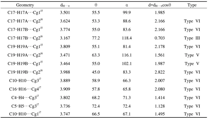
Geometry dH···π θ α d=dH···πcosθ Type
C17–H17A···Cg1i5 3.501 55.5 99.9 1.985
C17–H17A···Cg2i6 3.624 53.3 88.6 2.166 Type VI
C17–H17B···Cg1i5 3.774 55.0 83.6 2.166 Type VI
C17–H17B···Cg2i6 3.167 77.2 118.4 0.703 Type III
C19–H19A···Cg1i5 3.809 55.1 81.4 2.178 Type VI
C19–H19A···Cg2i6 3.471 63.3 116.1 1.561 Type V
C19–H19B···Cg1i5 3.464 55.0 102.1 1.987 Type V
C19–H19B···Cg2i6 3.988 45.0 83.3 2.822 Type VI
C10–H10···Cg3i7 3.889 58.9 66.3 2.007 Type VI
C16–H16···Cg4i7 3.909 57.8 65.8 2.080 Type VI
C4–H4···Cg3i7 3.802 68.2 71.3 1.414 Type VI
C5–H5···Cg3i7 3.736 72.4 72.4 1.128 Type VI
C10–H10···Cg1i7 3.747 66.5 67.1 1.495 Type VI
Table 3. C–H···π interactions geometry (Å, °)
molecules parallel to each other. Obviously π···π interactions were found between parallel molecules.
The centroid–centroid (C8/C10/C11/C12/O2/C9 and C11/C12/C13/C14/C15/C16) distance is 3.632(1) Å
O1 O2
H18B H18B
O2 O1
H19B O1 H5
Fig. 3. Partial packing diagram of the coumarin 6 with C---H···O interaction drawn as dashed lines.
H17A C17 H17B
H19B
H19A C19
C19 H19A
H19B
C17 H17B H17A
Fig. 4. Partial packing diagram of the title compound with π···π and C–H···π interaction drawn as dashed lines.
and the angle between the ring normal and the vectors of two interacted ring centroid is 67.4(1)°.
The shorter atom atom contact between the two parallel plane is 3.331(3) Å (C10 to C12i4, i4:
2-x, 1-y, 1-z), characteristic of a weak interaction9). Other π···π interactions are πC11-C16···πC1-C6 and π
C11-C16···πC8-C12/O2 with the centroid–centroid distances of 3.773(1) Å and 3.527(1) Å. The π···π interaction display the usual slipped stacking geometry, with the interacting π system parallel displaced. The distance between the adjacent parallel molecules are 3.326(2) Å and 3.474(2) Å, respectively.
There are thirteen C–H···π intermolecular inter- actions, involving eight hydrogen atoms and the π electron system of the four rings (Table 3).
A typical C–H···π interaction observed in the crystal is C17–H17B···Cg1i5(Cg1-centroid of ring C1/C6/S1/C7/N1, i5: -x, -y, 1-z). The hydrogen atom is directly above the centre of the hetero- cyclic ring, but the C–H bond points towards a ring carbon. The geometry of this interaction corresponds to type III10).
In another two bond, C19---H19A··· Cg2i6\ (Cg2- centroid of ring C1/C2/C3/ C4/C5/C6, i6: -x, 1-y, 1-z) and C19---H19B···Cg1i5, the hydrogen atoms interact with a carbon at the edge of the acceptor ring (typeV).
In the remaining C–H···π interactions, the direct of C–H bond indicates that it points away from the ring although the hydrogen is close to a carbon (typeVI).
(Symmetry codes i5: -x, -y, 1-z; i6: -x, 1-y, 1-z;
i7: x, y, z; Cg1-centroid of ring C1/C6/S1/C7/N1, Cg2-centroid of ring C1/C2/C3/C4/C5/C6, Cg3- centroid of ring C11/C12/C13/C14/C15/C16, Cg4-centroid of ring C9/C10/C11/C12/O2, definition of geometric parameters according to [10])
4. Conclusions
In conclusion, the single crystal of coumarin 6 was obtained and the structure was solved. Traditional π···π interactions and C–H···π interactions were established in the crystal, by which the molecules were packed parallel to each other. Unusual inter- actions, C–H···O interactions, were found in the crystal lattice, which is different from the tradi- tional hydrogen bonds of H···O. Only one kind of hydrogen bond was found. And the molecules
were joined into network by them.
Although parallel molecules are found in the crystal, the molecules are not packed layer by layer. The crystal is packed like the wall, in which the molecules is bricklike and they are bricked by various kinds of intermolecular interactions.
References
1. O. Kennedy, R. Thornes, and R. D., “Coumarins:
Biology, Applications and Mode of Action”, Wiley, Chichester, pp.15-30, 1997.
2. S. J. Lee, K. Sivakumar, W. S. Shin, F. Xie, and Q. Wang, Synthesis and Anti-angiogenesis Activity of Coumarin Derivatives, Bioorg. &
Med. Chem. Lett., 16(17), 4596-4599(2006).
3. J. P. Jasinski and E. S. Paight, 3-(2-ben- zothiazolyl)-7-(diethylamino)-Coumarin, Acta.
Crystallogr, C51, 531-533(1995).
4. G. M. Sheldrick, Phase Annealing in SHELX- 90: Direct Methods for Larger Structures, Acta. Crystallogr, A46, 467-473(1990).
5. G. M. Sheldrick, “SHELXS-97 Program for the Solution of Crystal Structures”, University of Götingen, Germany, pp.30-36, 1997.
6. Brandenburg, K. Diamond, “Visual Crystal Structure Information System Version 2 of CrystalImpact”, Bonn, Germany, pp.103-110, 1998.
7. D. J. Sutor, Evidence for the Existence of C–
H···O Hydrogen Bonds in Crystals, J. Chem.
Soc, 1105-1110(1963).
8. G. R. Desiraju, The C–H···O Hydrogen Bond in Crystals: What is it?, Acc. Chem. Res., 24, 290-296(1991).
9. C. Janiak, A Critical Account on π-π Stacking in Metal Complexes with Aromatic Nitrogen- containing Ligands, J. Chem. Soc. Dalton.
Trans., 21, 3885-3896(2000).
10. J. F. Malone, C. M. Murray, M. H. Charlton, R. Docherty, and A. J. Lavery, X–H···π (phenyl) Interactions Theoretical and Crystall- ographic Observations, J. Chem. Soc. Faraday Trans., 93, 3429-3436(1997).