www.ksfp.org
한국어병학회지 제32권 제1호 (2019) pISSN 1226-0819, eISSN 2233-5412
J. Fish Pathol., 32(1) : 045~048 http://dx.doi.org/10.7847/jfp.2019.32.1.045
서 론
흰반점병 (white spot disease, WSD)은 한국을 비 롯하여 중국, 일본, 동남아시아, 미국 등지에서 양 식되고 있는 새우에서 발생하는 질병으로 알려져 있다 (Egusa et al., 2006; OIE, 2017). WSD에 걸린 새우는 일반적으로 두흉갑에 흰반점이 형성되고, 병리조직학적으로는 외배엽과 중배엽 기원의 조 직에서 세포의 핵 비대와 핵내 봉입체가 관찰된다
(Egusa et al., 2006; OIE, 2017).
WSD의 원인 병원체인 흰반점증후군바이러스 (white spot syndrome virus, WSSV)는 Nimaviridae 과 Whispovirus 속에 속하는 크기 400×150 nm의 외막을 가진 막대모양의 바이러스로서 5개의 주 요 단백질 (외막: VP28, VP19, 뉴클래오캡시드:
VP26, VP24, VP15)과 약 300 kbp (dsDNA)의 핵산 으로 구성되어 있다 (Egusa et al., 2006; Vlak et al., 2005).
WSSV를 검사 또는 WSD를 진단하는 방법으로 는 유전자를 이용한 분자생물학적 방법(polymer- ase chain reaction (PCR), real time PCR 등), 항체를
White spot syndrome virus (WSSV)의 VP28에 대한 단클론 항체 생산
방지형·김위식·김춘섭*·김종오**·오명주†
전남대학교 수산생명의학과, *㈜엔바이로젠, **부경대학교 수산과학연구소
Production of monoclonal antibodies against VP28 of white spot syndrome virus (WSSV)
Ji-hyeong Bang, Wi-Sik Kim, Choon-sup Kim
*, Jong-Oh Kim
**and Myung-Joo Oh
†Department of Aqualife Medicine, Chonnam National University, Yeosu 59626, Korea
*EnbioGene, Yeosu 59771, Korea
**Institute of Fisheries Sciences, Pukyong National University, Busan 46041, Korea
We developed and subsequently characterized mouse monoclonal antibodies (MAbs) against recombinant VP28 structural protein (rVP28) of white spot syndrome virus (WSSV). We established six hybridoma clones secreting MAbs against rVP28: 15A11, 20G6, 31H2, 34H6, 38D1 and 43A1.
All six MAbs recognized the 25 kDa of protein in gill homogenates of WSSV-infected shrimp by western blot analysis, while no reactivity was observed in gill homogenates of normal shrimp.
Moreover, high enzyme-linked immunosorbent assay (ELISA) optical density (OD) values (0.8-2.68) were observed in the hemolymphs from WSSV-infected shrimp, while low OD values (less than 0.24) were recorded in the hemolymphs from normal shrimp, by using these six MAbs produced in this study. These results suggest that these six MAbs are useful for the detection of WSSV.
Key words: white spot syndrome virus, WSSV, monoclonal antibody, shrimp, VP28
†Corresponding author: Myung-Joo Oh Tel, Fax: +82-61-659-7173
E-mail: ohmj@jnu.ac.kr
단 보
46 방지형 · 김위식 · 김춘섭 · 김종오 · 오명주
이용한 면역학적인 방법(enzyme-linked immun- osorbent assay (ELISA), indirect fluorescent anti- body test, immunohistochemistry 등), 병리조직학적 방법 등이 사용되고 있다 (OIE, 2017). 이들 검사 방법 중, PCR과 병리조직학적 검사 방법은 국내 에서 WSSV를 검출하거나 WSD를 진단하는데 사 용되고 있다. 그러나 면역학적인 방법은 WSSV에 대한 특이 항체가 보급되어 있지 않아 사용되지 않고 있다. 본 연구에서는 양식 현장에서 신속한 진단에 용이하게 적용될 수 있는 면역학적 검사법 의 개선과 보급을 위한 목적으로 WSSV에 대한 단클론 항체(monoclonal antibody, MAb)를 제작 한 후 항체의 특성을 평가하였다. WSSV의 VP28은 다양한 면역학적 검사법에 유용하게 사용될 수 있 음이 보고되어 있어 (Sithigorngul et al., 2006;
Makesh et al., 2006), 본 연구에서는 MAb 제작을 위한 타겟 단백질로서 VP28을 선정하였다.
재료 및 방법
WSSV의 VP28에 대한 재조합 단백질 (rVP28) 을 제작한 후 마우스의 면역 항원으로 사용하여 MAb를 생산하였다.
2015년 인천에 위치한 새우양식장에서 WSSV 에 감염된 흰다리새우 (Litopenaeus vannamei)로 부터 DNA extraction kit (Bioneer, Korea)를 이용하 여 genomic DNA를 추출한 후, NdeI과 XhoI의 제 한효소서열을 추가하여 제작한 WSSV VP28 증폭 용 프라이머 (Forward: 5'-AAACATATGGATCTTT CTTTCACTCTTTC-3', reverse: 5'-GCTCTCGAGTT CCTCGGTCTCAGTGCC-3')를 이용하여 VP28 유전 자를 증폭하였다 (denaturation 95℃ 1분, annealing 58℃ 1분, extension 72℃ 1분, 35 cycle). PCR 산물 을 Qiaquick gel extraction kit (Qiagen, USA)를 이 용하여 정제한 후 T-blunt PCR cloning kit (Solgent, Korea)를 사용하여 클로닝하였다. 제한효소 NdeI 과 XhoI을 이용하여 VP28 부위를 잘라낸 후, pET- 23a(+) 발현 벡터 (Novagen, Germany)에 서브 클로 닝하였다. 재조합 VP28 단백질은 ExiProgen 단백 질 합성 시스템 (Bioneer, Korea)을 이용하여 제조 사의 매뉴얼을 따라 무세포단백질 발현 방법으로
생산하였다. 발현된 VP28 단백질은 Ni-affinity column chromatography (GE healthcare, USA)를 사 용하여 정제한 후 storage buffer (50 mM Tris-Cl (pH 7.6), 100 mM NaCl, 1 mM)에 넣어 실험에 사용 하기까지 -80℃에 보관하였다.
rVP28에 대한 MAb는 Jeong et al. (2017)의 방법 에 따라 제작하였다. rVP28 (100 ug)과 incomplete freund’s adjuvant를 동량으로 섞어 BALB/c 마우스 의 발바닥에 1차 접종하였고, 2주 후에 rVP28로 2차 접종하였다. 2차 접종 1주 후에 rVP28로 3차 면역을 하였다. 3일 후 마우스의 림프절을 분리한 후 polyethylene glycol (Roche, Germany)를 사용하 여 myeloma 세포 (SP2/0-Ag 14)와 융합시킨 후 fe- tal bovine serum이 10% 첨가된 hypoxanthine-ami- nopterin-thymidine (HAT) 배지 (Gibco, USA)로 현 탁시킨 후 96 well plate에 분주하여 37℃로 설정된 CO2 배양기에서 배양하였다. 양성 hybridoma는 rVP28를 항원으로 사용하여 ELISA로 선별하였 고, 제한 희석법으로 3회 클로닝 하였다 (Liddell and Cryer, 1991). 선별된 MAb의 isotyping은 Pierce rapid ELISA mouse mAb isotyping kit (Thermo, USA)를 사용하여 결정하였다.
제작된 MAb의 특이성을 조사하기 위해, west- ern blot과 ELISA를 실시하였다 (Laemmli, 1970;
Towbin et al., 1979; Kim et al., 2018). Western blot 는 rVP28과 WSSV에 감염된 새우와 정상새우의 아가미 조직 마쇄액을 사용하여 12% polyacryla- mide gel에 loading한 후 30 mA에서 전기영동 하였 다. 전기영동 후, gel에 있는 단백질을 transblot 장 치 (ATTO, Japan)를 이용하여 144 mA에서 1시간 동안 nitrocellulose membrane (Bio-Rad, USA)에 blotting하였다. 2% skim milk로 1시간 동안 block- ing한 후, 본 연구에서 제작한 MAb (1차 항체)와 alkaline phosphatase (AP)가 붙어있는 goat anti- mouse IgG (Sigma, USA, 2차 항체, 1,000배 희석액 사용)로 반응시키고 AP가 붙어 있는 substrate kit (Bio-Rad, USA)를 사용하여 발색하였다. 양성 대 조구로는 WSSV의 VP28을 인식하는 MAb (Aquatic diagnostics Ltd, Scotland)를 사용하였다.
ELISA는 WSSV에 감염된 새우와 정상새우의 혈림프를 사용하여 실시하였다. 증류수로 50배 희
47 White spot syndrome virus (WSSV)의 VP28에 대한 단클론 항체 생산
석한 혈림프를 96 well ELISA microplates (Greiner bioone, Germany)에 각각 50 ㎕/well 분주한 후 37
℃에서 overnight하여 항원을 코팅하였다. T-PBS (0.05% Tween-20/PBS (v/v))로 3회 세정하였고 5%
skim milk를 380 ㎕/well을 분주하여 25℃에서 1시 간 동안 blocking하였다. T-PBS로 3회 세정하고 1 차 항체로는 본 연구에서 생산한 MAb를 시료 당 2개의 well에 50 ㎕씩 분주하여 25℃에서 1시간 반 응하였다. T-PBS로 3회 세정한 후 HRP가 표식되 어 있는 goat anti-mouse IgG (Youngin, Korea)를 5% skim milk로 1,000배 희석하여 well 당 50 ㎕씩 분주하였으며, 25℃에서 1시간 반응하였다. T-PBS 로 5회 세정한 후 tetramethylbenzidine base (TMBC, Surmodics, USA)를 50 ㎕/well 분주하여 5분간 발 색하였다. 각 well에 2N H2SO4를 50 ㎕씩 넣어 발 색 반응을 중지시킨 후, microplate photometer (Mul- tiskan, USA)로 450 nm에서 흡광도 (opical density, OD)를 측정하였다. 양성 대조구로는 VP28을 인식 하는 MAb (Aquatic diagnostics Ltd, Scotland)를 사 용하였다. OD값은 ELISA를 2회 반복하여 평균값 으로 나타내었다.
결과 및 고찰
본 연구에서는 WSSV를 특이적으로 인식하는 MAb를 생산하고자 하였다. 무세포 단백질 합성 시스템으로 제작한 WSSV의 VP28 재조합 단백질 을 Ni-affinity column chromatography로 정제하여 SDS-PAGE를 실시한 결과, 약 27 kDa에서 단일 밴 드가 관찰되었다 (data not shown).
rVP28로 면역시킨 후 마우스의 림프절과 mye- loma 세포를 융합시켜 hybridoma를 제작하였다.
Hybridoma로부터 생성되는 항체를 rVP28 (항원) 을 사용한 ELISA법으로 선별한 후, 제한 희석법 으로 클로닝하여 최종적으로 6개의 MAb를 선별 하였다 (15A11, 20G6, 31H2, 34H6, 38D1, 43A1).
6개의 MAb와 rVP28를 사용하여 western blot을 실 시한 결과, 6개 항체 모두 약 27 kDa의 단백질을 강하게 인식하였다 (data not shown). WSSV에 감 염된 새우와 정상새우의 아가미 조직 마쇄액을 사 용하여 western blot을 실시한 결과에서는 6개 항
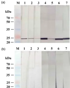
체 모두 WSSV에 감염된 새우의 아가미 조직 마 쇄액에서만 약 25 kDa에서 밴드가 관찰되었다 (Fig. 1). 34H6과 43A1 항체에서는 15A11, 20G6, 31H2, 38D1 보다 강한 반응을 보였다. 양성 대조 구에서도 WSSV에 감염된 새우의 아가미 조직 마 쇄액에서만 약 25 kDa에서 밴드가 관찰되었다. 이 상의 결과, 제작된 6개의 MAb는 western blot에서 WSSV에 특이적으로 반응하는 것이 확인되었다.
WSSV에 감염된 새우와 정상새우의 혈림프를 사용하여 ELISA를 실시한 결과, 6개의 MAb 모두 WSSV에 감염된 새우의 혈림프에 높은 OD값을 (0.8-2.68) 보였고, 정상 새우의 혈림프에는 0.24 이 하의 OD값을 보였다 (Fig. 2). 특히, 15A11, 20G6, 34H6 및 43A1 항체는 31H2과 38D1 보다 높은 OD 값을 보였다. 양성 대조구에서도 WSSV에 감염된 새우의 혈림프에서만 높은 OD값을 보였다. 이상 의 결과, 제작된 6 개의 MAb는 ELISA에서 WSSV 에 특이적으로 반응하는 것이 확인되었다. 제작된
(a)
(b)
Fig. 1. Western blot analysis using gill homogenates of WSSV-infected shrimp (a) and normal shrimp (b). M:
molecular marker, 1: MAb 15A11, 2: MAb 20G6, 3:
MAb 31H2, 4: MAb 34H6, 5: MAb 38D1, 6: MAb 43A1, 7: anti-WSSV VP28 MAb (positive control).
48 방지형 · 김위식 · 김춘섭 · 김종오 · 오명주
MAb의 isotype을 분석한 결과, H chain은 IgG1 (15A11, 34H6, 38D1, 43A1)과 IgG2a (20G6과 31H2) 로 나타났으며, L chain은 모두 kappa로 확인되었 다 (data not shown).
본 연구에서는 무세포 단백질 발현 방법으로 제작된 WSSV의 rVP28을 사용하여 총 6개의 MAb 를 생산하였다. 6개의 MAb는 western blot과 ELISA 상에서 WSSV에 감염된 새우와 정상새우를 뚜렷 이 구분할 수 있어 본 연구에서 제작된 항체는 WSSV를 검출하는데 유용하게 사용될 수 있을 것 으로 사료된다.
무세포 단백질 합성 시스템은 세포 배양과 같은 단계를 배제하면서 유전 정보를 특정 공간에 구애 받지 않고 번역할 수 있는 장점을 가지고 있다 (Gregorio et al., 2019). 본 연구를 통해 무세포 단 백질 합성 시스템으로 WSSV의 VP28 부위에 대 한 재조합 단백질을 생산할 수 있음이 확인되었다.
감사의 글
본 연구는 2017년 해양수산부 재원으로 한국해 양과학기술진흥원의 지원을 받아 수행되었습니 다(수산동물 바이러스 전염병 진단용 항체 생산).
References
Egusa, S., Wakabayashi, H. and Muroga, K.: Infectious
and parasitic diseases of fish and shellfish. Life Sci- ence Publishing Co. Seoul, pp. 54-58. (in Korean) Gregorio, N.E., Levine, M.Z. and Oza, J.P.: A user’s
guide to cell-free protein synthesis. Methods Protoc., 24: 1-34, 2019.
Jeong, H.N., Jang, M.S., Oh, M.J. and Kim, W.S.: Pro- duction of monoclonal antibodies against viral hem- orrhagic septicemia virus (VHSV, genotype IVa) from olive flounder. J. Fish Pathol., 30: 149-154, 2017. (in Korean)
Kim, W.S., Kim, S.W. and Oh, M.J.: Production of monoclonal antibodies against nervous necrosis vi- rus (NNV, RGNNV genotype). Korean J. Fish Aquat.
Sci., 51: 328-331, 2018. (in Korean)
Laemmli, U.K.: Cleavage of structural proteins during the assembly of the head of bacteriophage T4. Na- ture, 227: 680-685. 1970.
Liddell, J.E. and Cryer, A.: A practical guide to mono- clonal antibodies, pp. 90-104, John Wiley and Sons, Inc., New York, 1991.
Makesh, M., Koteeswaran, A., Daniel Joy Chandran, N., Murali Manohar, B. and Ramasamy, V.: Devel- opment of monoclonal antibodies against VP28 of WSSV and its application to detect WSSV using immunocomb. Aquaculture, 261: 64-71, 2006.
OIE (Office International des Epizooties).: Infection with white spot syndrome virus. Manual of Diag- nostic tests for Aquatic Animals. 2017.
Sithigorngul, W., Rukpratanporn, S., Pecharaburanin, N., Longyant, S., Chaivisuthangkura, P. and Sithigorn- gul, P.: A simple and rapid immunochromatographic test strip for detection of white spot syndrome virus (WSSV) of shrimp. Dis. Aquat. Org., 72: 101-106, 2006.
Towbin, H., Staehelin, T. and Gordon, J.: Electrophoretic transfer of proteins from polyacrylamide gels to ni- trocellulose sheets; procedure and some applications.
Proc. Natl. Acad. Sci., 76: 4350-4354, 1979.
Vlak, J.M., Bonami, J.R., Flegel, T.W., Kou, G.H., Lig- htner, D.V., Lo, C.F., Loh, P.C. and Walker, P.W.:
Family Nimaviridiae. In virus taxonomy, In Virus taxonomy: Eighth report of the international com- mittee on taxonomy of viruses, pp. 187-192, editors.
Fauquet, C.M., Mayo, M.A., Maniloff, J., Dessel- berger, U and Ball, L. A., Elsevier/Academic Press, United Kingdom, 2005.
Manuscript Received : Jun 10, 2019 Revised : Jun 15, 2019 Accepted : Jun 18, 2019 Fig. 2. ELISA analysis using hemolymphs from WSSV-
infected shrimp and normal shrimp and seven MAbs (15A11, 20G6, 31H2, 34H6, 38D1, 43A1, PC (positive control): anti-WSSV VP28 MAb).