Anti-inflammatory Effect of the Hedgehog Skin Extracts on LPS-Stimulated RAW 264.7 Cells
Tae Yeon Kim
1#, Na Young Jo
2, Kang Hyun Leem
3*1 : Department of Pathology, College of Korean Medicine, Semyung University, Republic of Korea 2 : Department of Acupuncture & Moxibustion, College of Korean Medicine, Semyung University,
Republic of Korea
3 : Department of Herbology, College of Korean Medicine, Semyung University, Republic of Korea
ABSTRACT
Objectives : Hedgehog skin is one of the animal medicines in Traditional Korean Medicine for hematochezia and hemorrhoids. In this study, we examined cytotoxicity and anti-inflammatory effects.
Methods : Cytotoxicity of hedgehog skin extracts was measured by MTT assay in vitro. We investigated the inhibition of lipopolysaccharide (LPS) stimulated nitric oxide (NO) production in RAW 264.7 cells. The phosphorylation of mitogen-activated protein kinases (MAPKs) was measured by western blot. And we observed the effect of hedgehog skin extracts on the expression of IL-6 genes using real time PCR.
Results : As a result of MTT assay for cytotoxicity, there were no significant differences between non-treatment group and hedgehog skin extracts treatment groups. 500 ㎍/㎖ of hedgehog skin extracts treatment significantly decreased nitric oxide production in comparison with non-treatment in LPS-induced RAW 264.7 cells. In measurement of the phosphorylation of MAPKs using western blot analysis, LPS stimulation increased the phosphorylation of MAPKs and 500 ㎍/㎖ of hedgehog skin extracts treatment decreased the phosphorylation of ERK1, ERK2 and p38 significantly.
But there were no significant differences the phosphorylation of JNK1 and JNK2. As a result of confirmation of the IL-6 mRNA gene expression using real time PCR, IL-6 mRNA gene expressions were significantly decreased in 50 ㎍/㎖, 100 ㎍/㎖ and 500 ㎍/㎖ hedgehog skin extracts treated groups by comparison with non-treatment group.
Conclusion : These results could provide a mechanistic explanation for the anti-inflammatory effects of the hedgehog skin.
1)
Key words : Hedgehog skin, Anti-inflammatory, Hemorrhoids, Mitogen-activated protein kinases (MAPKs), IL-6
Ⅰ. INTRODUCTION
Hedgehog skin is the dried skin of Erinaceus europaeus koreensis Mori and related species (Erinaceidae). It is one of the animal medicines in Traditional Korean Medicine and is known to have effects of hemostasis, calm the adverse-rising energy, stopping a pain and clearing a blood-heat. It has been commonly prescribed for regurgitation, nausea, abdominal pain, especially hematochezia and hemorrhoids
1). But Hedgehog skin
has not, so far, been noticed, nor has it been studied in detail.
Hemorrhoids are known to occur as a result of inflammation of the surrounding tissue of the anus
2). In Traditional Korean Medicine theory, hemorrhoids are known to be caused by blood-heat(血熱). Blood- heat(血熱) is major causes of inflammatory reactions.
Therefore Hedgehog skin is thought to be anti- inflammatory because it has been used in hemorrhoids by effects of clearing a blood-heat. However, its anti-
*Corresponding author : Kang Hyun Leem, Department of Herbology, College of Korean Medicine, Semyung University, 65 Semyung-ro, Jecheon-si, Chungcheongbuk-do, 27136, Republic of Korea.
·Tel : +82-43-649-1341 ·Fax : +82-43-649-1702 ·E-mail : [email protected]
#First author : Tae Yeon Kim, Department of Pathology, College of Korean Medicine, Semyung University, 65 Semyung-ro, Jecheon-si, Chungcheongbuk-do, 27136, Republic of Korea.
·Tel : +82-43-649-1339 ·Fax : +82-43-649-1702 ·E-mail : [email protected] ·Received : 18 March 2018 ·Revised : 4 May 2018 ·Accepted : 25 May 2018
inflammatory effect has not been examined to date.
Inflammation is a biological response to protect the body from pathogens, infections, or tissue damages.
But unusual or excessive inflammatory reactions may cause fever, allergic anaphylaxis, autoimmunity, cardiovascular disease, and tissue damages etc. This inflammation is caused by the expression of inflammatory mediators such as nitric oxide, cytokine, and prostanoids
3). Mitogen-activated protein kinases (MAPKs) are known to modulate the synthesis of such inflammatory mediators and has recently become the target of anti-inflammatory drugs
4).
In this study, we investigated the inhibitory effects of the hedgehog skin extracts on nitric oxide (NO) and MAPKs activation in RAW 264.7 cells to determine the anti-inflammatory effects.
Ⅱ. Materials and Methods
1. Samples
The dried hedgehog skin was purchased from BOZHOU JINGWAN CHINESE MEDICINE FACTORY (Anhui Sheng, China), identified by one of the authors (Prof. KH Leem), and a voucher specimen was kept in the College of Korean Medicine, Semyung University. 75 g of skins in 1,000 ㎖ distilled water was heated in a heating extractor for 3 hours. The extracts were filtered and concentrated by using the rotary evaporator. The hedgehog skin extracts were lyophilized by using freeze dryer (7.6 g).
2. Cell Culture
The mouse macrophage-like cell line RAW 264.7 was purchased from the American Type Culture Collection (Rockville, MD, USA) and maintained using Dulbecco's modified Eagle's medium (DMEM, Invitrogen, Grand island, NY, USA) containing 10% v/v fetal bovine serum (heat-inactivated, Invitrogen, Grand island, NY, USA) and 100 U/㎖ penicillin/streptomycin (Sigma-Aldrich, New York, NY, USA) in an incubator with a humidified atmosphere of 95% air and 5% CO
2, at 37 ℃.
3. Cytotoxicity
The cytotoxicity was assessed by the 3-(4,5-dimethyl- thiazol-2-yl)-2,5-diphenyl tetrazolium bromide (MTT, Sigma-Aldrich, St. Louis, MO, USA) assay. RAW 264.7 cells were seeded in a 96-well plate in a density of 1.0 × 10
4cells/well and incubated for 24 hours. The next day, the medium was changed to DMEM without FBS. The
following day, 0 (phosphate-buffered saline, PBS only), 10, 50, 100, and 500 ㎍/㎖ of hedgehog skin extracts were treated and incubated for 24 hours. The MTT was added and incubated for 4 hours. The formazan crystals that formed in the actively metabolizing cells were extracted with 200 ㎕ of dimethyl sulfoxide (DMSO, Sigma- Aldrich, St. Louis, MO, USA) and the absorbance at 595 ㎚ was measured by a Synergy 2 microplate ELISA reader (BioTek, Winooski, VT, USA).
4. NO Assay
Nitric oxide levels, induced by lipopolysaccharides (LPS, Sigma-Aldrich, St. Louis, MO, USA) in the culture supernatants, were measured by the Griess reaction.
In brief, RAW 264.7 cells were seeded in a 96-well plate in a density of 1.0 × 10
4cells/well and incubated for 24 hours. The next day, the medium was changed to DMEM without FBS. The following day, Blank (phosphate-buffered saline, PBS only), 0 (100 ng/㎖ LPS only), 10, 50, 100, and 500 ㎍/㎖ of hedgehog skin extracts with LPS were treated and incubated for 24 hours. The next day, 0.1 ㎖ of culture supernatants from each samples were mixed with the same volume of the Griess reagent. Absorbance values were measured at 540 nm using a microplate ELISA reader.
5. Western Blot
RAW 264.7 cells were seeded in 100-mm culture dishes (5.0 × 10
5cells/well) and incubated 24 hours.
The next day, the medium was changed to DMEM without FBS. After 24 hours, the cells were treated with 100, and 500 ㎍/㎖ of hedgehog skin extracts and incubated for 15 minutes. Proteins were extracted with PRO-PREP Protein Extraction solution (Intron biotechnology, Seongnam, Kyungi, Korea). The cells were washed twice with PBS and 300 ㎕ of PRO-PREP solutions containing phosphatase inhibitor cocktail 2 and 3 (Sigma-Aldrich, St. Louis, MO, USA) were added, and then incubated in -20℃ for 20 minutes. The cells were homogenized by pipetting and centrifuged at 13,000 rpm for 5 minutes at 4℃. The supernatant was transferred to a fresh tube. Protein concentrations were measured using DC Protein Assay kit (Bio-Rad, Hercules, CA, USA). 5 ㎕ of standards and protein samples were transferred to 96-well plate and 25 ㎕ of alkaline copper tartrate solution contains Reagent S.
Then 200 ㎕ of dilute Folin Reagent was added and incubated. After 15 minutes, the protein concentrations were measured at 750 nm using an ELISA reader.
Each protein was denatured with 5 X sample buffer
and boiled for 5 minutes. Each protein was then fractionated by electrophoresis through a 4-20% SDS polyacrylamide gel at 100 V for 1.5 hours, and the proteins were transferred onto PVDF membranes at 100 V for 50 minutes. Each membrane was blocked with TBST buffer (10 mM Tris-HCl, pH 7.4, 150 mM NaCl, 0.1% Tween-20) containing 5% BSA for 1 hours and then incubated with primary antibodies (anti-JNK, anti-ERK, anti-p38, anti-p-JNK, anti-p-ERK, and anti-p-p38 antibodies raised in rabbit) in TBST buffer containing 1% BSA at 4℃ overnight. The membranes were washed 3 times with TBST buffer and further incubated with anti-rabbit IgG secondary antibodies conjugated with horseradish peroxidase for 2 hours.
Each membrane was filmed by chemiluminescent imaging system (Fusion SL2, Vilber Lourmat, Marne-la-Vallée Cedex, France), and analyzed using Bio1D software (Vilber Lourmat, Marne-la-Vallée Cedex, France).
6. Realtime PCR
RAW 264.7 cells were seeded in 100-mm culture dishes (5.0 × 10
5cells/well) and incubated 24 hours
and the next day, the medium was changed to DMEM without FBS. After 24 hours, the cells were treated with 100, and 500 ㎍/㎖ of hedgehog skin extracts and incubated for 24 hours. Total RNAs were extracted using RNeasy® Protect Mini kit (Qiagen, Valencia, CA, USA) according to the manufacturer's protocol. Then cDNA was synthesized from mRNA using QuantiTect®
Reverse Transcription kit (Qiagen, Valencia, CA, USA).
Realtime PCR was performed using QuantiTect
TMSYBR®
Green PCR kit (Qiagen, Valencia, CA, USA) according to the manufacturer's protocol. In brief, an initial denaturation of 10 minutes at 94℃ was followed by cycles of 10 s at 94℃, 15 s at 58℃, 20 s at 72℃. A 30-minutes post-amplification dissociation protocol was conducted to assess primer specificity and product uniformity. During this step, products were slowly heated up to 95℃ and non-specific products were not detected. The PCR primer sequences are shown in Table 1. Analyses were performed using Rotor-Gene Q® (Qiagen, Valencia, CA, USA) and gene expression values were calculated based on the comparative ΔΔ CT method according to the manufacturer's protocol.
Sequence, 5' to 3'
mRNA Forward Reverse
GAPDH GCC ATT TGC AGT GGC AAA GTG G GAT GGG CTT CCC GTT GAT GAC AAG C
IL-6 GAG TTG TGC AAT GGC AAT TCT G GCA AGT GCA TCA TCG TTG TTC AT
GAPDH, glyceraldehyde 3‑phosphate dehydrogenase; IL-6, interleukin-6 Table 1. Primer Sequences Used for Realtime PCR
7. Statistical Analysis
The results were expressed as mean ± SE. One-way ANOVA followed by Tukey’s post hoc test was performed for statistical analysis (GraphPad prism ver. 6), and p-values of less than 0.05 (p < 0.05) indicated significant differences. All in vitro experiments were performed with triplicate independent samples.
Ⅲ. RESULTS
1. Cytotoxicity of hedgehog skin extracts
The cell proliferations of RAW 264.7 cells treated with PBS showed 100.0 ± 1.1% and 10, 50, 100, and 500 ㎍/㎖ of hedgehog skin extracts treated cells showed 103.7 ± 1.5%, 103.8 ± 1.6%, 100.0 ± 1.7%, and 99.0 ± 2.3% of cell proliferation. There were no significant differences among the 0, 10, 50, 100 and
500 ㎍/㎖ of hedgehog skin extracts treated groups (Figure 1).
Hedgehog skin extract Treatment (μg/ml)
Cell Viability (%)
0 10 50 100 500
0 50 100
Figure 1. Cell viability analysis of RAW 264.7 cells treated with 10, 50, 100, and 500 ㎍/㎖ of hedgehog skin extracts for 24h.
The amount of viable cells were determined by MTT assay. There were no significant differences between non-treated groups and hedgehog skin extracts treated groups.
2. The effects of hedgehog skin extracts on the production of nitric oxide
Nitric oxide production in RAW 264.7 cells stimulated with LPS showed 100.0 ± 1.6% and 10, 50, 100, and 500 ㎍/㎖ of hedgehog skin extracts treated cells showed 105.0 ± 1.6%, 103.5 ± 1.6%, 101.2 ± 2.2%, and 91.5 ± 1.1% of nitric oxide production, respectively. 500 ㎍/㎖
of hedgehog skin extracts treated groups significantly decreased nitric oxide production (Figure 2).
Hedgehog skin extract Treatment (μg/ml)
Blank 0 10 50 100 500
60 80 100 120
LPS (100 ng/ml)
*
Figure 2. The effects of hedgehog skin extracts on the production of nitric oxide in RAW 264.7 cells. The production of nitric oxide was assayed from culture medium of cells treated with 10, 50, 100, and 500 ㎍/㎖ of hedgehog skin extracts for 24h. NO production was determined by the Griess reaction. Data were presented as the means ± SEM of triplicate experiments (*p < 0.05).
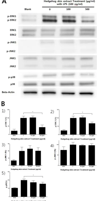
3. Effects of hedgehog skin extracts on the activation of mitogen-activated protein kinases (MAPKs)
The phosphorylation of ERK1 in RAW 264.7 cells stimulated with LPS showed 100.0 ± 11.4% and 100 and 500 ㎍/㎖ of hedgehog skin extracts treated cells showed 102.6 ± 9.6% and 44.0 ± 13.7% of the phosphorylation of ERK1. The phosphorylation of ERK2 in RAW 264.7 cells stimulated with LPS showed 100.0 ± 11.8% and 100 and 500 ㎍/㎖ of hedgehog skin extracts treated cells showed 103.1 ± 9.5% and 68.0 ± 8.4% of the phosphorylation of ERK2. The phosphorylation of JNK1 in RAW 264.7 cells stimulated with LPS showed 100.0 ± 26.9% and 100 and 500 ㎍/㎖ of hedgehog skin extracts treated cells showed 115.5 ± 21.9% and 101.2 ± 21.9%
of the phosphorylation of JNK 1. The phosphorylation of JNK2 in RAW 264.7 cells stimulated with LPS showed 100.0 ± 12.1% and 100 and 500 ㎍/㎖ of hedgehog skin extracts treated cells showed 97.1 ± 3.0% and 97.1 ± 8.1% of the phosphorylation of JNK2. The
phosphorylation of p38 in RAW 264.7 cells stimulated with LPS showed 100.0 ± 7.8% and 100 and 500 ㎍/㎖
of hedgehog skin extracts treated cells showed 86.6 ± 11.1% and 78.0 ± 10.0% of the phosphorylation of p38, respectively. LPS stimulation increased phosphorylation of MAPKs and 500 ㎍/㎖ of hedgehog skin extracts treated group decreased phosphorylation of ERK1/2 and p38 (Figure 3).
Figure 3. Effects of hedgehog skin extracts on the activation of mitogen-activated protein kinases (MAPKs). RAW 264.7 cells were treated with 100 and 500 ㎍/㎖ of hedgehog skin extracts for 30 min. The expressions of phosphorylated ERK, JNK, and p38 were measured by western blot analysis. Data were presented as the means ± SEM of triplicate experiments (*p < 0.05). A:
Western blot analysis of phosphorylated and total MAPks in RAW 264.7 cells. B: Densitometric quantification of western blot bands. 1) p-ERK-1 ; 2) p-ERK-2 ; 3) p-JNK-1 ; 4) p-JNK-2
; 5) p-p38.
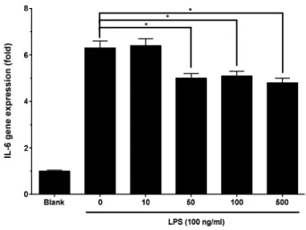
4. Effects of hedgehog skin extracts on the gene expression of IL-6
IL-6 gene expressions in RAW 264.7 cells stimulated with LPS increased 6.3 ± 0.3 fold over the normal group and 10, 50, 100, and 500 ㎍/㎖ of hedgehog skin extract treated cells showed 6.4 ± 0.3, 5.0 ± 0.2, 5.1 ± 0.2, and 4.8 ± 0.2 fold of IL-6 gene expressions. IL-6 gene expressions were significantly decreased in 50, 100 and 500 ㎍/㎖ treated groups (Figure 4).
Figure 4. Effects of hedgehog skin extracts on the gene expression of IL-6. RAW 264.7 cells were treated with 10, 50, 100, and 500 ㎕/㎖ of hedgehog skin extracts for 24h. The mRNA level of IL-6 was evaluated by realtime PCR. GAPDH was used as an internal control for realtime PCR. Data were presented as the means ± SEM of triplicate experiments (*p<0.05).