Kor J Fish Aquat Sci 47(6),757-764,2014 한수지 47(6), 757-764, 2014
Original Article
757
Copyright © 2014 The Korean Society of Fisheries and Aquatic Science pISSN:0374-8111, eISSN:2287-8815
서 론
염증의발병은외부자극에대한인체의정상적인방어시스 템이지만(De Heredia et al., 2012; Osborn and Olefsky, 2012) 염증에대한지속적이고과도한면역반응은오히려조직의손 상을촉진하고만성염증을유발하게된다(Brown et al., 2007).
이것이지속화되면관절염, 당뇨병, 동맥경화, 암, 노화및알츠 하이머병을포함하는퇴행성신경질환의원인이된다(De He- redia et al., 2012; Osborn and Olefsky, 2012) .
선천성면역과후천성면역에모두관여하는대식세포의주 작용은 reaction oxygen species (ROS)와 활성질소(reactive nitrogen species, RNS)를무기로사용하여세균, 바이러스, 암 세포에대해공격하며 T cell 활성화및 B cell 활성화에참여함 으로써면역계에서총체적이고핵심적인역할을하는세포이
다(Hakim et al., 2004). 또한대식세포는염증촉진성 cytokine 인 tumor necrosis factor-alpha (TNF-α), interleukin-6 (IL-6),
IL-1β 등과병원균의내독소로서여러염증세포들이생산하는
cytokine들의생산을촉진하는 lipopolysaccharide (LPS)에의 한자극으로활성화된다(Marriot et al., 1998). 활성화된대식 세포는 inducible NOS (iNOS), cyclooxygenase-2 (COX-2) 같은효소를생산하여 nitric oxide (NO) 및 prostaglandins E2 (PGE2)같은다양한염증매개체들을생성한다(Nathan., 1992).
염증성 cytokine 및 iNOS, COX-2의발현은전사인자인 nucle- ar factor-kappa B (NF-κB)에의해조절되는데 NF-κB 또한산 화적스트레스, LPS, cytokine등의외부자극에활성화되어핵 으로이동하여면역및염증반응에관여하는매개체들의발현 에관여한다(Choi et al., 2003). NF-κB는 p65, p50, Inhibitor kappa B (IκB) subunit의 trimer로구성되어있고산화적스트
방사무늬 김(Pyropia yezoensis) 추출물에 의한 RAW 264.7 대식세포의 항염증 효과
이지영·최정욱·이민경·김영민·김인혜·남택정*
부경대학교 식품영양학과
Anti-inflammatory Effects of Pyropia yezoensis Extract in LPS-stimulated RAW 264.7 cells
Ji Young Lee, Jeong Wook Choi, Min Kyeong Lee, Young Min Kim, In Hye Kim and Taek Jeong Nam*
Department of Food Science and Nutrition, Pukyong National University, Busan 608-737, Korea
Many researchers have studied algae as a source of material having potential biological activities, not least because many marine algae extracts have strong antioxidant properties. In this study, we investigated the anti-inflammatory effects of Pyropia yezoensis extract (PYE) on RAW 264.7 cells by measuring nitric oxide (NO), reactive oxygen species (ROS), superoxide dismutase (SOD), catalase activity, inducible NOS (iNOS), cyclooxygenase-2 (COX-2), nuclear factor-kappa B (NF-κB), interleukin-1β (1L-1β), and tumor necrosis factor-alpha (TNF-α). PYE decreased the production of intracellular ROS dose-dependently and increased SOD and catalase activity in lipopolysaccharide (LPS)-stimulated RAW 264.7 cells. PYE significantly suppressed the production of NO and reduced the expression of iNOS, COX-2, and NF-κB. PYE treatment also inhibited the production of IL-1β and TNF-α significantly and re- duced the phosphorylation of Akt and MAPK significantly in LPS-stimulated RAW 264.7 cells. These results suggest that PYE has potential anti-oxidant and anti-inflammatory activities.
Key words: Pyropia yezoensis, Anti -inflammation, Oxidative stress, NO, NF-κB
This is an Open Access article distributed under the terms of the Creative Commons Attribution Non-Commercial Licens (http://creativecommons.org/licenses/by-nc/3.0/)which permits unrestricted non-commercial use, distribution, and reproduction in any medium, provided the original work is properly cited.
http://dx.doi.org/10.5657/KFAS.2014.0757 Kor J Fish Aquat Sci 47(6) 757-764, December 2014
Received 2 October 2014; Revised 10 November 2014; Accepted 2 December 2014
*Corresponding author: Tel: +82. 51. 629. 5846 Fax: +82. 51. 629. 5842 E-mail address: [email protected]
이지영ㆍ최정욱ㆍ이민경ㆍ김영민ㆍ김인혜ㆍ남택정 758
레스에의해 IκB가분해되면 p65/p50 heterodimer가핵속으 로이동하여 DNA 결합을하는것으로알려져있다(Munoz et al., 1991). 이러한과정에서 IκB를분해시키는 IκB kinase의활 성화는 extracellular signal-regulated kinase (ERK), c-jun N- terminal kinase (JNK), p38, Akt 등의 kinase에의하여조절된 다(Nair et al., 2004).
생체는산소호흡으로인한대사과정에서산화적스트레스를 유발하는물질을생성하게되는데이것의방어기전으로우리 몸은 superoxide dismutase (SOD), catalase 등의항산화효소 를생성하여활성산소종으로부터생체기관을방어한다고알려 져있으며활성산소종과염증성 cytokine은이러한산화적스트 레스를유발하는기작과관련이있다(Cho et al, 2008).
홍조류에속하는김은예로부터한국을포함한중국, 일본등 아시아지역에서섭취하여왔으며영양학적측면으로열량은 낮지만비타민, 무기질, 식이섬유가풍부한특징을가지고있 다. 또한김은폴리페놀을함유하여항산화활성을가지는것으 로확인되었으며(Lee and Oh, 2002) 김에함유된 betaine은혈 중콜레스테롤을저하시키는 효과가있다고보고되었다(Jung et al., 2001) 특히건조김의 10% 내외로함유되어있는수용성 다당인포피란은항종양활성(Noda and Arashima, 1989)과항 산화활성(Zhang et al., 2004), 지질대사개선에대한효과(Lee et al., 2010) 등이확인되어있으나김의항염증효과와그에대 한분자기전에대한연구는미비한실정이다. 따라서본연구에 서는염증반응으로변화되는세포내부의지표물질과신호단 백질을측정하였으며, 김추출물이이에어떠한영향을미치는 지를확인하였다.
재료 및 방법 시료
본실험에사용한방사무늬김(Pyropia yezoensis)은 2012년 3월에기장군에서구입하였고, 수세한후염분을제거하고동결 건조한후마쇄하여김분말을제조하였다. 3구플라스크에증 류수 1 L를넣고김분말 40 g을취하여침지시킨후실온에서 4시간교반하여추출하였고추출액을원심분리(3,500 rpm, 20 min, 4℃)하여얻어진상층액에 95% 에탄올을첨가한뒤당을 침전시켰다. 침전된당을분리하기위해여과과정을거치고얻 은여액을감압여과하여동결건조한뒤 -70℃에보관하여실 험에사용하였다.
세포배양
실험에사용된 RAW 264.7 세포는 American Type Culture Collection (Manassas, VA, USA)에서구입하여사용하였다. 세포는 10% fetal bovine serum (Gibco BRL, Gaitherberg, MD, USA)과 5% penicillin/streptomyocin (Gibco BRL, Grand Island, NY, USA)이함유된 Dulbecco's modified Ea-
gle's medium (DMEM)배지에 37℃, 5% CO2가유지되는 in- cubator에서배양하였다. Cell culture plate에 RAW 264.7 세포 가 70-80% 정도자라면 PBS로씻어낸후계대배양하고배지는 2-3일마다교체하였다.
세포 생존율 측정
PYE의처리에따른 RAW 264.7 세포의증식에미치는영 향을알아보기위해 96-well plate에 96-well plate에 10×104
cells/well에 세포를동일하게 분주한 후 세포부착을 위하여
24시간배양하고, phenol red free-DMEM 배지에 12시간을 더 배양한후 PYE를 0, 5, 10, 20, 40 µg/mL씩농도별로처 리한후 24시간배양하였다. 3-(4,5-dimethylthiazol-2-yl)-5-(3- carboxymethoxyphenyl)-2-(4-sulphenyl)-2H-tetrazolium, in- ner salt/phenazine methosulfate. (MTS/PMS) solution (Pro- mega Co., Madison, WI, USA)을첨가하고 37℃에서 30분반 응시킨후 ELISA plate reader (Benchmark microplate reader;
Bio-Rad laboratories, Hercules, CA, USA)로 490 nm에서흡 광도를측정하였다.
Cytokine 발현 측정
RAW 264.7 세포를 100-mm dish에 배양하고 세포가 60- 80%정도증식하면 serum free media (SFM)으로교체하였다. 6시간뒤에 PYE (10, 20 µg/mL)가처리된 SFM으로교체하 고 4시간후에 1 µg/mL LPS를 20시간동안처리한후 phos- phate buffered saline (PBS)용액으로 세척하고 1 mL Lysis buffer [1% Igepal CA-360, 20 mM Tris- HCl (pH 8.0), 137 mM NaCl, 10% glycerol, 2 mM EDTA, 10 µg/mL aprotinin, 10 µg/mL leupeptin, 10 µg/mL pepstatin]로세포를취한다음 12,000 g, 5분동안원심분리하여상층액을취해 Mouse Cyto- kine Array kit (R&D system Inc., Minneapolis, MN, USA)를 사용하여제조사의방법에따라 TNF-α와 IL-1β의단백질양 을측정하였다.
Western Blot Analysis
단백질발현의분석은 RAW 264.7 세포세포를 100-mm dish 에 배양하고세포가 60-80%정도증식하면 SFM으로교체하 였다. 6시간뒤에 PYE (10, 20 µg/mL)가 처리된 SFM으로 교체하고 4시간후에 1 µg/mL LPS를 20시간동안처리한후 PBS용액으로세척하고 Lysis buffer [50 mM Tris-hydrochlo- ride (Tris-HCl), pH 7.4, 150 mM sodium chloride (NaCl), 1 mM ethylenediaminetetraacetic acid (EDTA), 1 mM sodium fluoride (NaF), 1% NP-40, 1 mM sodium orthovanadate (Na3VO4), 1 µg/mL aprotinin, 1 µg/mL leupeptin, 1 μg/mL pepstatin, 1 mM phenylsufonyl fluoride (PMSF), 0.25% Na- deoxycholate]를 radioimmunoprecipitation assay buffer [1%
NP-40, 0.25% sodium deoxycholate, 1 mM ethylene glycol tetraacetic acid (EGTA), 150 mM NaCl, 50 mM Tris-HCl, pH
김 추출물에 의한 대식세포의 항염증 효과 759
7.5]에넣어 cell lysate를회수하고 30분간방치한후원심분리 (12,000 rpm, 10 min, 4℃) 하여단백질농도를동일하게정량 하여 SDS-PAGE에전기영동한다음 PVDF membrane (Mil- lipore Co., Billerica, MA, USA)으로이동시켰다. 이때표준분 자량은 dual color marker를사용하였다. 전기영동시킨 mem- brane은 실온에서 1% bovine serum albumin/Tris-buffered saline-Tween 20 로 1시간 30분동안 blocking 시킨후각각 의 1차 antibody를 1:1,000으로희석하여 4℃에서 16시간반 응시키고 2차 antibody를 1:10,000 비율로희석하여 1시간 30 분간반응시킨후 Super Signal West Pico Stable Peroxide So- lution과 Super Signal West Pico Luminol/ Enhancer solution (Thermo Fisher Scientific Inc., Rockford, IL, USA)을사용하 여 KODAK X-ray film에감광시켜현상하여시각화하였다. NO 생성량 측정
RAW 264.7 세포를 5×10⁴ cells/well 농도로 96-well plate 에배양한후에 PYE를 0, 5, 10, 20, 40 µg/mL로처리하고 4시 간반응시킨후 LPS 1 µg/mL를처리하여 24시간배양한뒤세 포배양액 100 µL와 griess reagent (1% Sulfanilamide, 0.1%
Naphthlethy-diamine-dihydro-chloride in 2.5% phosphoric acid) 100 µL를혼합하여실온에서 10분동안반응시킨후 570 nm에서 ELISA plate reader (Benchmark microplate reader;
Bio-Rad laboratories, Hercules, CA, USA)로흡광도를 측정 하였다.
ROS 생성량 측정
세포내활성산소(ROS)를측정하기위하여활성산소와반응
하면형광을발산하는 DCF-DA (2′7′-dichloro-fluorescein di- acetate) (Sigma, St. Louis, MO, USA)을이용하여측정하였 다. RAW 264.7 세포를 5×10⁴ cells/well 농도로 96-well plate 에배양한후에 PYE를 0, 5, 10, 20, 40 µg/mL로 4시간처리 한다음 LPS 1 µg/mL농도로처리하여 24시간배양시킨후배 지를모두제거하고 PBS에 DCF-DA 첨가, 37℃, 30분반응한 뒤 excitation wavelength 485 nm, emission wavelength 530 nm에서 fluorescence microplate reader를이용하여활성산소 를확인하였다.
SOD 활성 측정
세포의 SOD측정은 superoxide dismutase assay kit (Cayman chemical, Ann arbor, MI, USA)를이용하여측정하였다. PYE 와 LPS가 처리된 RAW 264.7 세포를 extraction buffer (20 mM HEPES buffer, pH 7.2, 1 mM EGTA, 210 mM manni- tol, 70 mM sucrose)를이용하여회수하여원심분리(1,500 g, 5 min, 4℃)하고상층액을회수하였다. 상층액을 96-well plate 로옮기고 radical detector solution [50 mM Tris-HC1, pH 8.0, 0.1 mM diethylenetriaminepentaacetic acid, 0.1 mM hypo- xanthine, 0.0025 mg/mL tetrazolium salt solution]를첨가한
뒤실온에서 5분간반응시킨후 0.025 mg/mL xanthine oxidase 를넣고 20분간교반한후에 ELISA plate reader로 450 nm에서 흡광도를측정하였다.
Catalase 활성 측정
Catalase의측정은 catalase assay kit (Cayman chemical, Ann arbor, MI, USA)를사용하여측정하였다. PYE와 LPS가처리 된 RAW 264.7 세포를 extraction buffer (50 mM potassium phosphate, pH 7.0, 1 mM EDTA)를이용하여회수한뒤원심 분리 (1,500 g, 5 min, 4℃)하여상층액을회수하였다. 상층액 에 assay buffer (100 mM potassium phosphate, pH 7.0, 30%
methanol)를첨가하고실온에서 5분간반응시킨후 M hydro- gen peroxide를넣고 20분간교반하였다. 여기에 10 M potas- sium hydroxide를첨가하여반응을종료한후 0.5 M potassium periodate를첨가하여 ELISA plate reader로 540 nm에서흡광 도를측정하였다.
통계 처리
모든 실험의 분석 결과는 각각의 군별로 평균과 표준편차 (mean±S.D.)로 나타내었으며 실험군 간의 유의성은 SPSS 프로그램(Statistical Package for Social Science, SPSS Inc.
Chicago, IL, USA)을이용하여나타내었다. 반복측정에의한 ANOVA Test로검증한후, Duncan's multiple range test를통 하여 P<0.05 수준에서유의성을비교하였다.
결과 및 고찰
PYE가 세포 생존율에 미치는 영향
PYE가 RAW 264.7 대식세포의세포생존율에어떠한영향
을미치는지알아보기위해 MTS assay를이용하여분석하였
다. RAW 264.7 세포에 PYE를 5, 10, 20, 40 µg/mL의농도로 처리한후 24시간동안배양하였다. 그결과 Fig. 1에서나타난 바와같이 PYE는 RAW 264.7 세포의생존에영향을미치지않 는것으로나타났다.
PYE의 ROS 생성 저해효과
ROS는인체내산소의정상적인대사작용에의해서자연스 럽게생기고체내로유입되는세균이나바이러스를제거하는 면역체계로세포신호와항상성에중요한역할을하지만식품첨 가물, 방사선, 알콜, 공해, 자외선이나높은열에노출되는것처 럼환경적인스트레스또는지속적인염증반응에의해필요이
상으로증가하여질병과노화의주된요인이되고있다(Deva-
sagayam et al., 2004). PYE가 RAW 264.7 대식세포에 LPS 를처리하였을때생성되는 superoxide radical (O2-), hydrogen peroxide (H2O2), hydroxy radical (•OH) 등의 ROS 생성에미 치는영향을알아보았다. 대조군에비해 LPS를단독처리한군 에서 ROS가유의적으로증가하였고 PYE를농도별로처리한
이지영ㆍ최정욱ㆍ이민경ㆍ김영민ㆍ김인혜ㆍ남택정 760
군은 LPS 대조군에비해유의적으로감소하는경향을나타내
었다. 특히 PYE 농도가높을수록 ROS 농도가크게감소되는 것을볼수있었다(Fig. 2).
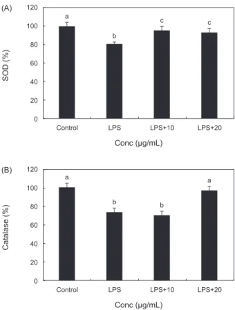
PYE의 항산화 효소 활성 효과
RAW 264.7 대식세포에서 LPS 처리후 PYE를농도별로처 리한결과 SOD와 catalase의활성에미치는효과는 Fig. 3에 서보는바와같다. Fig. 3 (A)를보면 LPS 단독처리후대조군 보다 SOD활성이유의적으로감소하였고 PYE를 10, 20 µg/
mL 농도로처리한군에서는 LPS단독처리군보다유의적으로
SOD 활성이증가하는것이확인되었다.
ROS인 O2- 는산소호흡을하는호기적세포에서생성되며산 화적스트레스로인한과다생성시암, 염증및여러질병의밀 접한관련이있다(Sun et al., 2004). 이렇게생성된 O2- 는생물 체내에가지고있는 SOD에의해제거된다. 대식세포의 LPS 처리는 SOD의활성을감소시켰으나 PYE를 LPS와동시투여 함으로서 SOD의활성이대조군수준으로회복시켰다. 이것은
PYE 투여가 LPS처리로인하여생긴내독소의산화적스트레
스에대한항산화작용이있는것으로사료된다. Fig. 3 (B)에 서 catalase의효소활성은 LPS만처리했을때는유의적으로 감소하였고 PYE 20 µg/mL처리시유의적으로증가하였다. 이 결과 PYE가 LPS를처리한대식세포에대해항산화작용을하 여 catalase 활성을증가시켜 ROS 생성을저해시켰음을확인 할수있었다. catalase는조직내의 SOD가효소반응에의해 O2- 를제거한후 TNF-α, IL-1β, IL-6 과같은염증성인자인 cytokine 생산의 2차 messenger 역할을하는 H2O2를물로분 Fig. 1. Effect of Pyropia yezoensis extract on cytotoxicity in RAW
264.7 macrophage.
RAW 264.7 cells were incubated with PYE for 24 h at the indi- cated concentration. The cell viability was determined by MTS as- say. Each value is expressed as mean±SD in triplicate experiment.
Values with different alphabets are significantly different at P<0.05 as analyzed by Duncan’s multiple range test.
- -
- +
10 +
20 +
iNos
COX-2
GAPDH PYE(μg/mL) LPS(1 μg/mL)
- -
- +
10 +
20 +
p-IκBα
IκBα
NF-κB
GAPDH PYE(μg/mL) LPS(1 μg/mL)
- -
- +
10 +
20 +
p-Akt
Akt
p-p38
p38
p-JNK
JNK
p-ERK
ERK
GAPDH
PYE(μg/mL) LPS(1 μg/mL)
a a a a
a 140
120 100 80 60 40 20 0
Control 5 10
Conc (μg/mL)
20 40
Cell viability (%)
a a
a a
a a
180 160 140 120 100 80 60 40 20 0
Control LPS LPS+5 Conc (μg/mL)
LPS+10 LPS+20 LPS+40
ROS (%)
a b
c cd d d
180 160 140 120 100 80 60 40 20
0 Control LPS LPS+5 Conc (μg/mL)
LPS+10 LPS+20 LPS+40
NO (%)
a
b b
a 120
100 80 60 40 20
0 Control LPS LPS+10
Conc (μg/mL)
LPS+20
Catalase (%)
(B)
a
b b
c 5,000
4,500 4,000 3,500 3,000 2,500 2,000 1,500 1,000 500 0
Control LPS LPS+10
Conc (μg/mL)
LPS+20
TNF-α (%)
B) - -
- +
10 +
20 +
TNF-α
IL-1β
PYE(μg/mL) LPS(1 μg/mL) A)
a
b
c c
120 100 80 60 40 20 0
Control LPS LPS+10
Conc (μg/mL)
LPS+20
SOD (%)
(A)
Fig. 2. Effect of Pyropia yezoensis extract on intracellular reac- tive oxygen species (ROS) levels in LPS stimulated RAW 264.7 macrophage.
RAW 264.7 cells were incubated with PYE for 24 h at the indi- cated concentration. Each value is expressed as mean±SD in trip- licate experiment. Values with different alphabets are significantly different at P<0.05 as analyzed by Duncan’s multiple range test.
- -
- +
10 +
20 +
iNos
COX-2
GAPDH PYE(μg/mL) LPS(1 μg/mL)
- -
- +
10 +
20 +
p-IκBα
IκBα
NF-κB
GAPDH PYE(μg/mL) LPS(1 μg/mL)
- -
- +
10 +
20 +
p-Akt
Akt
p-p38
p38
p-JNK
JNK
p-ERK
ERK
GAPDH
PYE(μg/mL) LPS(1 μg/mL)
a a a a
a 140
120 100 80 60 40 20 0
Control 5 10
Conc (μg/mL)
20 40
Cell viability (%)
a a
a a
a a
180 160 140 120 100 80 60 40 20
0 Control LPS LPS+5 Conc (μg/mL)
LPS+10 LPS+20 LPS+40
ROS (%)
a b
c cd d d
180 160 140 120 100 80 60 40 20 0
Control LPS LPS+5 Conc (μg/mL)
LPS+10 LPS+20 LPS+40
NO (%)
a
b b
a 120
100 80 60 40 20
0 Control LPS LPS+10
Conc (μg/mL)
LPS+20
Catalase (%)
(B)
a
b b
c 5,000
4,500 4,000 3,500 3,000 2,500 2,000 1,500 1,000 500 0
Control LPS LPS+10
Conc (μg/mL)
LPS+20
TNF-α (%)
B) - -
- +
10 +
20 +
TNF-α
IL-1β
PYE(μg/mL) LPS(1 μg/mL) A)
a
b
c c
120 100 80 60 40 20
0 Control LPS LPS+10
Conc (μg/mL)
LPS+20
SOD (%)
(A)
Fig. 3. Effect of Pyropia yezoensis extract on antioxidant enzyme activities in LPS stimulated RAW 264.7 cells.
RAW 264.7 cells were incubated with PYE for 24 h at the indicat- ed concentration. Each value is expressed as mean±SD in triplicate experiment. (A) SOD activity. (B) catalase activity. Values with different alphabets are significantly different at P<0.05 as analyzed by Duncan’s multiple range test.
- -
- +
10 +
20 +
iNos
COX-2
GAPDH PYE(μg/mL) LPS(1 μg/mL)
- -
- +
10 +
20 +
p-IκBα
IκBα
NF-κB
GAPDH PYE(μg/mL) LPS(1 μg/mL)
- -
- +
10 +
20 +
p-Akt
Akt
p-p38
p38
p-JNK
JNK
p-ERK
ERK
GAPDH
PYE(μg/mL) LPS(1 μg/mL)
a a a a
a 140
120 100 80 60 40 20 0
Control 5 10
Conc (μg/mL)
20 40
Cell viability (%)
a a
a a
a a
180 160 140 120 100 80 60 40 20
0 Control LPS LPS+5 Conc (μg/mL)
LPS+10 LPS+20 LPS+40
ROS (%)
a b
c cd d d
180 160 140 120 100 80 60 40 20
0 Control LPS LPS+5 Conc (μg/mL)
LPS+10 LPS+20 LPS+40
NO (%)
a
b b
a 120
100 80 60 40 20
0 Control LPS LPS+10
Conc (μg/mL)
LPS+20
Catalase (%)
(B)
a
b b
c 5,000
4,500 4,000 3,500 3,000 2,500 2,000 1,500 1,000 500 0
Control LPS LPS+10
Conc (μg/mL)
LPS+20
TNF-α (%)
B) - -
- +
10 +
20 +
TNF-α
IL-1β
PYE(μg/mL) LPS(1 μg/mL) A)
a
b
c c
120 100 80 60 40 20 0
Control LPS LPS+10
Conc (μg/mL)
LPS+20
SOD (%)
(A)
김 추출물에 의한 대식세포의 항염증 효과 761
해시켜생체를방어하는항산화효소이다(Yasui and Baba., 2006). 본연구의결과에서 LPS로인한산화적스트레스에대 해서 PYE가방어작용으로 SOD, catalase 등의항산화효소를 활성화시켜세포를보호하는효과를가지는것으로사료된다. PYE의 Nitric Oxide 생성 저해 효과
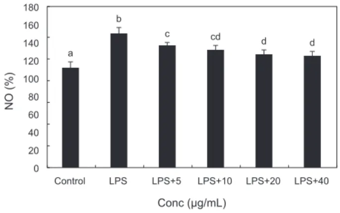
체내염증과정에서과량의 NO 및 PGE2등의염증인자는유도 형인 iNOS 및 COX-2에의해형성된다. NO를형성하는 NOS (NO synthase)는 L-arginine을 L-citrulline으로전환시키면서 NO를만들어내며 NO는 iNOS에의해생성된다(Schmidt and Walter., 1994). 이렇게만들어진 NO는염증반응을매개하는 역할을하고생체내에서빠르게 superoxide와반응하여강력 한산화제인 peroxynitrite를형성하여염증자극제로서대식세 포를활성화시켜(Ippouchi et al., 2002; Kang et al., 2000) NO 의생성을현저히증가시키고다른염증물질들과함께과도한 염증을유발시켜조직의손상을일으킨다(Nathan, 1992; Pan et al., 2011). LPS로자극된 RAW 264.7 대식세포에서생성되 는 NO에대한 PYE의억제효과를확인하였으며그결과는 Fig.
4에서보는바와같다. LPS 처리군은대조군에비하여 NO생성 이유의적으로증가하였고 LPS와 PYE를병행처리한군에서 는 NO생성이 LPS 처리군에비해농도유의적으로감소하는효 과가관찰되었다.
PYE의 iNOS, COX-2 발현 억제 효과
LPS로자극된 RAW 264.7 대식세포에서생성되는 COX-2, iNOS의생성에대한 PYE의효과를 Western blot으로분석하 였다. Fig. 5에서보는바와같이 iNOS의단백질발현은 LPS처
리군에비해 PYE를처리한군에서억제되는것을확인하였다. 이것은 PYE 처리에의한 NO 생성의감소가 iNOS 발현억제 에의한것임을의미한다. COX-2의단백질발현도 iNOS와마 찬가지로 LPS 처리군에비해 PYE를처리한군에서 COX-2의 단백질발현역시억제되는것을확인할수있었다. 염증은생 리적인보호기전으로방어기전을활성화시키거나손상을제 한하지만활성산소의파괴적성향으로인하여과다생성되면 세포와조직의손상을유도하고더나아가만성질환및노화 를일으킨다(Azard et al., 2008). 여기서생성되는 많은양의
염증유도 cytokine은대식세포의식세포작용을과도하게작
용시켜염증반응과산화적스트레스를증가시킨다(Blatteis et al., 2004; Fullerton et al., 2013). NOS는 type Ⅰ, Ⅱ, Ⅲ의 3 종류로구분되며 type Ⅲ인 iNOS는평소에는세포내에존재 하지않으나 LPS, cytokine 및박테리아독소같은자극이나
NF-κB활성에의해유도되어장시간동안다량의 NO를생성
한다(Nathan., 1992). 다른염증인자인 COX-2는 arachidonic acid를 prostaglandins로전환하는 cytokine, 자외선, 세균성내 독소및 TNF등과같은여러종류의 proinflammatory agent에 의해과다하게발현하여염증및각종퇴행성질환에관여한다 (Botting, 2006; Wu et al., 2009).
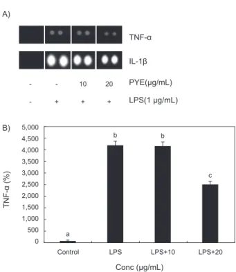
PYE의 염증성 Cytokines의 생성 억제 효과
RAW 264.7 대식세포에 LPS를처리한후 PYE를농도별로 처리하고 mouse cytokines array를이용하여각군별 TNF-α와 IL-1β의발현정도를비교하였다.
Fig. 6에서보는바와같이 TNF-α의경우 LPS 단독처리군에 비해 PYE를 20 µg/mL농도로처리하였을때발현이감소하였 고 IL-1β 역시 LPS 단독처리군에비해 PYE를처리하였을때 농도유의적으로감소하는것이관찰되었다.
LPS에의해자극된대식세포는 TNF-α와 IL-1β의생성을유 도하는데(Beutler and Ceramin., 1989) TNF-α의과잉생산은 T cell의수를감소시켜면역을떨어뜨리고(Abul et al., 2007) 혈액과조직사이의내피세포를자극하여여러백혈구들을염
Fig. 5. Effect of Pyropia yezoensis extract on iNOS and COX-2 protein expression in LPS stimulated RAW 264.7 cells.
RAW 264.7 cells were treated with LPS only or with different con- centration of Pyropia yezoensis extract for 24 h and lysated for western blot analysis.
- -
- +
10 +
20 +
iNos
COX-2
GAPDH PYE(μg/mL) LPS(1 μg/mL)
- -
- +
10 +
20 +
p-IκBα
IκBα
NF-κB
GAPDH PYE(μg/mL) LPS(1 μg/mL)
- -
- +
10 +
20 +
p-Akt
Akt
p-p38
p38
p-JNK
JNK
p-ERK
ERK
GAPDH PYE(μg/mL) LPS(1 μg/mL)
a a a a
a 140
120 100 80 60 40 20 0
Control 5 10
Conc (μg/mL)
20 40
Cell viability (%)
a a
a a
a a
180 160 140 120 100 80 60 40 20 0
Control LPS LPS+5 Conc (μg/mL)
LPS+10 LPS+20 LPS+40
ROS (%)
a b
c cd d d
180 160 140 120 100 80 60 40 20
0 Control LPS LPS+5 Conc (μg/mL)
LPS+10 LPS+20 LPS+40
NO (%)
a
b b
a 120
100 80 60 40 20 0
Control LPS LPS+10
Conc (μg/mL)
LPS+20
Catalase (%)
(B)
a
b b
c 5,000
4,500 4,000 3,500 3,000 2,500 2,000 1,500 1,000 500 0
Control LPS LPS+10
Conc (μg/mL)
LPS+20
TNF-α (%)
B) - -
- +
10 +
20 +
TNF-α
IL-1β
PYE(μg/mL) LPS(1 μg/mL) A)
a
b
c c
120 100 80 60 40 20 0
Control LPS LPS+10
Conc (μg/mL)
LPS+20
SOD (%)
(A)
Fig. 4. Effect of Pyropia yezoensis extract on nitric oxide (NO) level in LPS stimulated RAW 264.7 cells.
The cells were incubated with 1 µg/mL LPS only or with dif- ferent concentrations of Pyropia yezoensis extract for 24 h. The NO levels determined by Griess assay. Each value is expressed as mean±SD in triplicate experiment. Values with different alphabets are significantly different at P<0.05 as analyzed by Duncan’s mul- tiple range test.
- -
- +
10 +
20 +
iNos
COX-2
GAPDH PYE(μg/mL) LPS(1 μg/mL)
- -
- +
10 +
20 +
p-IκBα
IκBα
NF-κB
GAPDH PYE(μg/mL) LPS(1 μg/mL)
- -
- +
10 +
20 +
p-Akt
Akt
p-p38
p38
p-JNK
JNK
p-ERK
ERK
GAPDH
PYE(μg/mL) LPS(1 μg/mL)
a a a a
a 140
120 100 80 60 40 20 0
Control 5 10
Conc (μg/mL)
20 40
Cell viability (%)
a a
a a
a a
180 160 140 120 100 80 60 40 20
0 Control LPS LPS+5 Conc (μg/mL)
LPS+10 LPS+20 LPS+40
ROS (%)
a b
c cd d d
180 160 140 120 100 80 60 40 20 0
Control LPS LPS+5 Conc (μg/mL)
LPS+10 LPS+20 LPS+40
NO (%)
a
b b
a 120
100 80 60 40 20
0 Control LPS LPS+10
Conc (μg/mL)
LPS+20
Catalase (%)
(B)
a
b b
c 5,000
4,500 4,000 3,500 3,000 2,500 2,000 1,500 1,000 500 0
Control LPS LPS+10
Conc (μg/mL)
LPS+20
TNF-α (%)
B) - -
- +
10 +
20 +
TNF-α
IL-1β
PYE(μg/mL) LPS(1 μg/mL) A)
a
b
c c
120 100 80 60 40 20
0 Control LPS LPS+10
Conc (μg/mL)
LPS+20
SOD (%)
(A)