INTRODUCTION
The pulmonary lymphatic system plays an important role in the lung perfusion homeostasis. Congenital errors of lym- phatic vessel development lead to primary pulmonary lym- phatic disorders, including lymphangiomas, lymphangiec- tasis, lymphangiomatosis, and lymphatic dysplasia syndromes (1). Among them, pulmonary lymphangioma is a rare benign lesion thought to result from the development of abnormal- ly proliferating lymphatic vessels. The abnormal vessels may be capillary, cavernous or cystic in type. The basic defect has been considered to be an abnormality in the developmental lymphangiogenesis (2).
We present a case of intrapulmonary cystic lymphangioma involving the upper lobe of the right lung, which presented with dyspnea in a 2-month-old female infant, and review the literature on its pathogenesis, clinicopathologic features, and radiographic findings.
CASE REPORT
A 2-month-old Korean baby girl developed dyspnea and irritability about a week before admission to our hospital.
On physical examination at admission, her temperature was 36.2℃, blood pressure 80/60 mmHg, pulse 145/min, res- piratory rate 28/min, and weight 4,200 g. Breathing sound decreased in the right lung field and was accompanied by chest wall retraction. There was no edema signs in extremi-
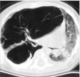
ties. Arterial blood gas analysis result was pH 7.26-pCO2 50.3%-pO297.7%-O2saturation 96.3%. Otherwise physical examination and laboratory results proved unremarkable. On birth history, delivery occurred spontaneously at 37 weeks gestation and her birth weight was 3,000 g. A chest radio- graph at admission showed a solitary cystic mass, entirely occupying the upper lobe of the right lung. High-resolution computed tomography (HRCT) of the chest depicted a well- circumscribed, multiseptate, air-filled cystic lesion in the upper lobe of the right lung, mimicking the feature of type I congenital cystic adenomatoid malformation (Fig. 1).
Under the preoperative diagnosis of congenital cystic ade- nomatoid malformation, she underwent bilobectomy of the upper and middle lobes of the right lung. Grossly, the large part of the upper lobe was replaced by multiple cystic spaces that contained some serous fluid and varied in size from 2-3 mm to 3.5 cm. Remained lung parenchyma is hemorrhagic and revealed emphysematous change. The middle lobe just showed emphysematous change and focal hemorrhage.
Histopathological examination of the upper lobe revealed a relatively circumscribed intrapulmonary cystic lesion com- posed of multiple cysts or cavities (Fig. 2). The cystic spaces were lined by a monolayer of flat or low cuboidal cells. They were interconnected each other. Their walls were supported by loose fibrous stroma with mild lymphocytic infiltrate (Fig.
3). Neither significant smooth muscle proliferation nor lym- phoid follicles were observed in the walls. Lung parenchyma outside the cystic lesion exhibited emphysematous change and some lymphangiectasia in the connective tissues of interlobu-
Chang Hun Lee, Young Dae Kim*, Kyun Il Kim�, Young Tak Lim�, Kyung Min Lee, Kyung Un Choi, Jin Suk Lee, Mee Young Sol
Departments of Pathology, *Thoracic Surgery,
�Diagnostic Radiology, and �Pediatrics, College of Medicine, Pusan National University, Busan, Korea
Received : 9 June 2003 Accepted : 8 August 2003
Address for correspondence Chang Hun Lee, M.D.
Department of Pathology, College of Medicine, Pusan National University, 1-10 Ami-dong, Seo-gu, Busan 602-739, Korea
Tel : +82.51-240-7718, Fax : +82.51-256-1780 E-mail : [email protected]
458 J Korean Med Sci 2004; 19: 458-61
ISSN 1011-8934
Copyright � The Korean Academy of Medical Sciences
Intrapulmonary Cystic Lymphangioma in a 2-month-old Infant
Lymphangioma is an abnormal collection of lymphatics that are developmentally isolated from the normal lymphatic system. Lymphangioma rarely presents as a solitary pulmonary lesion. We report a rare case of intrapulmonary cystic lymphan- gioma involving the upper lobe of the right lung, which presented with dyspnea in a 2-month-old infant. High-resolution computed tomography (HRCT) of the chest demonstrated a well-circumscribed, multiseptate, cystic lesion in the upper lobe of the right lung, mimicking the feature of type I congenital cystic adenomatoid mal- formation. The tumor was removed by bilobectomy of the upper and middle lobes of the right lung, and its pathologic examination confirmed the diagnosis of an intra- pulmonary cystic lymphangioma.
Key Words :Lung; Lymphangioma, Cystic; Infant
Intrapulmonary Cystic Lymphangioma 459
lar septa and subpleural zones. In the immunohistochemical staining the lining cells of the cystic walls were not reactive for epithelial membrane antigen and cytokeratin, but weak- ly positive for factor VIII-related antigen (Fig. 4). Intrapul- monary cystic lymphangioma was subsequently diagnosed.
The microscopic findings of the middle lobe were consistent with the gross finding.
The patient had an eventful recovery after operation. At
4-month follow up she remained well without evidence of recurrence.
DISCUSSION
Lymphangiomas are focal proliferation of well differentiat- ed lymphatic tissue that presents as multicystic or sponge-like
Fig. 1.High-resolution computed tomography of the chest depicts a well-circumscribed, multiseptate, air-filled cystic lesion in the upper lobe of the right lung.
Fig. 2.1:1 scaned photograph shows a large intrapulmonary cys- tic tumor (Ly), causing emphysematous change (Em) in surround- ing pulmonary parenchyma (H&E, ×1).
Fig. 3.The cystic walls (in the right top) of the tumor are support- ed by loose fibrous tissue and adjoin to the emphysematous lung tissue (in the left) (H&E, ×20).
Fig. 4.Immunohistochemical staining for factor VIII-related anti- gen shows weak positive reactions in the lining cells of the cystic walls (Streptavidin-biotin, ×100).
Em
Ly
460 C.H. Lee, Y.D. Kim, K.I. Kim, et al.
appearance. They are generally subdivided into three patho- logic categories: capillary lymphangiomas describe thin-walled lymphatic channels that occur as small well circumscribed cutaneous lesions; cavernous lymphangiomas are microscop- ic thin-walled lymphatic channels with associated stroma; and cystic lymphangiomas are large, well-circumscribed, multi- loculated cystic spaces lined by endothelium that contain a significant connective tissue component (2).
Their pathogenesis is still uncertain, but they may present as congenital or acquired forms. Congenital lymphangiomas probably represent embryologic remnants of lymphatic tis- sues that either failed to connect to efferent channels or arose from portions of lymph sacs that were sequestered during development (3). Acquired or secondary lymphangiomas develop in areas of chronic lymphatic obstruction related to surgery, chronic infection, or radiation.
Lymphangioma can occur in any region of the body in which there is lymphatic drainage. The single most common site of cystic occurrence is in the neck, where the lesion is referred to as a cystic hygroma. In the chest, lymphangiomas are most commonly found in the mediastinum, where they account for 0.7% to 4.5% of all mediastinal tumors (4). In a regard to intrapulmonary lymphangiomas they have been described but are extremely rare: approximately a dozen cases, with patient ages ranging from 6 months to 54 yr, are described in the English literature (1). They frequently occur in the lower lobe of the lung, which is suspected to be associated with a rich lymphatic supply noted in the part of the lung.
But we also found two case reports developed in the upper lobe (5, 6), as in our case. In a review of 20 patients with tho- racic lymphangioma (including mediastinal), Shaffer et al. (4) noted a slight predominance in women. But Wilson et al. (5) suggested that all adult patients with intrapulmonary lym- phangioma were men, including their case. Symptoms are known to vary widely depending on the age of the patient and the extent of disease. In neonates and infants, cystic lym- phangiomas often present with pneumothorax and respira- tory distress (4). In our case, she complained of dyspnea devel- oped recently, with the signs of respiratory distress. Radio- logically plain radiographs, ultrasound, CT scan, and mag- netic resonance imaging (MRI) have proven useful in deter- mining the number and extent of lesions. The most common CT appearance of lymphangiomas is that of a cystic mass with smooth margins, but in the childhood the CT and/or MRI findings do not appear to be specific to differentiate lymphan- giomas from other intrapulmonary cystic lesions, including congenital cystic adenomatoid malformation (CCAM), pleu- ropulmonary blastoma (PPB), bronchial atresia, bronchogenic cyst, and congenital lobar emphysema (7). Therefore histo- logical and roentgenographic correlation may be necessary for establishing a definite diagnosis.
Among the list of differential diagnosis, CCAM is a dis- order of embryonic bronchopulmonary development, char- acterized by a multicystic mass of pulmonary tissue with an
abnormal proliferation of bronchial structures. There are three types: Type I consists of a large single cyst; Type II of numer- ous small cysts; Type III appears as a solid mass. The prog- nosis is variable and depends on the size and the histologic type (8). PPB is a rare malignant dysontogenetic neoplasm affecting children, and is characterized histologically by a variably mixed blastematous and sarcomatous patterns. PPB has also known to have three pathological varieties, includ- ing predominantly cystic type I, cystic and solid type II, and predominantly solid type III (9). The solid areas of the types II and III PPB are composed of undifferentiated blastemal tissue which may overlap with spindle cell sarcomatous, rhab- domyosarcomatous, anaplastic, and chondrosarcomatous foci.
Bronchial atresia may be present as an asymptomatic mass in the newborn period or the lung may appear as a fluid-filled, pulmonary mass at birth. Later the fluid is replaced by air and the lobe becomes emphysematous (10). Bronchogenic cyst accounts for approximately 3% of mediastinal masses in the pediatric age group. Though the middle and posterior mediastinum are generally regarded as the commonest sites, the cysts can unusually show the intrapulmonary localization (11). When a communication persists between the tracheo- bronchial tree and the cyst, an air-fluid level is present as well as a history of multiple infections. A pseudostratifed, ciliated columnar epithelium, submucous glands, and isolated islands of cartilage are the composite of histologic findings for an unequivocal diagnosis of bronchogenic cyst. Congenital lobar emphysema or congenital lobar hyperinflation affects only one of the upper lobes or the right middle lobe of the lung. The main pathologic change consists of massive overdistention of the alveolar spaces, not accompanied by the destruction of the tissues. It is therefore not truly a cystic or an emphy- sematous process. In addition, intrapulmonary lymphangio- mas need to be differentiated from other primary pulmonary lymphatic disorders, including lymphangiectasis and lym- phangiomatosis (1). Lymphangiectasis is the pathologic dila- tion of lymphatics. Primary (congenital) and secondary forms have been described. The primary form presents in neonates and is usually fatal. Secondary forms of lymphangiectasis result from a variety of processes that impair lymph drainage and increase lymph production. Pathologically the lesion shows multifocal lymphatic dilations along the lymphatic routes of the interlobular septa and the bronchovascular bundles.
But it does not show the localized proliferation of anastomos- ing lymphatic channels which is characteristic of lymphan- giomas. Lymphangiomatosis literally describes the presence of multiple lymphangiomas. It is frequently associated with other lymphatic-related abnormalities and usually (in 75%
of cases) involves multiple organs. The histology of lymphan- giomatosis resembles a lymphangioma, but can appear to infiltrate tissues, raising concern for a more aggressive lesion.
Clinically it presents at a later age than solitary lymphan- giomas either because of an influence of hormonal factors or because a more subtle, albeit more widespread, defect requires
Intrapulmonary Cystic Lymphangioma 461
a longer period for growth.
The natural history of intrapulmonary lymphangiomas in infants is not well-known, but surgical resection should be considered in the light of the neoplastic nature of these lesions and of the difficulty in clinically differentiating them from other cystic lesions occurring in the age class (12). If the lesion is small and localized, the application of thoracoscopic surgery is becoming increasingly popular. Incomplete resection, how- ever, can result in recurrence at sites of surgical resection, because of their autonomous growth and also by their reac- cumulation of lymph fluid (1). Prior to the surgical explo- ration and excision, it is prudent to investigate for other lym- phangiomatous lesions and associated congenital anomalies.
In summary, we present a case of an intrapulmonary cys- tic lymphangioma causing dyspnea in a 2-month-old infant.
To the best of our knowledge this case is the second report in the Korean literature since 1997 (13). Despite of the scarci- ty, this lesion should be included in the differential diagno- sis of multicystic lung lesion noted during infancy.
REFERENCES
1. Faul JL, Berry GJ, Colby TV, Ruoss SJ, Walter MB, Rosen GD, Raf- fin TA. Thoracic lymphangiomas, lymphangiectasis, lymphangiomato- sis, and lymphatic dysplasia syndrome. Am J Respir Crit Care Med 2000; 161: 1037-46.
2. Enzinger FM, Weiss SW. Tumors of lymph vessels. In: Gay SM, edi- tor, Soft tissue tumors. Mosby, St. Louis. 1995; 679-700.
3. Tazelaar HD, Kerr D, Yousem SA, Saldana MJ, Langston C, Colby TV. Diffuse pulmonary lymphangiomatosis. Hum Pathol 1993; 24:
1313-22.
4. Shaffer K, Rosado-de-Christenson ML, Patz EF Jr, Young S, Farver CF. Thoracic lymphangioma in adults: CT and MR imaging features.
AJR Am J Roentgenol 1994; 162: 283-9.
5. Wilson C, Askin FB, Heitmiller RF. Solitary pulmonary lymphan- gioma. Ann Thorac Surg 2001; 71: 1337-8.
6. Takemura T, Watanabe M, Takagi K, Tanaka S, Aida S. Thoraco- scopic resection of a solitary pulmonary lymphangioma: report of a case. Surg Today 1995; 25: 651-3.
7. Heij HA, Ekkelkamp S, Vos A. Diagnosis of cystic adenomatoid malformation of the lung in new born infants and children. Thorax 1990; 45: 122-5.
8. Stocker JT, Madewell JE, Drake RM. Congenital cystic adenoma- toid malformation of the lung. Classification and morphologic spec- trum. Hum Pathol 1977; 8: 155-71.
9. Dehner LP. Pleuropulmonary blastoma is the pulmonary blastoma of childhood. Semin Diagn Pathol 1994; 11: 141-51.
10. Schuster SR, Harris GB, Williams A, Kirkpatrick J, Reid L. Bronchial atresia: a recognizable entity in the pediatric age group. J Pediatr Surg 1978; 13: 682-9.
11. Ramenofsky ML, Leape LL, McCauley RG. Bronchogenic cyst. J Pediatr Surg 1979; 14: 219-24.
12. Vogt-Moykopf I, Rau B, Branscheid D. Surgery for congenital mal- formations of the lung. Ann Radiol (Paris) 1993; 36: 145-60.
13. Jeon MY, Chi JG. Intrapulmonary cystic lymphangioma-A case re- port. Korean J Pathol 1997; 31: 492-4.