Case Report
원고 접수일 2010년 12월 24일, 게재 확정일 2011년 2월 24일 책임저자 김수관
(501-759) 광주시 동구 서석동 375번지, 조선대학교 치과대학원 구강악안면외과학 교실
Tel: 062-220-3819, Fax: 062-228-7316, E-mail: [email protected]
RECEIVED December 24, 2010, ACCEPTED February 24, 2011 Correspondence to Su-Gwan Kim
Department of Oral and Maxillofacial Surgery, School of Dentistry, Chosun University
375, Seosuk-dong, Dong-gu, Gwangju 501-759, Korea
Tel: 82-62-220-3819, Fax: 82-62-228-7316, E-mail: [email protected]
CC This is an open access article distributed under the terms of the Creative Commons Attribution Non-Commercial License (http://creativecommons.org/licenses/
by-nc/3.0) which permits unrestricted non-commercial use, distribution, and reproduction in any medium, provided the original work is properly cited.
하악 전치부 임플란트 식립 후에 발생한 과다출혈: 증례보고
조지호ㆍ김수관ㆍ문성용ㆍ오지수ㆍ김정선1
조선대학교 치의학전문대학원 구강악안면외과학교실, 1광주보건대학교 치위생과
Abstract
Excessive Bleeding after Implant Placement in the Anterior Mandible: Case Report
Ji-Ho Jo, Su-Gwan Kim, Seong-Yong Moon, Ji-Su Oh, Jeong-Sun Kim
1Department of Oral and Maxillofacial Surgery, School of Dentistry, Chosun University,
1
Department of Dental Hygiene, Gwangju Health College University
Implant placement on the anterior mandible is considered a common and safe surgical procedure. However, severe hemorrhage can occur if branches of the sublingual artery, which run through the lingual cortical plate of the mandible, are damaged.
Excessive hemorrhage caused by injury to the sublingual artery can result in life-threatening problems such as airway obstruction.
A 54-year old male patient without any generalized systemic conditions was referred due to active bleeding after implant placement in the anterior mandible. Gauze compression with surgicel and bosimin were performed and hemostasis was achieved.
The patient was discharged after 3 days without any supplementary bleeding.
Key words: Complication, Implant, Postoperative hemorrhage, Sublingual artery
서 론
최근 골유착 임플란트는 무치악부의 치료로서 일반화된 술식으 로, 고정성 보철물이나 가철성 보철물에 우선하여 사용되고 있으 며, 특히, 하악 전치부 임플란트 식립은 총의치의 안정과 유지를 위해 널리 이용되고 있는 방법이다. 다른 외과적 술식과 마찬가지 로 하악 전치부의 임플란트 식립 역시 합병증이 동반될 수 있으며, 출혈, 인접 조직의 손상, 감염, 통증, 부종, 공기색전 등이 있다.
그 중 설하동맥(sublingual artery)의 손상에 따른 출혈 및 구강저
의 혈종은 생명을 위협할 수 있는 합병증이다[1-3]. 설하동맥은 이하동맥(submental artery)과 함께 구강저의 주요한 동맥이며, 이 두 동맥의 말단 가지는 이설골근에 평행하게 하악 정중부의 설측까지 주행하며 서로 문합한다[4]. 이 중 설하동맥의 종말 가지 는 하악 설측 피질골판을 뚫고 하악골 내부까지 이르는 경우가 있으며, 이는 임플란트 식립 중 심각한 출혈의 원인이 될 수 있다 [5].
임플란트의 식립 시 하악전방부의 출혈은 드물지만, 설하동맥
의 손상으로 인해 하악골 설측의 과도한 출혈이 발생한 경우
Fig. 2. Initial clinical examination.
Marked swelling was noted on sub- mental area.
Fig. 1. Hematoma in the floor of the mouth resulting in elevation of the tongue.
본 증례는 54세 남자 환자로, 하악 전치부의 만성치주염으로 개인 치과에서 양측 하악 중절치와 측절치를 발거하고 양측 측절 치 부위에 즉시 임플란트 식립술(Osstem GS III 직경 3.5 mm,
인으로 국소마취한 후 설측 피판을 거상하고 출혈부위를 확인하였
고, 피판의 찢어짐과 함께 맥동성 출혈이 관찰되었다. Surgicel과
bosmin으로 압박지혈을 시행하였으며, 약 30분 후 지혈이 확인
Fig. 3. Initial neck computed tomog- raphy show sublingual hematoma, slight upper air way obstruction due to the elevation of tongue.
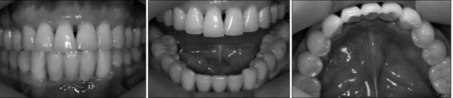
Fig. 5. On postoperative 4 months clinical examination.
Fig. 4. On postoperative 3 days clinical examination, swelling in the floor of the mouth was subsided and active bleeding was not observed.
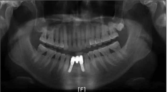
되어 일차 봉합을 시행하였으나, 삼출성 출혈 및 혈종이 다시 관찰되어 혈종을 제거한 후 penrose drain을 이용하여 배출 통로 를 확보하고 봉합하였다. 입원 후 전신 검사와 경부 컴퓨터 단층촬 영을 시행하였으며(Fig. 3), 전신 검사상 특별한 이상은 발견되지 않았다. 입원 후 출혈은 관찰되지 않았으며, 구강저의 혈종 역시 감소되어 환자는 입원 3일 후 퇴원하였고(Fig. 4), 4개월 후 임플 란트 보철을 완성한 상태로 본원에 내원하였으며 임상 검사 상 양호한 치유소견을 보였다(Fig. 5, 6).
고 찰
무치악 부위의 치료로서 치과 임플란트 식립은 일반화된 술식
이며, 간단한 외과적 과정을 통해 교합을 회복할 수 있는 비교적
안전한 술식이라 생각되어 왔지만, 하악 전치부 임플란트의 식립
시 때로 생명을 위협하는 출혈이 발생할 수 있다. 출혈은 임플란트
식립 과정 중에 발생할 수 있고, 임플란트 식립 후 몇 시간 후에
지연출혈의 형태로 발생할 수도 있다. Mason 등[6]은 임플란트
Fig. 6. Panoramic radiograph with prosthesis set up 16 weeks after the surgery.
식립 과정 중 하악 설측 피질골 천공에 의한 4∼5시간 후의 지연 출혈 및 혈종, 그로 인한 상기도폐쇄의 위험성을 증례를 통해 보고한 바 있으며, 지연출혈의 원인에 대해 혈관 수축제의 작용 소실이 한 원인이 될 수 있다고 보고하였다. 물론, 이러한 형태의 지연출혈은 장기간 항응고제를 복용한 환자의 경우 그 가능성이 커진다고 하였다[7,8].
구강저의 혈종은 임플란트 식립뿐 아니라 발치, 전정 성형술 (vestibuloplasty), 하악골 골절단술(mandibular osteotomy), 구강저의 낭종 절제술 등을 비롯한 하악 전치부의 다양한 외과적 술식에 의해서 발생할 수 있으며[9-12], 주요한 원인은 하악 전치 부 설측 피질골의 천공, 골막의 열상이나 설하동맥의 가지의 손상 등이다[1]. 설하동맥은 이하동맥과 함께 구강저의 주요 공급동맥 으로 이설골근에 평하게 주행하다가 하악 정중부에 이르는데, 때로 하악골 설측 피질골을 뚫고 하악골 내부에까지 이르는 경우 가 있으며, 본 증례에서도 설측 골막의 박리중 발생한 골막의 열상과 함께 설측 천공 동맥의 손상이 출혈의 원인으로 생각되었 다. Longoni 등[5]은 100개의 건조 두개골과 100명의 환자 CT를 통한 연구에서 각각 80%, 60%에서 하악 전치부에 적어도 1개의 설측 천공동맥이 관찰되었다고 보고하였으며, 설측 천공동맥은 전체 건조 두개골의 약 51%가 정중부에서 관찰되었고, CT에서는 55%가 정중부에서 천공동맥이 관찰되었다고 보고한바 있다. 또 한, 이미 많은 연구들에서 설하 동맥의 말단 가지에 의한 하악 전치부의 설측 천공동맥의 존재는 확인된 바 있다[13-16]. 또한, Niamtu[1]는 하악골 설측 피질골의 천공과 천공동맥의 손상으로 인한 구강저의 혈종이 생명을 위협할 수 있음을 보고한바 있으며, Goldstein[17] 역시 구강저의 혈종으로 인한 상기도 폐쇄가 생명 을 위협할 수 있음을 보고한 바 있다. 따라서, 하악 전치부 임플란 트 식립이 계획되었다면, 술자는 CT 촬영을 통해 하악골 설측의 천공동맥 유무 및 그 위치에 대한 평가를 시행해야 할 것이다.
하악 전치부 임플란트 식립과 관련된 출혈 환자의 관리에 있어 가장 중요한 것은 기도의 확보이며, 기도가 확보된 후에 출혈의 원인을 찾고, 지혈의 과정이 필요할 것이다. 지혈의 방법에는
시행 하였으나, 다시 혈종 및 삼출성 출혈이 발생하여 배출관을 삽입하였다. 구강저의 출혈과 혈종에 대해 배출관을 삽입하는 것이 좋은지 혹은 삽입하지 않는 것이 좋은 지에 대한 명확한 근거나 연구가 확립되어 있지 않지만, 본 증례에서처럼 조기에 지혈이 달성되었다면, 외래 환자에 있어 삼출성 출혈로 인한 혈종 의 형성과 그로 인한 급성 상기도 폐쇄의 위험성을 고려할 때 배출관의 삽입은 적절한 방법이라고 생각된다.
하악 전치부의 임플란트 식립 후 발생하는 즉시 출혈 혹은 지연 출혈이 때로 생명을 위협할 수 있음을 본 증례와 많은 문헌을 통해 확인할 수 있다. 특히, 임플란트 식립 도중 발생하는 하악 설측 골막의 열상이나 설하 동맥의 하악 설측 천공가지의 손상은 심한 출혈의 원인이 될 수 있다. 따라서, 하악골 설측의 동맥 손상의 위험성을 최소화하기 위해서는 술 전 CT촬영을 통한 정확 한 분석이 선행되어야 하며, 술 중에 판막의 거상시 골막의 열상이 발생하지 않도록 하며, 천공동맥의 유무를 확인하고, 천공동맥이 존재할 경우 전기소작술 등을 이용하여 출혈이 유발되지 않도록 처치하며, 임플란트의 식립시에도 설측 피질골을 천공시키지 않 도록 주의하는 것이 필요할 것이라 생각된다.
References