TPA로 유도된 마우스 귀 부종 동물모델에서 소목추출물의 항염증 효과
음원식
1
*․이광재2
*․김대원1
․임순성3
․강일준3
․박진서1
․최수영1†
1
한림대학교 바이오메디컬학과 및 생명공학연구소
2
강원도농업기술원
3
한림대학교 식품영양학과 및 RIC 센터
Anti-Inflammatory Effects of Extracts from Caesalpinia sappan L.
on Skin Inflammation
Won Sik Eum
1*
, Kwang-Jae Lee2*
, Dae Won Kim1
, Soon Sung Lim3
, Il-Jun Kang3
, Jinseu Park1
, and Soo Young Choi1†
1
Dept. of Biomedical Science and Research Institute of Bioscience and Biotechnology, Hallym University2
Gangwon Agricultural Research and Extention Services, Gangwon 200-822, Korea3
Dept. of Food Sciences and Nutrition and RIC Center, Hallym University, Gangwon 200-702, KoreaAbstract
This study investigated the anti-inflammatory effects of extracts from Caesalpinia sappan L. (CSL) on 12-O-tetradecanoylphorbol-13-acetate (TPA)-induced ear edema in mice. Skin inflammation was detected by immunohistochemistry and the protein and mRNA expression levels of cyclooxygenase-2 (COX-2) and cytokines (IL-6, IL-1β and TNF-α) detected by Western blotting and RT-PCR. The activation of nuclear factor-kappa B (NF-κB) and mitogen-activated protein kinase (MAPK) were analyzed by Western blotting. CSL extracts markedly inhibited the TPA-induced expression of COX-2 and pro-inflammatory cytokines. Also, CSL extracts significantly reduced the activation of NF-κB and MAPK. These results suggest that CSL extracts may serve as therapeutic agents against skin diseases related to inflammation.
Key words: inflammation, TPA, Caesalpinia sappan L., NF-κB, MAPK
*
The first two authors contributed equally to this work.
†
Corresponding author. E-mail: [email protected]
†
Phone: 82-33-248-2112, Fax: 82-33-241-1463
서 론
염증반응은 자극에 대한 생체조직의 방어반응의 하나로 다양한 질병의 생리 및 병리학적인 과정에 관여한다. 여러 가지 염증매개인자들이 생성되는 염증으로 인하여 발열, 홍 반, 부종 등의 증상이 나타난다. 또한 지속적인 염증반응은 다양한 난치성 질환들(치매, 심혈관질환, 암, 비만, 관절염, 당뇨병 및 동맥경화증 등)의 원인이 된다고 보고되고 있다 (1-6). 생체 내 염증반응을 촉진하는 효소 및 인자들로서 cyclooxygenase-2(COX-2), tumor necrosis factor-α(TNF- α ), interleukin(IL)-6, IL-1β과 신호전달경로인 nuclear fac- tor kappa-B(NF-κB) 및 mitogen-activated protein kin- ase(MAPK)가 잘 알려져 있으며, 이들은 염증반응뿐 아니 라 다양한 난치성질환에서도 관여하기 때문에 이들 효소 및 인자들을 억제하는 물질은 염증성 질환에 대한 항염증제로 개발될 가능성이 매우 높다(7-10).
소목(
Caesalpinia sappanL.)은 인도, 말레이사아 등 아시 아에 분포하는 낙엽 관목의 콩과(Leguminosae)식물로 약용
및 염료로 이용되고 있으며, 소목추출물은 항통증, 항경련, 항산화, 면역조절, 항바이러스 효과(11-15) 등이 보고되었 다. 소목의 주요성분으로 brazilin, campesterol, stigmaster- ol, hematein, sappanin 등이 밝혀졌으며, 이중 brazilin은 무 색의 flavonoid 구조를 가지고 있으며 공기 중에 산화되어 brazilein이 된다(16-18). 한방에서 brazilin은 어혈, 월경통 및 출산 후 다양한 증상에 사용해 왔다(19,20). 지금까지 소 목추출물의 생리활성에 관한 많은 연구가 보고되었으나, 염 증동물모델에서 소목추출물의 생리활성에 관한 연구는 미 비한 실정이다. 따라서 본 연구에서는 소목추출물의 항염증 효능과 기전을 TPA로 유도한 마우스 귀 부종 동물모델에서 규명하고자 하였다.
재료 및 방법
실험재료
수컷 ICR 마우스(6주령)은 한림대학교 실험동물센터에서
구입하여 사용하였다. COX-2, β-actin, p65, Lamin B 항체
는 Santa Cruz Biotechnology(Santa Cruz, CA, USA)에서 구입하였고, p38, p-p38, ERK, p-ERK 항체는 Cell Signal- ing Technology(Beverly, MA, USA)에서 구입하였다. 그 밖 에 사용된 시약들은 Sigma-Aldrich(St. Louis, MO, USA) 의 특급시약을 사용하였다.
소목추출물 제조
실험에 사용한 소목은 춘천 대광약업사에서 구입하여 사 용하였다. 건조된 소목 100 g을 70% 에탄올 1 L에 넣어 3시 간 동안 환류추출 하였으며, 이를 3회 반복하였다. 추출 후 Whatman No.2 filter paper(GE Healthcare, Little Chalfont, UK)를 이용하여 여과한 후 감압회전 농축기(Rotavapor R- 220, Buchi, Flawil, Switzerland)를 이용하여 에탄올을 제거 하였고, 이를 동결건조 하여 분말 상태의 소목추출물을 획득 하여 실험에 사용하였다(21).
실험동물
실험동물은 6주령 ICR 수컷 마우스 21마리를 한림대학교 실험동물센터에서 구입하여 사용하였으며, 온도 23
oC, 습도 60%, 12시간 명암조건에서 사육하였다. 실험동물은 구입 후 1주일 동안 순화한 다음, 각 군당 7마리씩을 배치하여 총 3군 으로 실험군을 분류하였다. 본 연구에 사용된 마우스는 한림 대학교 동물실험윤리위원회의 사전심의와 윤리 규정을 준 수하여 수행되었다.
염증동물모델
염증동물모델은 12-
O-tetradecanoylphorbol-13-acetate (TPA)를 마우스 귀에 도포하여 귀 부종을 유발하였다(22).
TPA(1 μg)를 아세톤 20 μL에 녹인 후 마우스 귀에 하루 한 번씩 3일 동안 도포하였다. 실험동물은 정상군(Control)과 TPA만 처리한 대조군(TPA), 그리고 TPA 처리 후
Caesal- pinia sappanL.(CSL)을 같이 처리한 군(CSL)의 3군으로 구성하였다. TPA 처리 후 하루 한번씩 1시간 동안 CSL 시 료(2 mg/kg)를 처리하였다. 마지막으로 TPA와 시료를 처 리한 다음, 24시간 후 직경 5 mm 펀칭을 이용하여 귀 조직을 얻어 귀의 무게와 두께를 측정하고, 면역조직염색 및 염증인 자 발현 분석을 수행하였다.
면역조직염색
실험동물은 질소와 산소 혼합가스가 들어있는 3% 이소플 루레인(isoflurane)으로 마취시킨 후 관류 세척하였다. 관류 세척이 끝난 후, 적출된 장기는 파라포름알데히드를 함유한 0.1 M PBS(phosphate buffered saline, pH 7.4)로 고정하고 고정이 끝난 조직은 조직을 떼어내어 4% 파라포름알데히드 용액에 담가 보존하였다. 조직을 임배딩 배지(Reichert- Jung, Nußloch, Germany)로 포매하고 동결박절기(cryo- stat, Reichert-Jung)를 이용하여 5 μm 절편으로 만든 다음 사용하였다.
조직 절편은 0.01 M PBS로 10분간 완충시킨 후 다시 증류
수로 10분간 세척하였다. PBS로 7분씩 3회 세척한 후 비특 이적 반응을 방지하기 위하여 조직을 PBS에 들어있는 5%
정상 염소 혈청에 30분간 반응시킨 다음 통상적인 헤마토자 일린 및 에오신(hematoxyline and eosin) 염색을 하였다. 그 다음 현미경 관찰 후 사진촬영(axioscope microscope, Carl Zeiss, Jena, Germany) 하였다(23,24).
Western blot analysis
귀 조직 시료를 분쇄하여 sodium dodecyl sulfate-poly- acrylamide gel electrophoresis(SDS-PAGE)에 의해 분리 된 단백질을 polyvinylidene difluoride(PVDF) membrane (Millipore, Bedford, MA, USA)으로 이동시켰다. Membrane 은 5% milk-TBST(tris buffered saline, pH 7.5)로 1시간 incubation한 후, TBST로 10분간 3회 세척하였다. 각각의 1차 항체를 5% TBST로 희석(1:1,000)한 후, membrane을 1시간 incubation하고 TBST로 10분간 3회 세척하였다. 2차 항체로 anti-rabbit IgG를 TBST에 희석(1:10,000)하여 membrane을 1시간 incubation하고 TBST로 10분간 3회 세 척하였다. 항체에 결합된 단백질들은 chemiluminescence 방 법을 통하여 가시화하였다(23,24).
Reverse transcription-polymerase chain reaction (RT-PCR)
귀 조직에서 total RNA는 TRIzol
®reagent kit(Invitro- gen, Carlsbad, CA, USA)를 이용하여 분리하였다. Oligo dT 와 reverse transcriptase를 이용하여 template RNA로부터 cDNA를 합성하고 TNF-α, IL-6, IL-1β의 primer를 사용하 여 PCR를 수행하였다. PCR product는 1% agarose gel에서 전기영동하고 ethidium bromide를 사용하여 가시화하였다 (23,24). TNF-α, IL-6, IL-1β 각각의 primer는 다음과 같다.
TNF-α sense primer:
5'-AAGTTCCCAAATGGCCTCCC-3' TNF-α antisense primer:
5'-TGGCACCACTAGTTGGTTGTCTTT-3' IL-6 sense primer:
5'-CAAGAAAGACAAAGCCAGAGTCCTT-3' IL-6 antisense primer:
5'-TGGATGGTCTTGGTCCTTAGCC-3' IL-1β sense primer:
5'-TGCAGAGTTCCCCAACTGGTACATC-3' IL-1β antisense primer:
5'-GTGCTGCCTAATGTCCCCTTGAATC-3' 통계분석
실험결과는 평균±표준편차로 나타내었고, 유의성을 검
정하기 위하여 SAS program(v9.1, SAS Institute Inc, Cary,
NC, USA)의 one-way ANOVA를 p<0.05 수준으로 실시하
였으며, 군 간의 변화값 비교분석은 Student’s t-test로 0.05
유의수준에서 검증하였다.
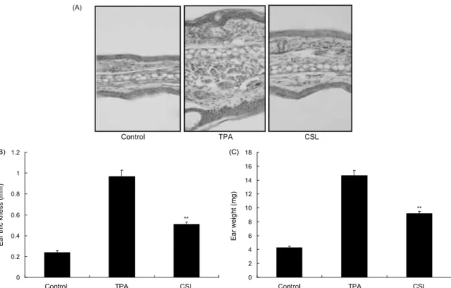
(A)
Control TPA CSL
0 0.2 0.4 0.6 0.8 1 1.2
Ear thic kness (mm) .
Control TPA CSL
**
(B)
0 2 4 6 8 10 12 14 16 18
Ear weight (mg) .
(C)
**
Control TPA CSL
Fig. 1. Inhibitory effect of Caesalpinia sappan L. (CSL) extracts on TPA-induced ear edema. The ears of mice were treated with TPA (1 μg/ear) once a day for 3 days. CSL topically applied to mice ears 1 h after TPA treatment for 3 days. For histological analysis, ear skin sections were prepared and then stained with hematoxylin and eosin (A). Inhibition of TPA-induced ear edema by topically application of CSL was analyzed by measuring changes in ear thickness (B) and ear weight (C).
**p<0.01 compared with mice treated with TPA alone.
결과 및 고찰
소목추출물의 귀 부종 억제 효능
마우스의 귀에 반복적인 TPA 처리는 만성적인 피부염을 유발하기 때문에 TPA로 유도한 마우스 귀 부종은 오랫동안 염증동물모델로 사용되어 왔다(22). 따라서 본 연구에서는 TPA로 유도한 마우스 귀 부종 동물모델에서 소목추출물의 염증억제 효능을 조사하였다. 하루에 한번씩 3일 동안 TPA 를 마우스 귀에 처리하였을 경우 염증반응에 의한 귀의 무게 와 두께가 크게 증가되었다. 같은 조건하에 TPA를 처리한 다음 소목추출물을 같이 처리한 경우 마우스 귀의 무게와 두께가 현저히 감소되는 것을 확인하였다(Fig. 1). 이러한 결과는 소목추출물이 귀 부종에 대한 염증억제 효능이 있음 을 의미한다. 따라서 소목추출물에 의한 염증반응 관련 단백 질인 COX-2와 염증반응에 관여하는 cytokine의 mRNA 발 현억제 및 염증작용 기전 실험을 진행하였다.
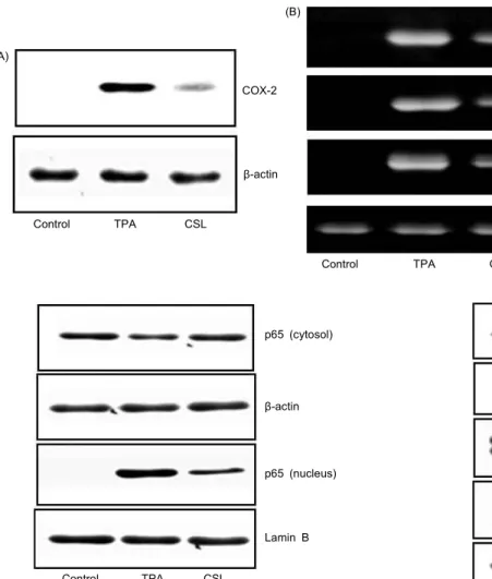
소목추출물의 COX-2 및 cytokine 발현에 미치는 영향 COX는 2가지 isoform(COX-1, COX-2)으로 존재한다.
COX-1과 달리 COX-2의 발현은 성장인자, cytokine 등 pro- inflammatory agent에 의하여 과대발현 되어 염증뿐 아니라 퇴행성 질환에서도 중요한 역할을 하는 것으로 알려져 있다 (25,26). 따라서 소목추출물이 TPA에 의한 COX-2 단백질 발현에 대한 억제효과를 Western blot을 통해 확인한 결과
COX-2 단백질의 발현이 TPA 처리에 의해 현저히 증가하 였으며, 소목추출물 처리에 의해 감소되었다(Fig. 2A). 또한 TPA에 의한 pro-inflammatory cytokine인 IL-6, TNF-α 그리고 IL-1β의 mRNA(27,28) 수준을 RT-PCR로 확인한 결과 TPA 처리에 의해 cytokine의 mRNA 발현 수준이 현저 히 증가하였고, 소목추출물에 의해 증가된 cytokine의 mRNA 발현 수준이 유의성 있게 감소하였다(Fig. 2B). 위 결과는 소목추출물이 염증반응에 관여하는 단백질 및 cytokine의 mRNA 발현 수준을 억제하므로 염증반응 관련 다양한 질병 에도 치료물질로 이용될 수 있을 것으로 사료된다.
소목추출물이 신호전달에 미치는 영향
염증반응에서 중요한 역할을 NF-κB는 cytokine의 합성
을 조절하는 전사인자로 정상적인 세포에서는 p50과 p65의
heterodimer 형태로 cytosol에 존재하며, IκBα와 결합되어
있어 전사인자로의 작용을 하지 못한다. 그러나 외부의 자극
에 의해 IκBα가 인산화되어 degradation되면 NF-κB는 핵
안으로 이동하여 전사인자로 작용하여 COX-2 및 염증관련
cytokine을 합성한다(29-33). 또한 염증반응은 MAPK의 활
성을 유도하며, MAPK는 extracellular signal-regulated
protein kinase(ERK), c-Jun terminal kinase(JNK), 그리고
p38이 있다(34,35). 소목추출물이 NF-κB 및 MAPK signal-
ing을 통하여 염증관련 인자들을 조절하는지 확인하기 위해
Western blot를 수행하였다. TPA에 의해 cytosol에 존재하
(A)
COX-2
β-actin
Control TPA CSL
(B)
IL-6
TNF-α
IL-1β
β-actin
Control TPA CSL
Fig. 2. Inhibitory effect of Cae- salpinia sappan L. (CSL) on TPA-induced COX-2 expre- ssion and pro-inflammatory cytokines in mice ears. Mice were stimulated with TPA and treated with CSL. Ear biopsies were homogenized. Mice ear ex- tracts were prepared and analyzed for COX-2 protein expression (A).
The production of cytokine (IL-6, TNF-α, IL-1β) and β-actin mRNA was analyzed by RT-PCR using specific primers (B).
p38
p-p38
ERK
p-ERK
β-actin
Control TPA CSL
Fig. 4. Inhibitory effect of TPA-induced MAPK activation by Caesalpinia sappan L. (CSL) in mice ears. The ears of mice were treated with TPA (1 μg/ear) once a day for 3 days. CSL topically applied to mice ears 1 h after TPA treatment for 3 days.
Extracts from ear biopsies were prepared and analyzed for MAPK protein activation by Western blotting.
p65 (cytosol)
β-actin
p65 (nucleus)
Lamin B
Control TPA CSL
Fig. 3. Inhibitory effect of TPA-induced NF-κB activation by Caesalpinia sappan L. (CSL) in mice ears. The ears of mice were treated with TPA (1 μg/ear) once a day for 3 days. CSL topically applied to mice ears 1 h after TPA treatment for 3 days.
The activity of NF-κB was analyzed by Western blotting.
는 p65가 감소하였고, 핵에서 p65가 증가하는 것을 확인하였 다(Fig. 3). 또한 p-p38과 p-ERK가 TPA에 의해 현저히 증 가되었고, 증가된 p-p38과 p-ERK은 소목추출물에 의해 감 소하는 것을 확인하였다(Fig. 4). 이러한 결과는 소목추출물 이 NF-κB 및 MAPK signaling을 통하여 염증관련 인자들 (COX-2 및 cytokine)의 단백질 및 mRNA 발현을 억제하는 항염증 효능이 있음을 의미한다.
요 약
본 연구를 통하여 TPA로 유도한 마우스 귀 부종 염증반 응에 대한 소목추출물의 항염증 효능과 기전을 확인하였다.
소목추출물은 TPA로 유도한 마우스 귀 부종을 억제하였으 며, TPA에 의한 염증관련 단백질인 COX-2 발현 및 cyto- kine(IL-6, TNF-α 그리고 IL-1β)의 mRNA 발현을 현저히 감소시켰다. 또한 TPA에 의한 NF-κB 및 MAPK의 활성을 억제하였다. 본 연구 결과, 소목추출물은 NF-κB 및 MAPK
의 신호전달을 억제함으로서 항염증 효능을 나타내었다.
감사의 글
본 연구는 농촌진흥청(PJ907105) 및 교육과학기술부의 재원으로 한국연구재단의 중점연구소사업(2009-0093812) 에 의해 수행되었으며, 이에 감사드립니다.
문 헌