331
©The Korean Society of Food Science and Technology
β-Glucosidase 처리에 의한 치자추출물의 항염증 활성 증진
손동화*·최대운·김미혜
한국식품연구원Improvement of Anti-Inflammation Activity of Gardeniae fructus
Extract by the Treatment of β-Glucosidase
Dong-Hwa Shon*, Dae-Woon Choi, and Mi-Hye Kim Korea Food Research Institute
Abstract In this study, we selected Gardeniae fructus (GF) as an anti-inflammatory functional material and improved the biological activity of GF through the treatment of β-glucosidase. For the simple evaluation of anti-inflammatory activity, the inhibitory activity of GF extract (GFE) on the production of NO by RAW264.7 cells in the presence of LPS was
examined. β-glucosidase originating from Aspergillus niger or Aspergillus fumigatus has effectively improved the
anti-inflammatory activity of GFE. The enzyme treatment raised the activity of GFE by more than 10 times. The optimum
conditions for the enzyme reaction were at pH 4.6, 45oC, and 20 U/mL for 24 h with agitation. In addition, in vitro
production of cytokines (IL-1β, IL-6, TNF-α), COX-2, and the NF-κB activation of RAW264.7 cells decreased more in the presence of GFE treated with β-glucosidase originating from Aspergillus niger (GFAN) than in the presence of GFE. These results suggest that enzyme-treated GFE might be a potential candidate for natural anti-inflammatory food materials. Keywords: RAW264.7 cells, anti-inflammatory, Gardeniae fructus, β-glucosidase
서
론
염증반응은 조직의 손상을 비롯하여 외부의 물리적, 화학적 자 극 및 다양한 감염원에 대한 방어 반응으로서 손상된 조직을 수 복하고 재생하려는 기전이다(1-3). 그러나 지속적으로 또는 과도 하게 발생된 만성 염증반응은 조직의 손상을 유발하여 위, 대장, 방광 그리고 전립선암으로의 진행을 유도하며, 류마티스 관절염, 만성 감염 등 다양한 질환의 원인이 된다(4-6). 염증반응이 일어나면 대식세포와 같은 염증세포들은 nitric oxide (NO), prostagladin E2(PGE2), tumor necrosis factor-α(TNF-α), interleukin-1β(IL-1β) 등 염증 매개물질을 분비한다(7). 염증반응의 대표적인 예로, 대식세포는 pathogen associated molecular patterns (PAMPs)라고 불리는 염증성 매개물질 중 하나인 lipopolysaccharide (LPS)를 toll like receptor 4(TLR4)의 heterodimerization 형성으로 인식하며(8), TLR4에 의하여 인식된 LPS는 세포내 전사요소인 nuclear factor-κB(NF-κB)의 활성화를 유도한다(9,10). 핵으로 이동 한 NF-κB는 염증성 cytokine, inducible nitiric oxide synthase (iNOS), cyclooxygenase-2(COX-2)의 유전자 발현을 유도하며(11), iNOS에 의하여 발생된 nitric oxide(NO)는 염증발생을 심화시킨 다(12). 이러한 염증반응은 식품에 함유된 항염증 성분에 의하여 억제되며 염증성 질병의 발생이 감소된다고 보고되었다(13).치자나무(Gardenia jasminoides)는 꼭두서니과(Rubiaceae)에 속 하는 상록활엽 관목으로 항염증 활성성분이 함유되어있으며, 주 요성분으로는 iridoid 화합물인 genipin, geniposide, geniposidic acid, gardenoside, gardoside, shanzhiside, methyl deacetyl asperu-loside 그리고 quinic acid와 그 유도체, vanillic acid 4-O-β-D-(6'-sinapoyl) glucopyranoside, caffeic acid 등이 있다(14).
생리활성을 효과적으로 개선하기위한 방법으로 천연물 중에 흔 하게 존재하는 배당체(glycoside)들의 당쇄를 효소로 절단하여 비 배당체(aglycone)를 생성시키는 방법이 연구되고 있다. 그 대표적 인 예로서 인삼사포닌의 저분자화를 이용한 기능성식품 제조방 법과(15), Bifidobacterium lactis 및 이 미생물에서 생산된 β-glu-cosidase를 이용한 대두의 이소플라본 비배당체 생성방법 등이 있 다(16-18). 본 연구에서 치자 추출물의 항염증 활성 증진을 개선하고자 치 자 추출물에 β-glucosidase처리하였다. 즉, 최적의 효소를 선발하 고, 이 효소의 최적 처리조건을 확립하였으며, 효소 처리한 치자 추출물의 항염증 활성 변화를 분석하였다.
재료 및 방법
추출물의 제조 본 실험에 사용된 치자는 서울 경동시장에서 구입하였으며, 치 자의 건조 분쇄물의 효과적인 유용성분 추출을 위해 2,450 MHz 주파수에 환류냉각관이 장치된 상압형 마이크로파 추출장치(MAP, Soxwave-100, Prolabo, France)를 사용하였다. 마이크로웨이브 추 출조건은 건물 중량 10 g의 20배에 해당하는 추출용매 200 mL로 50% ethanol을 사용하였고, 마이크로 파워는 90 W, 추출시간은 5 분으로 하였다. 이렇게 얻어진 추출물을 Whatman filter paper에 *Corresponding author: Dong-Hwa Shon, Korea Food ResearchInstitute, Seongnam, Gyeonggi 463-746, Korea Tel: 82-31-780-9133
Fax: 82-31-709-9876 E-mail: [email protected]
Received December 25, 2011; revised March 30 2012; accepted April 1, 2012
332 한국식품과학회지 제 44 권 제 3 호 (2012) 거른 후 회전 감압 증발기(Ratavapor R-123, Buchi, Flawil,
Swiz-erland)로 감압 농축하여 50 mL의 증류수로 정용하여 치자 추출 물을 얻었다.
치자열매 추출물의 β-glucosidase 및 HCl 처리
본 실험에는 Aspergillus niger(A. niger, Fluka, Buchs, Germany), 일본 Amano사의 Aspergillus fumigatus(A. fumigatus) 그리고 almond 유래의 β-glucosidase 사용하였다. 각각의 β-glucosidase (in sodium acetate buffer, pH 4.6)을 50% 에탄올 추출물(GFE) 5 mL 에 처리하여 45oC에서 24시간 동안 반응시켜 준비하였다. 또한
GFE 5 mL에 반응시간(0, 1, 3, 6, 12, 18, 24 h) 및 효소의 양(0, 0.5, 1, 2, 5, 10, 20 U/mL)에 변화를 주어 시료의 항염증 활성 변 화를 측정하였다. 그리고 GFE 5 mL에 A. niger 유래의 β-glucosi-dase(10 U/mL)를 처리하여 시간에 따른 효소활성 변화를 측정하 였다.
세포배양 및 처리
본 실험에 사용된 murine macrophage cell line RAW264.7 세 포는 한국세포주은행(KCLB, Korean Cell Line Bank, Seoul, Korea)에서 분양 받아 사용하였다. 세포배양 배지는 Dulbecco’s modified Eagle’s medium(DMEM)에 10% fetal bovine serum, 100 U/mL penicillin, 100µg/mL streptomycin 그리고 L-glutamine (2 mM)을 첨가하여 사용하였으며, 37oC, 5% CO2의 조건에서 배 양하였다.
NO 생성물 측정
본 실험은 RAW264.7 세포를 사용하여 Green 등의 방법(19)을 변형하여 측정하였다. 10% (v/v) FBS, 100 U/mL penicillin, 100 µg/mL streptomycin을 포함하는 DMEM에 1×10⁶ cells/mL의 농도
로 96-well에 분주하여 2시간 동안 배양한 후 시료를 농도별로 처리하여 1시간 동안 배양하고, LPS(1 µg/mL)를 처리하여 24시 간 동안 37oC, 5% CO 2 incubator에서 배양하였다. 양성대조군으 로써 resveratrol을 사용하였으며, NO assay를 위하여 세포배양 시 생성된 NO의 양을 측정하였다. 세포배양 상징액 50 µL를 취하여 Griess 시약 50 µL와 5분간 반응 시킨 후, microplate reader(EMax Endpoint ELISA Microplate Reader, Sunnyvale, CA)를 사용하여 540 nm에서 흡광도를 측정하였다. 이때 표준품으로 NaNO2를 사 용하였다.
시료추출물의 세포독성
MTT assay에 의하여 세포독성을 평가하였다. RAW264.7 세포 를 1×10⁶ cells/mL의 농도로 96-well plate에 분주하여 2시간 동안
배양한 후 LPS(1 µg/mL) 및 시료를 농도별로 처리하여 3일간 배 양하였다(20). 다음으로 MTT 시약(5 mg/mL)을 각각의 well에 20 µL씩 처리한 후, 4시간 동안 배양하여 10% SDS(in 0.01 M HCl) 를 100 µL씩 첨가하였으며, 12시간 배양한 후 microplate reader 를 사용하여 595 nm에서 흡광도를 측정하였다.
염증성 cytokine IL-6, IL-1β 및 TNF-α의 분석
Cytokine의 생성정도를 분석하기 위하여 12-well plate에 RAW 264.7 세포를 1×10⁶ cells/mL의 농도로 분주하였으며, 2시간 동안 배양한 후 각각의 시료를 농도별로 처리하여 1시간 동안 배양하 였다. 그 후 LPS(1 µg/mL)를 처리하여 24시간 동안 37oC, 5%
CO2 incubator에서 배양하였다. 이때 얻어진 상징액을 시료로 하 였고, 분석 kit로는 BD OptEIATM MOUSE ELISA(enzyme-linked
immunosorbent assay) set(BD Biosciences, San Diego, CA, USA) 을 사용하였으며, kit에 명시된 실험방법에 준하여 pro-inflammatory cytokine인 IL-6, IL-1β, TNF-α의 생성량을 sandwich-ELISA법으로 분석하였다.
Western blot analysis
COX-2의 생성정도를 분석하기 위해 6-well plate에 RAW264.7 세포를 1×10⁶ cells/mL의 농도로 분주하여 2시간 동안 배양한 후
각각의 시료를 농도별로 처리하여 1시간 동안 배양 하였으며, 다 음으로 LPS(1 µg/mL)를 처리하여 24시간 동안 37oC, 5% CO
2
incubator에서 배양하였다. 배양된 세포를 RIPA lysis buffer로 처 리하여 세포의 단백질을 추출한 후 원심분리하여 상등액을 취하 였다. 상등액을 Bradford 시약을 사용해 단백질 농도를 정량하여 40µg의 단백질을 취했다. 추출된 단백질은 10%의 SDS-polyacry-lamide gel에 전기영동한 후 gel의 단백질을 nitrocellulose mem-brane으로 transter시켰다. 5% skim milk로 하루 밤 동안 blocking 한 후 1:500의 비율로 COX-2 antibody를 4시간 동안 상온에서 방치한 후 15분 간격으로 2회 세척하고, 1:1,000의 비율로 희석 한 secondary antibody를 1시간 동안 상온에서 방치 시켰다. 다음 으로 15분 간격으로 3회 세척한 후 ECL(GE Healthcare, Piscat-away, NJ, USA)로 현상하였다.
Luciferase assay를 이용한 NF-κB 억제활성
NF-κB의 발광효소 유전자 분석은 선행연구에서 실행된 방법 을 변형하여 실시하였다(21,22). NF-κB 억제 활성을 평가하기 위 하여 E. coli에 대량증식시켜 endofree plasmid maxi kit(Qiagen, Venlo, Netherland)로 정제하여 얻은 firefly luciferase plasmid와 renilla luciferase plasmid(Promega, Madison, WI, USA)를 각각 0.5, 0.005µg/well이 첨가되도록 RAW264.7 세포에 co-transfection 시켰다. 이때 세포는 전날 1×10⁵ cells/well의 밀도로 96-well에 분
주하여 배양을 해둔 것을 사용하였다. Transfection 6시간 후 한 차례 complete media로 배지교환을 하였다. 그 후 18시간 배양하 여 농도별로 시료를 처리하고, 1시간 후에 LPS(1 µg/mL)를 처리 하여 24시간 배양하였다. 다음으로 Dual-Glo luciferase assay sys-tem(Promega)에 따라 시약을 처리하여 나타나는 발광정도를 lumi-nometer로 측정하고 그 결과를 relative response로 나타내었다. 통계처리 실험 결과는 3회 반복 측정 후 평균±표준편차로 나타냈으며, 시료가 포함되지 않은 대조구와 비교하여 Student’s t-test로 p<0.05 수준에서 검정하였다.
결과 및 고찰
치자 추출물의 NO생산 억제활성 치자 추출물(GFE) 준비는 50% 에탄올을 용매로하여 단 시간 에 효율적인 추출이 가능한 microwave로 추출하였다(23). 먼저, GFE의 항염증 활성의 조사하고자 LPS 존재하에 RAW264.7 세포가 생산하는 NO의 변화를 조사하였다. 그 결과, LPS만을 처리한 대조구에서 생산된 NO의 양은 57.8 µM이었으 며, resveratrol을 46.5 µg/mL농도로 처리한 양성대조구는 28.0 µM 로 확연히 줄어들었다. GFE를 500 µg/mL의 농도로 처리한 경우 에 생산된 NO의 수치는 41.8 µM로 비교적 낮은 활성을 나타냈 다(Fig. 1A). 따라서, 다음 연구에서는 효소 및 화학적 처리를 통 한 GFE의 항염증 활성 증진 방안을 모색하였다.처리에 의한 치자 추출물의 NO생산 억제활성
GFE에 A. niger에서 유래된 β-glucosidase를 처리한 시료(GFAN) 와 almond에서 유래된 β-glucosidase가 처리된 치자 추출물(GFA) 그리고 HCl이 처리된 치자 추출물(GFH)의 NO생산 억제활성을 확인한 결과, GFH는 1,000 µg/mL의 높은 농도에서도 NO생산 억제활성은 미약하게 나타났다. 그러나 GFAN과 GFA는 250 µg/ mL 농도에서 각각 77.5%와 24.9%의 NO생산 억제활성을 나타냈 으며, GFAN의 활성이 상대적으로 높았다(Fig. 1B). 결론적으로 GFE의 활성을 증진 시키는 방법은 A. niger유래 효소처리 > almond유래 효소처리 > 산(HCl) 처리의 순으로 나타났으며, 곰팡 이 유래 β-glucosidase가 식물에서 유래된 β-glucosidase보다 항염 증 활성 증진에 효과적이라는 것을 보여준다.
선행 연구(24)를 바탕으로 치자 추출물을 lactic acid bacteria로 발효하여 NO생산 억제활성을 조사해 본 결과, β-glucosidase를 직 접적으로 처리한 경우와 비교하여 그 활성은 미비하였으며(자료 생략), GFE에 β-glucosidase를 처리하는 방법이 치자의 항염증 활 성 증진에 더욱 효과적이었다.
다음으로 치자 성분 중 스틸벤류 화합물인 piceid, piceatannol, rhaponticin, resveratrol 그리고 rhapontigenin의 함량과 효소처리에
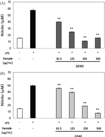
의한 성분들의 변화를 조사하였다. 분석 결과, 치자 추출물 중 piceid(57.7 mg/100 g), piceatannol(27.9 mg/100 g), rhaponticin (1.49 g/100 g), resveratrol(17.0 mg/100 g) 그리고 rhapontigenin(51.4 mg/100 g)이 확인되었지만, β-glucosidase 처리에 의한 각 성분의 변화는 확인되지 않았다(결과 생략). 그러나, β-glucosidase의 처리 로 치자의 성분 중 geniposide가 genipin으로 전환되어 항염증 활 성이 증진되는 것이 확인되었기에(25), 본 연구에서도 장내 흡수 율 및 세포투과도가 높은 비배당체가 형성되어 항염증 활성이 증 진되었다고 사료된다. 곰팡이 유래 β-glucosidase 처리에 의한 치자 추출물의 항염 증 활성 증가 앞의 결과를 바탕으로 A. niger 유래 효소와 A. fumigatus 유래 효소 활성을 비교하고자 하였으며, GFAN과 GFE에 A. fumigatus 유래 β-glucosidase를 처리한 추출물(GFAF)의 효과를 비교하였 다. 그 결과, GFAN와 GFAF는 모두 높은 NO생산 억제 활성을 나타냈다. 하지만 125 µg/mL 농도에서 GFAN은 높은 NO생산 억 제 활성을 나타낸 반면(Fig. 2A), GFAF의 활성은 상대적으로 미 약하였다(Fig. 2B). 또한, GFAN과 GFAF의 IC50(50% inhibitory
concentration)값은 각각 71.7±0.57 µg/mL과 161.1±0.70 µg/mL로 Fig. 2. The effect of (A) GFAN and (B) GFAF on LPS-induced NO production in RAW264.7 cells. Cells were preincubated with indicated concentrations of GFAN and GFAF for 2 h and then incubated with LPS (1µg/mL) for 24 h. GFAN, Gardeniae fructus 50% ethanol extracts (GFE) treated with A. niger β-glucosidase(20 U/mL) for 24 h; GFAF, GFE treated with A. fumigatus β-glucosidase (20 U/mL) for 24 h. Control is treated with only LPS. The values represent mean±SD (n=3) significantly different from the values of control (**p<0.01).
Fig. 1. Effect of (A) Gardeniae fructus 50% ethanol extracts (GFE), (B) GFAN, GFA and GFH on LPS-induced NO production in RAW264.7 cells. Cells were preincubated with indicated concentrations of GFE for 2 h and then incubated with LPS (1 µg/mL) for 24 h. RV, Resveratrol; GFAN, GFE treated with A. niger β-glucosidase (25 U/mL) for 24 h; GFA, GFE treated with almond β-glucosidase (25 U/mL) for 24 h; GFH, GFE treated with HCl (0.1 N) for 24 h. Control is treated with only LPS. The values represent mean±SD (n=3) significantly different from the values of control (*p<0.05, **p<0.01).
334 한국식품과학회지 제 44 권 제 3 호 (2012)
측정되었으며, 치자의 항염증 활성 증진에는 A. niger 유래 β-glucosidase가 더욱 효과적이라고 판단되었다. 그럼으로 다음 실 험부터는 A. niger 유래 β-glucosidase를 중심으로 분석하였다.
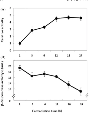
다음으로, GFE의 활성을 증진시키는 A. niger 유래 β-glucosi-dase의 처리 조건을 확립하고자 효소처리 시간의 변화에 따른 NO 생산 억제 활성을 조사하였다. β-glucosidase를 1-24시간 동안 효 소와 반응시킨 결과, 효소처리 시간과 비례하여 NO생산 억제 활 성이 높아졌지만, 12시간 이 후에는 활성의 변화가 거의 없었다 (Fig. 3A). 또한, 24시간 동안 β-glucosidase의 활성은 시간의 변화 와 비례하여 저하되었다(Fig. 3B). 다음으로, A. niger 유래 β-glucosidase를 농도별로 처리하여 추 출물의 항염증 활성의 변화를 확인하였다. GFE에 β-glucosidase 를 0.5-20 U/mL의 농도로 24시간 동안 반응시켰으며, 각각의 추 출물을 125 µg/mL의 농도로 처리하여 NO생산 억제 활성을 비교 한 결과, 효소 농도에 비례하여 NO생산이 강하게 억제되었다(Fig. 4). 이러한 결과를 바탕으로 20 U/mL의 효소 농도로 24시간 이상 충분한 반응시간을 주는 것이 GFE의 항염증 활성 증진을 위한 최적조건으로 판단하였으며, 다음 실험부터는 이와 같은 조건으 로 GFE에 A. niger 유래 효소를 처리하여 실시하였다. 효소처리한 치자 추출물의 세포독성 검토 MTT assay에 의하여 세포독성을 평가하고자 하였다. 즉, 치자 추출물에 효소를 처리한 것과 처리하지 않은 시료의 농도를 달 리하여 실험을 실시하였다. 그 결과, LPS만을 처리한 대조구와 비교하여 GFE 및 GFAN의 세포 생존율은 낮았지만, 그 수치는 대략 10% 전후로 큰 차이를 나타내지는 않았으며, 처리 농도에 따른 세포 생존율의 변화는 없었기 때문에 GFE와 GFAN은 세포 독성의 문제가 없는 것으로 판단된다. 또한, GFE와 GFAN은 동 일한 농도에서 대체로 유사한 세포 생존율을 나타내었는데, 이것 은 GFAN의 높은 NO생성 억제활성(Fig. 2A, 4)이 세포 독성에 기인한 결과가 아님을 의미한다(Fig. 5).
염증성 cytokine생산 억제 활성
LPS의 자극에 의하여 RAW264.7 세포가 생산하는 cytokine인 Fig. 4. The effect of β-glucosidase concentration on nitrite inhibition activity of GFAN. RAW264.7 cells were preincubated with indicated concentration of GFAN for 2 h and then incubated with LPS (1µg/mL) for 24 h. GFAN, Gardeniae fructus 50% ethanol extracts was treated with indicated concentration of A. niger β-glucosidase for 24 h. Control is treated with only LPS. The values represent mean±SD (n=3) significantly different from the values of control (*p<0.05).
Fig. 5. The effects of GFE and GFAN on LPS-induced RAW264.7 cell death. Cells were preincubated with indicated concentrations of GFE and GFAN for 2 h and then incubated with LPS (1µg/mL) for 72 h. GFE, Gardeniae fructus 50% ethanol extracts; GFAN, GFE was treated with A. niger β-glucosidase (20 U/ mL) for 24 h. The values represent mean±SD (n=3) significantly different from the values of control (*p<0.05).
Fig. 3. Anti-inflammatory effect of fermentated Gardeniae fructus 50% ethanol extracts (GFE) in a time dependent manner. (A) Inhibition activity of LPS induced NO production in RAW264.7 cells by GFE treated with 20 U/mL of β-glucosidase originated from A. niger at pH 4.6, 45oC for 1, 3, 6, 12, 18, and 24 h. Results were expressed as relative inhibitory activity of LPS-induced NO production in RAW264.7 cells. Cells were preincubated with samples for 2 h and then incubated with LPS (1µg/mL) for 24 h. (B) Remaining β-glucosidase activity (U/mL) in Gardeniae fructus extracts treated with 10 U/mL of the enzyme from A. niger at pH 4.6, 45oC for 1, 3, 6, 12, 18, and 24 h. Water extract of GFE were used for the enzyme treatment. The values represent mean±SD (n=3).
IL-1β, IL-6 그리고 TNF-α를 분석하였다. 즉, GFE와 GFAN의 염 증성 cytokine생산 억제 활성을 측정하였다. IL-1β의 생성은 GFE 를 250 µg/mL 농도로 처리하였을 때 대조구와 비교하여 감소하 였고, GFAN는 500 µg/mL의 농도에서 IL-1β의 생산이 억제되었 다(Fig. 6A). IL-6는 대조구와 비교하여 GFAN을 250 µg/mL 이상 의 농도로 처리하였을 경우 83%이상 감소된 반면에, 동일한 농 도로 GFE를 처리한 경우는 27%의 억제율을 나타냈다(Fig. 6B). 또한, TNF-α의 생성은 GFAN을 500 µg/mL의 농도로 처리하였을
경우 현저하게 억제되었지만, GFE의 효과는 거의 나타나지 않았 다(Fig. 6C). 이러한 결과는 GFE가 A. niger 유래 β-glucosidase의 처리로 염증성 cytokine생산 억제활성이 증진되었음을 의미한다. COX-2생산 억제
COX는 COX-1과 COX-2 두 가지 isoform 형태로 존재하며, 염 증반응에는 COX-2가 작용을 하는데(26), 이러한 COX-2의 생산 을 억제하는 효과를 확인하고자 하였다. 그 결과, LPS로 자극된 RAW264.7 세포에서는 다량의 COX-2가 생산되었으며, GFE는 500µg/mL에서 미약하게 억제하였지만, GFAN의 경우 500 µg/mL 농도에서 상대적으로 높은 억제효과를 나타냈다(Fig. 7). 따라서 GFE에 대한 β-glucosidase 처리로 COX-2생산 억제활성이 증진되 며 세포내에 유발되는 일련의 염증반응이 억제되었음을 의미한다. 효소처리한 치자 추출물의 NF-κB 활성 억제 효과 염증유발 물질에 의하여 활성화된 NF-κB가 핵내로 이동하여 염증성 cytokine과 효소 등의 생산을 유도함으로써 궁극적으로 염 증반응이 유발된다(11). 따라서 본 실험에서는 효소처리에 의하 여 증진된 NF-κB 억제활성을 조사하였다.
Fig. 7. Effect of GFE and GFAN on LPS induced activation of COX-2. RAW 264.7 cells were preincubated with indicated concentrations of GFAN and GFE for 2 h and then incubated with LPS (1µg/mL) for 24 h. Cell lysates were subjected to Western blotting with a COX-2 antibody. GFE, Gardeniae fructus 50% ethanol extracts; RV, Resveratrol; GFAN, GFE treated with A. niger β-glucosidase (20 U/mL) for 24 h.
Fig. 8. Effect of GFE and GFAN treated on LPS induced activation of NF-κB. RAW264.7 cells were transfected with NF-κB luciferase reporter plasmid and preincubated with indicated concentrations of GFE and GFAN for 1 h and then incubated with LPS (1µg/mL) for 24 h. GFE, Gardeniae fructus 50% ethanol extracts; GFAN, GFE treated with A. niger β-glucosidase (20 U/mL) for 24 h. The values represent mean±SD (n=3) significantly different from the values of control (*p<0.05).
Fig. 6. Effects of GFE and GFAN on LPS-induced (A) IL-1β, (B) IL-6 and (C) TNF-α production in RAW264.7 cells. Cells were preincubated with indicated concentrations of GFAN and GFE for 2 h and then incubated with LPS (1µg/mL) for 24 h. Cytokines in the conditioned media were measured using the IL-1β, IL-6 and TNF-α assay kit. GFE, Gardeniae fructus 50% ethanol extracts; GFAN, GFE treated with A. niger β-glucosidase (20 U/mL) for 24 h. Control is treated with only LPS. The values represent mean±SD (n=3) significantly different from the values of control (*p<0.05, **p<0.01).
336 한국식품과학회지 제 44 권 제 3 호 (2012) 그 결과, GFAN는 125 µg/mL 이상의 농도에서 대조구와 비교 하여 65% 이상 감소하였지만, GFE를 처리한 경우에는 NF-κB 억제활성이 거의 나타나지 않았다(Fig. 8). 결론적으로, A. niger 유래 효소처리로 비배당체가 생성되어 NF-κB 억제 활성이 증진 되었다고 판단된다. 이상의 결과를 종합하면, GFE의 미약한 항염증 활성은 A. niger 유래 β-glucosidase처리로 현격히 증진되며, GFAN이 LPS 자극으 로 RAW264.7 세포에서 유발되는 NF-κB의 활성화를 억제하여 염증성 cytokine, COX-2 그리고 NO생산을 효과적으로 억제시켰 음을 의미한다.
요
약
본 연구에서는, 치자 열매를 항염증 소재로 선택하여, 치자 추 출물(GFE)에 β-glucosidase를 처리함으로써 항염증 활성을 증진하 고자 하였다. 항염증 활성의 비교를 위해 추출물과 LPS를 함께 처리하여 RAW264.7 세포가 생산하는 NO의 양을 측정하였다. Lactic acid bacteria, 식물 그리고 곰팡이 유래 β-glucosidase를 처 리한 결과, 곰팡이(A. niger, A. fumigatus)에서 유래된 효소를 처 리하는 것이 GFE의 NO생산 억제활성 증진에 가장 효과적이었 다. 그리고 GFE에 대한 A. niger 유래 β-glucosidase의 최적 처리 조건은 pH 4.6, 45oC 그리고 20 U/mL의 효소 농도로 24 h 반응 시키는 것이었다. 이와 같은 조건으로 GFE에 효소를 처리한 GFAN은 GFE와 유사한 세포 생존율을 나타냈고, 세포독성은 나 타나지 않았다. 또한 GFAN의 처리에 의하여 RAW264.7 세포의 염증성 cytokine생산, COX-2생산 그리고 NF-κB활성이 현저히 줄 어들었다. 본 연구에서 GFAN의 항염증 활성 증진은 치자의 성 분 중 주로 geniposide가 β-glucosidase처리에 의하여 genipin으로 전환되었기 때문이라 생각된다. 이상의 결과는 β-glucosidase를 처 리한 치자 추출물(GFE)을 이용하여 안전하고 효과적인 항염증 기능성 식품 소재 개발이 가능함을 제시하였다.문
헌
1. Zamora R, Vodovotz Y, Billiar TR. Inducible nitric oxide syn-thase and inflammatory diseases. Mol. Med. 6: 347-373 (2000) 2. Mariathasan S, Monack DM. Inflammasome adaptors and
sen-sors; intracellular regulators of infection and inflammation. Nat. Rev. Immunol. 7: 31-40 (2007)
3. Lee HN, Lim DY, Lim SS, Kim JD, Park HY. Anti-inflammatory effect of ethanol extract from Eupatorium japonicum. Korean J. Food Sci. Technol. 43: 65-71 (2011)
4. Hofseth LJ and Ying L. Identifying and defusing weapons of mass inflammation in carcinogenesis. Biochim. Biophys. Acta 1765: 74-84 (2006)
5. Yun HY, Dawson VL, Dawson TM. Neurobiology of nitric oxide. Crit. Rev. Neurobiol. 10: 291-316 (1996)
6. Stuehr DJ, Cho HJ, Kwon NS, Weise MF, Nathan CF. Purifica-tion and characterizaPurifica-tion of the cytokine-induced macrophage nitric oxide synthase: An FAD- and FMN-containing flavoprotein. P. Natl. Acad. Sci. USA 88: 7773-7777 (1991)
7. Guha M, Mackman N. LPS induction of gene expression in human monocytes. Cell Signal. 13: 85-94 (2001)
8. Miyake K. Innate recognition of lipopolysaccharide by Toll-like receptor 4-MD-2. Trends Microbiol. 12: 186-192 (2004)
9. Youn HS. Anti-inflammatory effects of resveratrol,
epigallocat-echin-3-gallate, and curcumin by the modulation of toll-like receptor signaling pathways. Korean. J. Food Sci. Technol. 39: 481-487 (2007)
10. Youn HS. Anti-inflammatory effects of phytochemicals having michael addition acceptors by the modulation of toll-like receptor signaling pathways. Korean. J. Food Sci. Technol. 41: 477-482 (2009)
11. Noh KH, Jang JH, Min KH, Chinzoring R, Lee MO, Song YS. Suppressive effect of green tea seed coat ethyl acetate fraction on inflammation and its mechanism in RAW264.7 macrophage cell. J. Korean Soc. Food Sci. Nutr. 40: 625-634 (2011)
12. Weisz A, Cicatiello L, Esumi H. Regulation of the mouse induc-ible- type nitric oxide synthase gene promoter by interferon-γ, bacterial lipopolysaccharide, and NG-monomethyl-L-arginine. Bio-chem. J. 316: 209-215 (1996)
13. Lee CH, Youn Y, Song GS, Kim YS. Immunostimulatory effects of traditional Doenjang. J. Korean Soc. Food Sci. Nutr. 40: 1227-1234 (2011)
14. Yang HJ, Park MJ, Lee HS. Antioxidative activities and como-nents of Gardenia jasminoides. Korean J. Food Sci. Technol. 43: 51-57 (2011)
15. Han JY, Kim MH, Tie J, Solihin BW, Ryu GH. Extrusion of gin-seng root in twin screw extruder: Pretreatment for hydrolysis and saccharification of ginseng extrude. J. Food Sci. Nutr. 11: 318-322 (2006)
16. Donkor ON, Shah NP. Production of β-glucosidase and hydrolysis of isoflavone phytoestrogens by Lactobacillus acidophilus, Bifido-bacterium lactis, and Lactobacillus casei in soymilk. J. Food Sci. 73: M15-M20 (2008)
17. Ismail B, Hayes K. Beta-glucosidase activity toward different gly-cosidic forms of isoflavones. J. Agr. Food Chem. 53: 4918-4924 (2005)
18. Yang SO, Chang PS, Lee JH. Isoflavone distribution and β-glu-cosidase activity in cheonggukjang, a traditional Korean whole soybean-fermented food. Food Sci. Biotechnol. 15: 96-101 (2006) 19. Green LC, Wagner DA, Glogowski J. Analysis of nitrate, nitrite,
and [15N] nitrate in biological fluids. Anal. Biochem. 126: 131-138 (1982)
20. Yang HM, Lim SS, Lee YS, Shin HK, Oh YS, Kim JK. Compar-ison of the anti-inflammatory effect of the extracts from Rubus coranus and Rubus occidentakis. Korean J. Food Sci. Technol. 39: 342-347 (2007)
21. Youn HS, Kang SA, Hwang D. Anti-cancer and anti-inflamma-tory effects of curcumin by the modulation of Toll-like receptor 2, 3, and 4. Korean J. Food Sci. Technol. 39: 175-180 (2007) 22. Lee JY, Zhao L, Youn HS, Weatherill AR, Tapping R, Feng L,
Lee WH, Fitzgerald KA, Hwang DH, Saturated fatty acid acti-vates but polyunsaturated fatty acid inhibits Toll-like receptor 2 dimerized with Toll-like receptor 6 or 1. J. Biol. Chem. 279: 16971-16979 (2004)
23. Jeong HJ, Kim SN, Kwon JH, Kim HY. Physiological activities of Gardeniae fructus extracts by microwave-assisted extraction as affected by solvents. J. Korean Soc. Food Sci. Nutr. 37: 282-287 (2008)
24. Tsangalis D, Ashton JF, Stojanovska L, Wilcox G, Shah NP. Development of an isoflavone aglycone-enriched soymilk using soy germ, soy protein isolate, and bifidobacteria. Food Res. Int. 37: 301-312 (2004)
25. Yang JH, Lee NH. Skin permeation and anti-inflammatory effects of hydrolyzed products of Gardeniae fructus extracts. J. Korean Pharm. Sci. 34: 115-123 (2004)
26. Surh YJ, Chun KS, Cha HH, Han SS, Keum YS, Park KK, Lee SS. Molecular mechanisms underlying chemopreventive activities of anti-inflammatory phytochemicals: Down-regulation of COX-2 and iNOS through suppression of NF-κB activation. Mutat. Res. 480-481: 243-268 (2001)