pISSN 1738-1843 © 2012 The Korean Society of Pathologists/The Korean Society for Cytopathology
4,4´-Diaminodiphenyl sulfone (dapsone) is widely used to treat a variety of infectious, immune, and hypersensitivity dis-orders, with indications ranging from Hansen’s disease to in-flammatory diseases and insect bites.1 However, a potential
se-vere side-effect, known as dapsone-induced hypersensitivity syndrome (DHS), may occur. DHS is typically characterized by fever, skin rashes, and multiple lymphadenopathy. Furthermore, occasionally severe systemic reactions can be combined, for ex-ample, hepatitis with splenomegaly, cholangitis, pneumonitis, colitis, thyroiditis, or myocarditis.2 In these situations,
physi-cians should be cautious and carefully rule out the actual cause of unusual systemic reactions. Here, we report a case of cervical lymphadenopathy mimicking T-cell lymphoma with hepatitis and splenomegaly after dapsone treatment.
CASE REPORT
A 36-year-old woman presented at our institution (Gachon University Gil Hospital) with erythematous confluent maculo-patches on her face, trunk, and palms; jaundice; fatigue; pruri-tus; and aggravation of skin lesions over the previous week. She had been previously diagnosed as having erythema multiforme 2 weeks earlier at a private dermatology clinic and had also been administered dapsone with oral antihistamines (mequitazine and cetirizine) and topical corticosteroid for 8 months.
On admission, her vital signs were a blood pressure of 110/70 mm Hg, heart rate of 78 beats/min, respiratory rate of 18 breaths/ min, and body temperature of 38.8˚C. She reported no night sweats, loss of weight, or arthralgia. A physical examination conducted at presentation revealed bilateral, multiple enlarged
Cervical Lymphadenopathy Mimicking Angioimmunoblastic T-Cell
Lymphoma after Dapsone-Induced Hypersensitivity Syndrome
Min Young Rim · Junshik Hong Inku Yo · Hyeonsu Park
Dong Hae Chung1· Jeong Yeal Ahn2 Sanghui Park3· Jinny Park
Yun Soo Kim · Jae Hoon Lee Departments of Internal Medicine, 1Pathology,
and 2Laboratory Medicine, Gachon University Gil
Hospital, Gachon University of Medicine and Science, Incheon; 3Department of Pathology,
Ewha Womans University School of Medicine, Seoul, Korea
A 36-year-old woman presented with erythematous confluent macules on her whole body with fever and chills associated with jaundice after 8 months of dapsone therapy. Her symptoms had developed progressively, and a physical examination revealed bilateral cervical lymphadenopathy and splenomegaly. Excisional biopsy of a cervical lymph node showed effacement of the normal architecture with atypical lymphoid hyperplasia and proliferation of high endothelial venules com-patible with angioimmunoblastic T-cell lymphoma. However, it was assumed that the cervical lymphadenopathy was a clinical manifestation of a systemic hypersensitivity reaction because her clinical course was reminiscent of dapsone-induced hypersensitivity syndrome. A liver biopsy revealed drug-induced hepatitis with no evidence of lymphomatous involvement. Intravenous glucocorticoid was immediately initiated and her symptoms and clinical disease dramatically im-proved. The authors present an unusual case of cervical lymphadenopathy mimicking angioim-munoblastic T-cell lymphoma as an adverse reaction to dapsone.
Key Words: Pseudolymphoma; Dapsone therapy Received: October 24, 2011
Revised: January 9, 2012 Accepted: February 1, 2012 Corresponding Author Sanghui Park, M.D.
Department of Pathology, Ewha Womans University School of Medicine, 1071 Anyangcheon-ro, Yangcheon-gu, Seoul 158-710, Korea Tel: +82-2-2650-5822
Fax: +82-2-2650-5822 E-mail: [email protected]
lymph nodes on both sides of the neck with splenomegaly. All of the nodes were non-tender, firm, and measured 0.5 cm to 1.0 cm in diameter. Laboratory tests results were as follows: Hb 11.2
g/dL, white blood cell 13,070/mm3 (segmented neutrophils
24%, lymphocytes 66.3%, and eosinophils 0.1%), platelets
245,000/mm3, total bilirubin 5.4 mg/dL, direct bilirubin 3.8
mg/dL, aspartate aminotransferase/alanine aminotransferase (AST/ALT) 233/366 U/L, lactate dehydrogenase 1,610 IU/L (normal range, 200 to 485 IU/L), and an international normal-ized ratio of 1.25. She was negative for hepatitis B surface anti-gen, anti-hepatitis C virus Ab, and anti-human immunodefi-ciency Ab, but positive for hepatitis B surface antibody; and positive for anti-nuclear Ab, but negative for rheumatoid factor, anti-neutrophil cytoplasmic Ab, anti-Ro (SS-A, B) Ab, and an-ti-smith Ab. Serology tests showed no evidence of active Ep-stein-Barr virus (EBV) infection and no bacterial growth was found during a blood culture study. There was no hypergam-maglobulinemia.
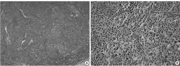
An excisional biopsy of a 1.0 cm cervical node showed diffuse polymorphic lymphoid hyperplasia with effacement of the nor-mal architecture and abundant high endothelial venules; the cellular infiltrate was polymorphic, with abundant transformed lymphocytes and plasma cells (Fig. 1). Sinuses were frequently recognizable, but reactive germinal centers were not found.
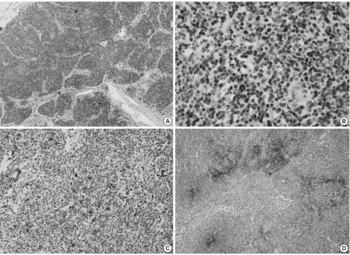
Immunohistochemically, the majority of infiltrating atypical lymphocytes were CD3+ T-cells (Fig. 2A, B), and some activat-ed large cells were reactive for CD30 (Fig. 2C). Follicular den-dritic cell meshworks expressing CD21 were expanded (Fig. 2D), and infiltrating atypical T-cells were negative for CD10,
BCL-6, and EBV by in situ hybridization. For a definitive diag-nosis, a molecular study on the rearrangement of the T-cell re-ceptor (TCR)-gamma gene was performed and showed polyclo-nality (Fig. 3). An IgH gene rearrangement study also showed polyclonality.
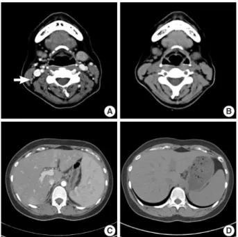
An abdomino-pelvic computed tomography (CT) scan re-vealed splenomegaly (14 cm), an enlarged portocaval lymph node (1.7 cm), and gallbladder wall edema (Fig. 4A, C). No ev-idence of lymphomatous involvement was found by chest CT or a bone marrow trephine biopsy.
Because aggravation of the skin eruption persisted and the liver function test remained abnormal, the possibility of DHS (exfoliative dermatitis, acute hepatitis, and multiple lymphade-nopathy) was raised. The findings of a transjugular liver biopsy were compatible with drug-induced hepatitis manifesting as granulomatous inflammation (Fig. 5). No evidence of lympho-matous hepatic involvement was found. In view of her skin le-sions, medication history, and age, we concluded that the cervi-cal lymphadenopathy was actually pseudolymphoma. Accord-ingly, low dose prednisolone (30 mg/day) per os was started after transjugular liver biopsy to relieve the dapsone-induced skin le-sions and hepatitis. Gradually, the skin lele-sions improved and the cervical lymph node enlargement regressed. Her total bili-rubin, AST, and ALT values also normalized after 3 weeks of prednisolone, which was then tapered over a month. Follow-up CT scans conducted 3 and 6 months after presentation showed that the size of the cervical lymph nodes and spleen had normal-ized, and produced no other findings suggestive of lymphoma progression (Fig. 4B, D).
A B
Fig. 1. Histologic findings of the cervical lymph node. (A) At low power, the pattern is diffuse, with loss of most normal topographic markings. (B) At high power, rich vascularity, resulting from an increased number of high endothelial venules with polymorphic cell components is evi-dent. In addition, small lymphocytes with regular nuclear contours, immunoblasts, and sparse plasma cells are also observed.
A B
C D
Fig. 2. Immunohistochemical results of the cervical lymph node. (A) A CD3 stain shows predominantly T-cell proliferation. (B) At high power, atypical small and large cells are reactive for CD3. (C) Scattered immunoblasts and Reed-Sternberg-like cells are highlighted (CD30). (D) Ir-regularly shaped, disorganized clusters of follicular dendritic cells (CD21).
Fig. 3. T-cell receptor (TCR)-gamma gene rearrangement study. A gene rearrangement study for TCR using polymerase chain reac-tion single-strand conformareac-tion polymorphism analysis shows a polyclonal band. Lane M, 100 base pair DNA ladder marker; lane v1-8, Vγ1-8 region; lane v9, Vγ9 region; lane v10, Vγ10 region; lane v11, Vγ11 region; (-), negative control; (+), positive control.
(-) (+)
M v1-8 v9 v10 v11
DISCUSSION
Pseudolymphoma is a rare but interesting phenomenon be-cause of its diagnostic and therapeutic implications. It was first identified in the early 1940s following the introduction of hy-dantoin and its derivatives for the treatment of convulsive dis-orders.3 Several drugs other than hydantoins, such as tamoxifen,
amlodipine, carbamazepine, and valsartan, have also been
re-ported to cause pseudolymphoma.4-7
Pathologically, lymph nodes of pseudolymphoma show oblit-eration of the normal architecture, along with hyperplasia of the reticulum cells and other elements, with frequent mitoses. They also show eosinophilic leukocyte infiltration, focal necroses, and phagocytosis, but no Reed-Sternberg cells.8
Clinically, systemic reactions are usually combined with pseu-dolymphoma, fever, skin eruption, hepatitis, and less frequently with hepatosplenomegaly.8 The differential diagnosis of
multi-system illness in our patient included: drug reaction with eo-sinophilia and systemic symptom (DRESS) syndrome and its variants, vasculitis (Churg-Strauss syndrome), hypereosinophilic syndrome, toxic epidermal necrolysis syndrome (TENS) and Stevens-Johnson syndrome. DRESS syndrome presents as a drug rash, eosinophilia, and systemic symptoms. Churg-Strauss syn-drome is a medium and small vessel autoimmune vasculitis, which leads to necrosis, and mainly involves the blood vessels of the lungs, gastrointestinal system, and peripheral nerves, though it can also affect the heart, skin, and kidneys. Hypereosinophilic syndrome is characterized by a persistently elevated eosinophil count (≥1,500 eosinophils/mm³) in the blood for at least six months without any recognizable cause. TENS and Stevens-Johnson syndrome are characterized by diffuse erythematous or purpuric macules with involvement of more than 30% of the body surface area with epidermal necrosis with mucosal mem-brane involvement.
DHS is a systemic hypersensitivity response to the drug. The incidence of DHS ranges from 0.5% to 3%, and the median la-tency before symptom onset can be as little as 2 to 6 hours in previously sensitized patients, to as late as 6 months.9 The
mech-anism of DHS has not been clearly defined.9 However, a few
mechanisms have been proposed, for example, DHS might be a combination of type I, type IV, and perhaps type III Gel and Coombs hypersensitivity reactions,10 or alternately, it could be a
modified form of graft-versus-host disease mediated by
activat-ed T-lymphocytes.10 It is worth noting that although dapsone
hepatotoxicity is a dose-dependent effect, DHS is not.10 The
classic triad of DHS consists of fever, eruption, and internal or-gan involvement, although hepatitis, exfoliative dermatitis, lym-phadenopathy, and hemolytic anemia might be observed in vary-ing combinations and sequences.11 In addition, cholangitis has
also been described as a component of DHS.11 Cutaneous
le-sions can range from erythematous papules, as in our patient, to plaques, pustules, and eczematous lesions. Some patients may also develop severe dermatitis and complications, such as
Ste-vens-Johnson syndrome or toxic epidermal necrolysis.12
How-ever, the severity of cutaneous changes is not correlated with the severity or extent of internal organ involvement.12
The histologic findings of the cervical lymph node were com-patible with angioimmunoblastic T-cell lymphoma, and with-out clinical information, it is difficult to exclude this possibility. However, a diagnosis of dapsone-associated lymphadenopathy was made because of a definite medication history capable of causing a systemic hypersensitivity reaction, and because the lymphadenopathy occurred only after the systemic hypersensi-tivity reaction. Moreover, both TCR-γ and IgH gene rearrange-ment studies showed polyclonality and infiltrating atypical T-cells and no aberrant expression of BCL-6 or CD10. For poly-merase chain reaction (PCR) amplification of the TCR-γ locus, DNA was prepared by standard proteinase K digestion and phe-nol/chloroform extraction. PCR followed by single-stranded
con-Fig. 4. Radiologic findings of cervical lymph node and spleen. (A) Contrast-enhanced neck computed tomography (CT) image show-ing enlarged cervical lymph nodes (arrow). (B) Non-contrast-en-hanced neck CT image showing normally sized cervical lymph no-des. (C) Contrast enhanced abdomino-pelvic CT image showing splenomegaly (14 cm). (D) Non-contrast enhanced abdomino-pel-vic CT image showing a normally sized spleen (9 cm).
A
C
B
D
Fig. 5. Histologic finding of the liver biopsy. The liver biopsy shows granulomatous inflammation, compatible with dapsone-induced hepatitis.
formational polymorphism analysis were performed.13
Thus, in view of the clinical and pathologic characteristics, we concluded that the manifestations were compatible with DHS. Furthermore, we presumed that the hepatitis with hepa-tosplenomegaly was due to a reactive change induced by DHS rather than lymphomatous involvement, and the lymphade-nopathy was actually a pseudolymphoma rather than true an-gioimmunoblastic T-cell lymphoma (AITL), which is common-ly encountered in the seventh decade of life.14 In fact, the
find-ings of a transjugular liver biopsy were compatible with drug-induced hepatitis.
The pathologic findings of the liver biopsy were granuloma-tous inflammation, which is observed in a large number of drug-associated injury cases.15 In the case described here, had we
ad-opted a combination chemotherapy, such as cyclophosphamide, adriamycin, vincristine and prednisolone (CHOP) for AITL, the unnecessary treatment may have caused further hepatic im-pairment.
To our knowledge, this is the first report of dapsone-induced lymphadenopathy mimicking AITL. Our experience of this case cautions physicians that lymph node biopsy results suggestive of lymphoma in the situation of drug-induced systemic hyper-sensitivity reaction should be an interpretation of the biopsy before deciding on the optimal treatment.
Conflicts of Interest
No potential conflict of interest relevant to this article was reported. REFERENCES 1. Kosseifi SG, Guha B, Nassour DN, Chi DS, Krishnaswamy G. The Dapsone hypersensitivity syndrome revisited: a potentially fatal multisystem disorder with prominent hepatopulmonary manifes-tations. J Occup Med Toxicol 2006; 1: 9. 2. Zhu KJ, He FT, Jin N, Lou JX, Cheng H. Complete atrioventricular block associated with dapsone therapy: a rare complication of dap-sone-induced hypersensitivity syndrome. J Clin Pharm Ther 2009; 34: 489-92. 3. Bocquet H, Bagot M, Roujeau JC. Drug-induced pseudolymphoma and drug hypersensitivity syndrome (Drug Rash with Eosinophilia and Systemic Symptoms: DRESS). Semin Cutan Med Surg 1996; 15: 250-7. 4. Gatti FR, Pires MC. Pseudolymphoma as adverse reaction to tamo-xifen. J Eur Acad Dermatol Venereol 2008; 22: 1004-5. 5. Kabashima R, Orimo H, Hino R, Nakashima D, Kabashima K, To- kura Y. CD30-positive T-cell pseudolymphoma induced by amlo-dipine. J Eur Acad Dermatol Venereol 2008; 22: 1522-4. 6. Sawada Y, Yoshiki R, Kawakami C, et al. Valsartan-induced drug eruption followed by CD30+ pseudolymphomatous eruption. Acta Derm Venereol 2010; 90: 521-2. 7. Yeo W, Chow J, Wong N, Chan AT, Johnson PJ. Carbamazepine-in- duced lymphadenopathy mimicking Ki-1 (CD30+) T-cell lympho-ma. Pathology 1997; 29: 64-6. 8. Saltzstein SL, Ackerman LV. Lymphadenopathy induced by anti- convulsant drugs and mimicking clinically pathologically malig-nant lymphomas. Cancer 1959; 12: 164-82. 9. Pavithran K, Bindu V. Dapsone syndrome: hepatitis-B infection a risk factor for its development? Int J Lepr Other Mycobact Dis 1999; 67: 171-2. 10. Knowles SR, Shapiro LE, Shear NH. Reactive metabolites and ad- verse drug reactions: clinical considerations. Clin Rev Allergy Im-munol 2003; 24: 229-38. 11. Itha S, Kumar A, Dhingra S, Choudhuri G. Dapsone induced chol- angitis as a part of dapsone syndrome: a case report. BMC Gastro-enterol 2003; 3: 21. 12. Camus P, Bonniaud P, Fanton A, Camus C, Baudaun N, Foucher P. Drug-induced and iatrogenic infiltrative lung disease. Clin Chest Med 2004; 25: 479-519. 13. Ko YH, Ree HJ, Kim WS, Choi WH, Moon WS, Kim SW. Clinico-pathologic and genotypic study of extranodal nasal-type natural killer/T-cell lymphoma and natural killer precursor lymphoma among Koreans. Cancer 2000; 89: 2106-16. 14. Bal M, Gujral S, Gandhi J, Shet T, Epari S, Subramanian PG. Angio- immunoblastic T-cell lymphoma: a critical analysis of clinical, mor- phologic and immunophenotypic features. Indian J Pathol Micro-biol 2010; 53: 640-5. 15. Burt AD, Portmann BC, Ferrell LD. MacSween’s pathology of the liver. 5th ed. Edinburg: Churchill Livingstone, 2007; 665.