continued stimulus of the affected articular disc, injury, and inflammation are considered possible causes. Lesions caused by embryological factors present as rather aggressive, while those caused by an injury or inflammation are chronic and less aggressive3,4.
Clinical symptoms displayed by patients with synovial chondromatosis resemble common symptoms of TMJ disor-der such as pain and swelling around the TMJ, mouth open-ing limitation, and joint sounds; abnormal occlusion with crossbite and facial asymmetry are present in more severe cases5.
We report rare cases of synovial chondromatosis that in-vaded the joints and skull in the upper direction and present a literature review.
II. Cases Report
1. Case 1
A 26-year-old female patient visited the Department of Neurology at our institution with a chief complaint of head-ache and was referred to our department after lesions in the
I. Introduction
Synovial chondromatosis is a rare, intra-articular, benign bone lesion that leads to the formation of a large number of cartilaginous or osteochondromatous nodes within the syno-vial fluid and intra-articular loose bodies1. It most frequently
develops in the knee, elbow, wrist, and shoulder2. When
occurring at the temporomandibular joint (TMJ), synovial chondromatosis usually develops in the superior joint space, and infiltration of the skull is very rare3.
While the causes of synovial chondromatosis are not clear-ly known, embryological causes, degenerative joint lesions,
Jong-Ki Huh
Department of Oral and Maxillofacial Surgery, Gangnam Severance Hospital, Yonsei University College of Dentistry, 211 Eonju-ro, Gangnam-gu, Seoul 06273, Korea
TEL: +82-2-2019-4560 FAX: +82-2-3463-4052 E-mail: [email protected]
ORCID: http://orcid.org/0000-0002-7381-3972
This is an open-access article distributed under the terms of the Creative Commons Attribution Non-Commercial License (http://creativecommons.org/ licenses/by-nc/4.0/), which permits unrestricted non-commercial use, distribution, and reproduction in any medium, provided the original work is properly cited.
CC
Temporomandibular joint synovial chondromatosis extending to
the temporal bone: a report of two cases
Dae-Hoon Kim1, Eun Hee Lee1, Eunae Sandra Cho2, Jae-Young Kim1, Kug-Jin Jeon3,4, Jin Kim2, Jong-Ki Huh1
1Department of Oral and Maxillofacial Surgery, Gangnam Severance Hospital, Yonsei University College of Dentistry, 2Department of Oral Pathology, Oral Cancer Research Institute, Yonsei University College of Dentistry,
3Department of Oral and Maxillofacial Radiology, Yonsei University College of Dentistry, Seoul, 4Department of Dentistry, Yongin Severance Hospital, Yongin, Korea
Abstract(J Korean Assoc Oral Maxillofac Surg 2017;43:336-342)
Synovial chondromatosis is a rare benign lesion originating from the synovial membrane. It presents as adhesive or non-adhesive intra-articular carti-laginous loose bodies. Although the causes of synovial chondromatosis have not been fully elucidated, inflammation, external injury, or excessive use of joints have been suggested as possible causes. Synovial chondromatosis has been reported to occur most frequently at large joints that bear weights, with a rare occurrence at the temporomandibular joint (TMJ). When synovial chondromatosis develops at TMJ, clinical symptoms, including pain, joint sounds, and mouth opening may common. Moreover, synovial chondromatosis rarely spreads to the mandibular condyle, glenoid cavity, or ar-ticular eminence of TMJ. The goal of this study was to discuss the methods of surgery and other possible considerations by reviewing cases of patients who underwent surgery for synovial chondromatosis that extended to the temporal bone.
Key words: Synovial chondromatosis, Temporal bone, Temporomandibular joint
[paper submitted 2017. 6. 30 / revised 2017. 8. 9 / accepted 2017. 9. 11]
Copyright © 2017 The Korean Association of Oral and Maxillofacial Surgeons. All rights reserved.
A hole 2 mm in diameter was observed in the middle of the glenoid cavity. A lesion that extended to the skull through the pore contained loose bodies.(Fig. 3) The dura mater was ex-posed following the removal of the lesion, but no cerebrospi-nal fluid (CSF) leak occurred. The exposure site was closed with Tachocomb (Nycomed International Management, Zu-rich, Switzerland), a type of collagen matrix barrier.
Biopsy results showed clusters of synovial fibroblasts (Fig. On the MRI scans, the lesions were surrounded by a film
that measured 11×11×7 mm and was positioned above the right temporal bone joint and TMJ. Within the lesion, a mix of high intensity and low intensity signals were observed on both T1 and T2 images.(Fig. 1) Erosion and communication with the right TMJ were observed in the base of the right temporal bone.(Fig. 2. A, 2. B)
After inducing anesthesia through nasotracheal intubation,
A B C
D E F
Fig. 1. Case 1. Preoperative magnetic resonance imaging (MRI). Cranial extension of lesion with well-defined margin (arrows) and heterog-enous signal intensity (arrows) on T1- and T2-weighted MRI was observed on right temporomandibular joint. A. T1-weighted MRI of axial view. B. T1-weighted MRI of coronal view. C. T1-weighted MRI of sagittal view. D. T2-weighted MRI of axial view. E. T2-weighted MRI of coronal view. F. T2-weighted MRI of sagittal view.
value of 50 mm at 6 months after the surgery. Symptoms that caused discomfort, including tinnitus and decreased sensa-tions, were also relieved.
CT scans obtained before and after the surgery showed complete removal of the lesion in the right cranial base.(Fig. 2) CT scans obtained 7 days after the surgery showed that approximately 2 mm of the area left by lesion removal was 4. A) and typical cartilage cells (Fig. 4. B). The patient was
finally diagnosed with synovial chondromatosis.
No important symptoms other than decreased sensations in the ipsilateral side and tinnitus were observed, and the patient no longer complained of a headache. Two weeks after the open surgery of the TMJ, a maximum jaw range of motion (ROM) of 25 mm was noted, which returned to the normal
A B C
D E F
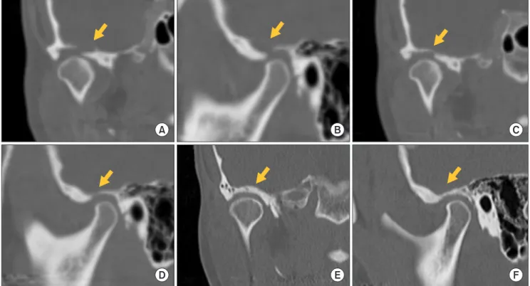
Fig. 2. Case 1. Coronal and sagittal images of computed tomography (CT). A, B. Preoperative CT image (A: coronal, B: sagittal). C, D. Postoperative day (POD) 3 months CT image (C: coronal, D: sagittal). E, F. POD 2 years and 6 months CT image (E: coronal, F: sagittal). Lesion was removed completely. And well bone healing process (arrows) was observed.
Dae-Hoon Kim et al: Temporomandibular joint synovial chondromatosis extending to the temporal bone: a report of two cases. J Korean Assoc Oral Maxillofac Surg 2017
A B
Fig. 3. Case 1. An approximate 13×8 mm sized cystic lesion (A) and loose bodies (B) were removed.
with a high-intensity signal and low-intensity signals possibly originating from loose bodies above the glenoid fossa and ar-ticular eminence.(Fig. 5) CT scans showed condylar erosion in the left TMJ and no calcification.(Fig. 6. A)
The patient was diagnosed with synovial chondromatosis of the left TMJ based on these examination results, and sur-gery was performed accordingly. A preauricular approach extending to the left temporal region in to the glenoid cavity was used after inducing general anesthesia. A flat, soft, beige mass from the superior joint space was weakly attached to the middle of the glenoid cavity and was easily removed. A bone defect formed in the upper direction from the center of filled with air, which confirmed that neither perforation of the
meninges nor brain herniation occurred. CT scans obtained at 3 months and at 2.5 years after the surgery showed reossifi-cation and satisfactory recovery of the defects caused by the lesion.(Fig. 2. C-F)
2. Case 2
A 31-year-old female patient visited the clinic with a chief complaint of pain in the TMJ. The maximum jaw ROM was 45 mm, and the patient complained of pain in the left TMJ when opening her mouth. T2 scans in MRI showed a lesion
A B
C
F
F F
Fig. 4. Case 1. Histopathologic examination of the specimen. A. Cartilage nodules (C) and fibrous tissue (F) arranged in a pseudocystic pattern. There is no true cystic epithelium, only fibrous tissue (arrows; inset) lining the pseudocystic lumen (H&E staining, ×40). The speci-men was diagnosed as middle stage synovial chondromatosis. B. Upon high power inspection of the cartilage nodules, chondrocytes with small eccentric nuclei are seen seated in the lacunae within a pink to blue hyaline matrix (H&E staining, ×200).
Dae-Hoon Kim et al: Temporomandibular joint synovial chondromatosis extending to the temporal bone: a report of two cases. J Korean Assoc Oral Maxillofac Surg 2017
A B
Fig. 5. Case 2. Preoperative magnetic resonance imaging (MRI). High-intensity signal and low-intensity signals (arrows) possibly originat-ing from loose bodies above the glenoid fossa and articular eminence. A. T2-weighted MRI of sagittal view. B. T2-weighted MRI of coronal view.
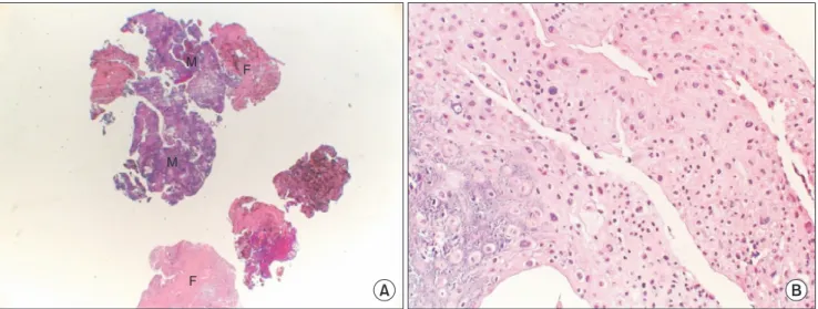
fibroblasts and cartilage cells.(Fig. 8) The patient was finally diagnosed with synovial chondromatosis. No symptoms of relapse were observed during the follow-up for 6 months af-ter the surgery.(Fig. 6. B)
III. Discussion
Synovial chondromatosis is a benign lesion that develops in large joints of the limbs such as the knee, hipbone, shoul-der, and elbow, and rarely in jaw joints. While the causes of the glenoid cavity measured 5 mm in diameter and 1.5 mm
in depth. Another defect located at the articular tubercle mea-sured 10 mm in diameter and 7 mm in depth and was filled with a grayish-brown mass of soft tissue.(Fig. 7) No hole was observed in the glenoid cavity. The articular disc was relatively easily restored. It was confirmed that there was no functional deformity of the articular disc after securing the space via eminoplasty. After sufficiently rinsing the inside of the glenoid cavity, a 0.02-inch silastic sheet was placed in the glenoid cavity and sutured. Biopsy results showed synovial
A B
C D
Fig. 6. Case 2. Computed tomography (CT). A, B. Preoperative CT image (A: sagittal, B: coronal). C, D. Postopera-tive day 3 months CT image (C: sagit-tal, D: coronal). Lesion was removed completely. And well bone healing pro-cess (arrows) was observed.
Dae-Hoon Kim et al: Temporomandibular joint synovial chondromatosis extending to the temporal bone: a report of two cases. J Korean Assoc Oral Maxillofac Surg 2017
Fig. 7. Case 2. A. Soft mass in joint space. B. Grayish mass in intrabony lesion.
Dae-Hoon Kim et al: Temporomandibular joint synovial chondromatosis extending to the temporal bone: a report of two cases. J Korean Assoc Oral Maxillofac Surg 2017
lation of synovial fluid within the glenoid cavity and loose bodies can be observed. In the final stage, loose bodies can still be observed, but the synovial fluid exhibits normal char-acteristics15.
For differentiation of synovial chondromatosis from chon-drosarcoma, a histological examination is necessary. Once the absence of necrosis, mixed cartilage, activation of nuclear differentiation, and fusiform cells is confirmed, the lesion can be confirmed benign16.
Cases of progression of synovial chondromatosis to a malignant lesion in knee joints have been reported, but are very rare. As the recurrence of a lesion in these areas is also reported at low rates17, it is considered a benign lesion with a
good prognosis.
Of the 47 patients who were diagnosed with synovial chon-dromatosis of the TMJ and had the lesion removed at our institution between 1990 and 2016, 43 had lesions confined to the superior joint space, and 2 had lesions confined to the inferior joint space; only 2 cases (4.25%) showed extension into the skull and erosion of the base of the temporal bone.
Synovial chondromatosis usually develops in the supe-rior joint space, rarely extends to the skull, and gives rise to symptoms associated with TMJ disorder18. When it occurs in
the inferior joint space, it can extend to the mandibular con-dyle and surrounding structures. In this case, additional surgi-cal procedures such as a condylectomy might be required19.
When synovial chondromatosis extends to the skull, the dura mater can become exposed, and a CSF leak can occur; synovial chondromatosis have not been clearly identified, the
occurrence can be due to metaplasia of the synovial mesen-chymal cells, injury or joint disease6.
Synovial chondromatosis developing in a jaw joint can cause a variety of clinical symptoms, such as swelling and pain in the affected region, crepitation when opening the mouth, and mouth opening limitation. However, in some cases, no unusual symptoms are observed around the jaw joints, as occurred in the first patient discussed in this study. In addition, due to chronic, gradual swelling, synovial chon-dromatosis can be misdiagnosed as dysplasia of the mandibu-lar condyle, osteochondromatosis, tumor in the preauricumandibu-lar region, or lesions in the parotid gland7-9.
Synovial chondromatosis exhibits CT characteristics simi-lar to osteoarthritis in terms of ensimi-largement of intra-articusimi-lar spaces, soft tissue swelling, irregularity of joint surface, hy-perostosis of the glenoid cavity, and osteosclerosis. A unique characteristic of synovial chondromatosis is that loose bod-ies, which are an end product of cartilaginous metaplasia, can be observed. In some cases, these loose bodies can be calcified. Exudation and accumulation of synovial fluid can be observed on MRI and can help determine whether or not a lesion is of synovial origin. Therefore, it is recommended to perform both CT and MRI before surgery10-14.
Synovial chondromatosis can be histologically differenti-ated into 3 stages. In the early activation stage, it is character-ized as a lesion within the synovia that develops without the formation of any loose bodies. In the middle stage,
accumu-A B
F
Fig. 8. Case 2. Histopathologic examination of the specimen. A. Histopathologic examination of the specimen reveals cellular masses (M) and areas of fibrous tissue (F) (H&E staining, ×10). B. High power inspection of the masses shows large round chondroblastic cells with significant nuclear atypia (H&E staining, ×200). These’s features are not considered as a malignant transformation in metaplastic lesions confined in the synovial space. The specimen was diagnosed as middle stage synovial chondromatosis.
collaborative treatment with a department of neurosurgery must be considered5. Dura mater repair might be necessary in
some cases.
In the first patient discussed in the present case report, although the lesion invaded the skull, no unusual symptoms were noted other than a headache on the contralateral side in the first medical examination. The second patient complained of pain around the TMJ area ipsilateral to the lesion when opening the mouth, a symptom that did not significantly dif-fer from typical symptoms of TMJ disorder. By the end of the follow-up that lasted 2.5 years after the lesions were removed through open surgery on the TMJ, the aforementioned symp-toms were relieved for both patients, and no recurrence oc-curred.
Conflict of Interest
No potential conflict of interest relevant to this article was reported.
ORCID
Dae-Hoon Kim, http://orcid.org/0000-0001-8778-4095 Eun Hee Lee, http://orcid.org/0000-0001-6814-421X Eunae Sandra Cho, http://orcid.org/0000-0002-0820-3019 Jae-Young Kim, http://orcid.org/0000-0002-9423-438X Kug-Jin Jeon, http://orcid.org/0000-0002-5862-2975 Jin Kim, http://orcid.org/0000-0001-5398-8989 Jong-Ki Huh, http://orcid.org/0000-0002-7381-3972
References
1. Akhtar M, Mahajan S, Kott E. Synovial chondromatosis of the temporomandibular joint. J Bone Joint Surg Am 1977;59:266-7. 2. von Lindern JJ, Theuerkauf I, Niederhagen B, Bergé S, Appel T,
Reich RH. Synovial chondromatosis of the temporomandibular joint: clinical, diagnostic, and histomorphologic findings. Oral Surg Oral Med Oral Pathol Oral Radiol Endod 2002;94:31-8.
3. Karlis V, Glickman RS, Zaslow M. Synovial chondromatosis of the temporomandibular joint with intracranial extension. Oral Surg Oral Med Oral Pathol Oral Radiol Endod 1998;86:664-6.
4. Ardekian L, Faquin W, Troulis MJ, Kaban LB, August M. Synovial chondromatosis of the temporomandibular joint: report and analy-sis of eleven cases. J Oral Maxillofac Surg 2005;63:941-7. 5. Quinn PD, Stanton DC, Foote JW. Synovial chondromatosis with
cranial extension. Oral Surg Oral Med Oral Pathol 1992;73:398-402.
6. Simon GT, Kendrick RW, Whitlock RI. Osteochondroma of the mandibular condyle. Case report and its management. Oral Surg Oral Med Oral Pathol 1977;43:18-24.
7. Lustmann J, Zeltser R. Synovial chondromatosis of the temporo-mandibular joint. Review of the literature and case report. Int J Oral Maxillofac Surg 1989;18:90-4.
8. Hammodeh N, Nasser NA. Synovial chondromatosis of the tem-poromandibular joint, presenting as parotid mass. J Laryngol Otol 2006;120:e40.
9. Cannon CR. Osteochondrosis of the temporomandibular joint presenting as an apparent parotid mass. Ann Otol Rhinol Laryngol 1987;96:330-2.
10. Allias-Montmayeur F, Durroux R, Dodart L, Combelles R. Tu-mours and pseudotumorous lesions of the temporomandibular joint: a diagnostic challenge. J Laryngol Otol 1997;111:776-81. 11. Balliu E, Medina V, Vilanova J, Peláez I, Puig J, Trull JM, et al.
Synovial chondromatosis of the temporomandibular joint: CT and MRI findings. Dentomaxillofac Radiol 2007;36:55-8.
12. Wong WC, Cheng PW, Chan FL. MRI appearance of synovial chondromatosis in the temporomandibular joint. Clin Radiol 2001;56:773-4.
13. Koyama J, Ito J, Hayashi T, Kobayashi F. Synovial chondromatosis in the temporomandibular joint complicated by displacement and calcification of the articular disk: report of two cases. AJNR Am J Neuroradiol 2001;22:1203-6.
14. Herzog S, Mafee M. Synovial chondromatosis of the TMJ: MR and CT findings. AJNR Am J Neuroradiol 1990;11:742-5.
15. Kim HG, Park KH, Huh JK, Song YB, Choi HS. Magnetic reso-nance imaging characteristics of synovial chondromatosis of the temporomandibular joint. J Orofac Pain 2002;16:148-53.
16. Milgram JW. Synovial osteochondromatosis: a histopathological study of thirty cases. J Bone Joint Surg Am 1977;59:792-801. 17. Hallam P, Ashwood N, Cobb J, Fazal A, Heatley W. Malignant
transformation in synovial chondromatosis of the knee? Knee 2001;8:239-42.
18. Lieger O, Zix J, Stauffer-Brauch EJ, Iizuka T. Synovial chondrom-atosis of the temporomandibular joint with cranial extension: a case report and literature review. J Oral Maxillofac Surg 2007;65:2073-80.
19. Huh JK, Park JY, Lee S, Lee SH, Choi SW. Synovial chondroma-tosis of the temporomandibular joint with condylar extension. Oral Surg Oral Med Oral Pathol Oral Radiol Endod 2006;101:e83-8.