Ha, Yangsoo Jang, Byung-Chul Chang and Namsik Chung
Yun-Hyeong Cho, Eui-Young Choi, Se-Jung Yoon, Jeehyun Lee, Young-Jin Kim, Jong-Won
The Use of 2 Contrast Filling Patterns in the Diagnosis of a Giant Coronary Aneurysm
Print ISSN: 0009-7322. Online ISSN: 1524-4539
Copyright © 2007 American Heart Association, Inc. All rights reserved.
is published by the American Heart Association, 7272 Greenville Avenue, Dallas, TX 75231
Circulation
doi: 10.1161/CIRCULATIONAHA.106.683805
2007;115:e452-e454
Circulation.
http://circ.ahajournals.org/content/115/19/e452
World Wide Web at:
The online version of this article, along with updated information and services, is located on the
http://circ.ahajournals.org/content/suppl/2007/05/21/115.19.e452.DC1.html
Data Supplement (unedited) at:
http://circ.ahajournals.org//subscriptions/
is online at:
Circulation
Information about subscribing to Subscriptions:
http://www.lww.com/reprints
Information about reprints can be found online at: Reprints:
document.
Permissions and Rights Question and Answer
this process is available in the
click Request Permissions in the middle column of the Web page under Services. Further information about Office. Once the online version of the published article for which permission is being requested is located,
can be obtained via RightsLink, a service of the Copyright Clearance Center, not the Editorial
Circulation
in
Requests for permissions to reproduce figures, tables, or portions of articles originally published Permissions:
at CONS KESLI on June 2, 2014
http://circ.ahajournals.org/
Downloaded from http://circ.ahajournals.org/ at CONS KESLI on June 2, 2014 Downloaded from
The Use of 2 Contrast Filling Patterns in the Diagnosis of a
Giant Coronary Aneurysm
Yun-Hyeong Cho, MD; Eui-Young Choi, MD; Se-Jung Yoon, MD; Jeehyun Lee, MD;
Young-Jin Kim, MD; Jong-Won Ha, MD, PhD; Yangsoo Jang, MD, PhD;
Byung-Chul Chang, MD, PhD; Namsik Chung, MD, PhD
A
75-year-old woman was admitted for exertional dys-pnea and chest pain that had been present for several months. She denied any past history of medical illness such as hypertension or diabetes, as well as any past history of significant trauma. On physical examination, no definite cardiac murmur was auscultated. A chest x-ray showed a bulging silhouette at the right border of the heart. Electrocar-diographic findings showed no significant abnormalities. Two-dimensional echocardiography showed an abnormal echolucent large mass lesion at the right atrial side, suggest-ing an intraright atrial mass, pericardial cyst, or aneurysm. To determine the exact location of the mass and its relation to the right atrium, a contrast echocardiogram with intravenous agitated saline injection was performed. This revealed an extracardiac mass compressing the right atrium without contrast filling (Figure 1, Movie I). To clarify the communi-cation with the left-side chamber, perfluorocarbon-exposed sonicated dextrose albumin, a pulmonary circulation passing contrast agent, was injected via an antecubital vein. Contrast echocardiogram with perfluorocarbon-exposed sonicated dextrose albumin showed contrast filling in the mass after opacification of the left ventricular cavity, suggesting a coronary aneurysm (Figure 2, Movies II and III). Addition-ally, an abnormal continuous flow entering into the main pulmonary artery was noted, suggesting the drainage of a giant coronary aneurysm (Figure 3, Movie IV). With this in mind, multislice computed tomography and conventionalangiography were performed to confirm the diagnosis. Sim-ilar to the echocardiographic findings, a huge coronary aneurysm feeding from the right coronary artery and draining to the main pulmonary artery was detected (Figure 4). The patient underwent removal of the aneurysm, combined with bypass grafting surgery. The patient was later discharged without any complications.
In contrast to agitated saline, a perfluorocarbon-based contrast agent can pass through microcirculation, such as a pulmonary capillary, and can then opacify the left-side cardiac chambers and arterial structures.1This case
demon-strates that the combined use of these 2 types of contrast agents can provide vital information in the clarification of intra- or extracardiac mass characteristics.
Disclosures
None.
Reference
1. Hundley WG, Kizilbash A, Afridi I, Franco F, Peshock RM, Grayburn PA. Administration of an intravenous perfluorocarbon contrast agent improves echocardiographic determination of left ventricular volumes and ejection fraction: comparison with cine magnetic resonance imaging.
J Am Coll Cardiol. 1998;32:1426 –1432.
From the Divisions of Cardiology (Y.-H.C., E.-Y.C., S.-J.Y., J.L., J.-W.H., Y.J., N.C.), Diagnostic Radiology (Y.-J.K.), and Cardiovascular Surgery (B.-C.C.), Yonsei University College of Medicine, Seoul, South Korea.
The online-only Data Supplement, consisting of Movies I through IV, is available with this article at http://circ.ahajournals.org/cgi/content/ full/115/19/e452/DC1.
Correspondence to Eui-Young Choi, MD, Cardiology Division, Yonsei University College of Medicine, Yonsei Cardiovascular Center, CPO Box 8044, Seoul, South Korea 120 –752. E-mail choi0928@yumc.yonsei.ac.kr
(Circulation. 2007;115:e452-e454.) © 2007 American Heart Association, Inc.
Circulation is available at http://www.circulationaha.org DOI: 10.1161/CIRCULATIONAHA.106.683805 e452
Images in Cardiovascular Medicine
at CONS KESLI on June 2, 2014
http://circ.ahajournals.org/
Figure 1. A, Apical 4-chamber view showing an echolucent large mass lesion in the right atrial side. B, Contrast echo with intravenous agitated saline, showing an extra–right atrial compressing mass without contrast filling (Movie I). LA indi-cates left atrium; LV, left ventricle; RV, right ventricle.
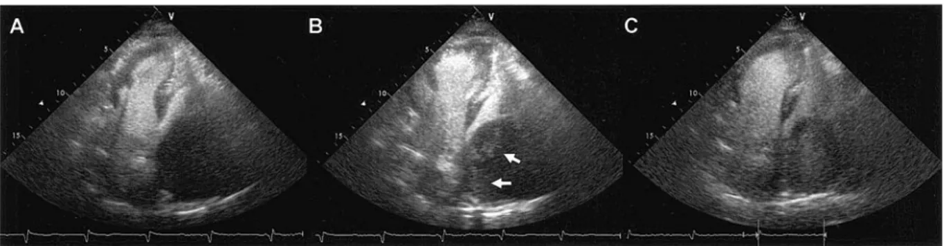
Figure 2. Sequential filling of the perfluorocarbon-exposed sonicated dextrose albumin (PESDA). A, Contrast echo with pulmonary cir-culation passing contrast agent, PESDA, showing contrast filling in the left ventricle cavity without filling of the right atrial mass. B, PESDA entering the mass (arrow heads) after initial opacification of the left ventricular cavity (demonstrated by Movie II). C, Full-contrast filling of the right atrial mass (Movie III).
Figure 3. Echo (A) and color Doppler (B) findings showing an abnormal flow entering the main pulmonary artery (arrows) (Movie IV). C, Pulsed-wave Doppler finding showing the nature of its continuous flow.
Cho et al Diagnosis of a Giant Coronary Aneurysm e453
at CONS KESLI on June 2, 2014
http://circ.ahajournals.org/
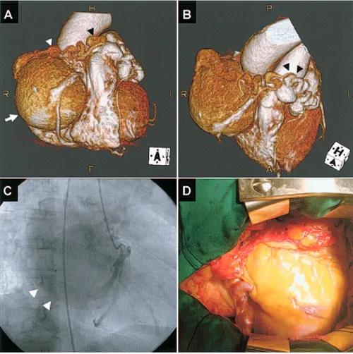
Figure 4. A and B, Multislice com-puted tomography findings showing a giant coronary aneurysm originating from the right coronary artery (white arrows) and draining into main pul-monary artery (arrow heads). C, Coro-nary angiography showing a giant right coronary aneurysm (arrow heads) draining into the pulmonary artery, similar to the multislice com-puted tomography findings. D, Surgi-cal view of the 10-cm giant right cor-onary artery aneurysm compressing the right atrium.
e454 Circulation May 15, 2007
at CONS KESLI on June 2, 2014
http://circ.ahajournals.org/