Effect of Pelvic Compression Belt on Abdominal Muscle Activity, Pelvic Rotation and Pelvic Tilt During Active Straight Leg Raise
Eun-young Jo1, MS, PT, Duk-hyun An2, PhD, PT
1Department of Physical Therapy, Graduate School, Inje University
2Department of Physical Therapy, College of Healthcare Medical Science and Engineering, Inje University
Abstract
1)Background: Uncontrolled lumbopelvic movement leads to asymmetric symptoms and causes pain in the lumbar and pelvic regions. So many patients have uncontrolled lumbopelvic movement. Passive support devices are used for unstable lumbopelvic patient. So, we need to understand that influence of passive support on lumbopelvic stability. It is important to examine that using the pelvic belt on abdominal muscle activity, pelvic rotation and pelvic tilt.
Objects: This study observed abdominal muscle activity, pelvic rotation and tilt angles were compared during active straight leg raise (ASLR) with and without pelvic compression belt.
Methods: Sixteen healthy women were participated in this study. ASRL with and without pelvic compression belt was performed for 5 sec, until their leg touched the target bar that was set 20 ㎝ above the base. Surface electromyography was recorded from rectus abdominis (RA), internal oblique abdominis (IO), and external oblique abdominis (EO) bilaterally. And pelvic rotation and tilt angles were measured by motion capture system.
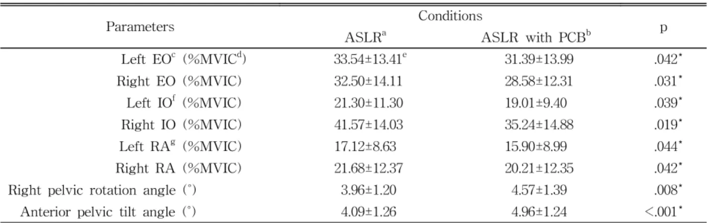
Results: There were significantly less activities of left EO (p=.042), right EO (p=.031), left IO (p=.039), right IO (p=.019), left RA (p=.044), and right RA (p=.042) and a greater right pelvic rotation angle (p=.008) and anterior pelvic tilt angle (p<.001) during ASLR with pelvic compression belt.
Conclusion: These results showed that abdominal activity was reduced while the right pelvic rotation angle and anterior pelvic tilt angle were increased during ASLR with a pelvic compression belt. In other words, although pelvic compression belt could support abdominal muscle activity, it would be difficult to control pelvic movement. So pelvic belt would not be useful for controlled ASLR.
Key Words: Active straight leg raise; Pelvic compression belt; Pelvic movement angles.
Introduction
Pelvic and lumbar stabilization during limb move- ment is acquired via external and internal stabiliza- tion (Kisner and Colby, 2007; Oh et al., 2007; Park et al., 2013). The external oblique (EO), internal oblique (IO), and rectus abdominis (RA) muscles are very important for lumbar internal stabilization (Kisner and Colby, 2007; Noh et al., 2014). Muscle recruitment and pelvic stability is associated in the pelvic pain and groin pain (Sawle et al., 2013). And lumbopelvic in- stability makes limited controlling lumbar rotation and
extension and pelvic anterior tilt and rotation during low limb movement (Jeon et al., 2016a; Sahrmann, 2001). If lumbopelvic movement is increased, it makes abnormal stress, microtrauma and injury in lumbar spine tissue. So restriction of the lumbopelvic move- ment makes decrease pain (Sadeghisani et al., 2015;
Sahrmann, 2001). Therefore lumbopelvic movement pattern test is important clinical test during low limb movement (Hoffman, 2011; Sadeghisani et al, 2015).
A therapeutic belt and strap can produce external stabilization that makes it possible to sustain the body segment (Oh et al., 2007). With the external
Corresponding author: Duk-hyun An [email protected]
Figure 1. Elastic pelvic belt (SFD20FT, Needs Co., Japan).
stabilization, a pelvic belt causes force closure which is helpful to stabilizing muscle activity by creating internal stabilization. and it has been used for pelvic instability in females. (Hu et al., 2010; Park et al., 2013). In the pain and functions, non elastic pelvic compression belt is important to improve the pain during ASLR in clinical test performance. Because it makes force closure (Lee, 2004; Mens et al., 2006;
Sawle et al., 2013). In the present study of Kim et al (2013), wearing a non elastic pelvic compression belt decreased muscle activity during a single-leg hold in the hook-lying position on a round foam roll. Because pelvic belt improves abdominal muscle control and lumbopelvic stability. Jung et al. (2013) confirmed that if sacroiliac joint pain patients using the elastic pelvic compression belt, the hip extensor muscle ac- tivity patterns were changed during one-leg standing.
The active straight leg raise (ASLR) test involves raising the lower limb in knee extension about 20 ㎝ above the ground (Linek et al., 2015). It is used to examine patients with impaired lumbopelvic muscu- lature and as an abdominal exercise in fit individuals (Noh et al., 2014). During the ASLR, the load is transferred between the lower limbs and spine via the pelvis. Accordingly, the best movement pattern is to maintain a neutral pelvic and lumbar posture, while activating stabilizer muscles (Jeon et al., 2016b). If the lumbar, pelvic, and hip regions are not stabilized during the ASLR, the lumbar region will undergo immoderate movement and pelvic rotation (Lee, 2004).
Comerford and Mottram (2012) identified ipsilateral lumbar and pelvic rotation in the transverse plane during leg movement. Uncontrolled lumbopelvic rota- tion leads to asymmetric symptoms and causes pain in the lumbar and pelvic regions (Jeon et al., 2016b).
Consequently, lower back pain recurs with continuous repeated lumbar and pelvic rotation during ASLR (Jeon et al., 2016b; Noh et al., 2014; Park et al., 2013). During ASLR, decreasing lumbar and pelvic motion is a good training method in patients with lower back pain (Hoffman et al, 2011; Sahrmann,
2001; Scholtes et al., 2010). It is very important com- ponent to physical therapy treatment (Hoffman et al., 2011). Many studies were explain that pelvic com- pression belt is effect for muscles, so it is affect for lumbopelvic stability. In the clinically, pelvic com- pression belt is used by management of pelvic pain, groin pain, sacroiliac pain and increased the sacroiliac joint stability (Hu et al., 2010; Sawle et al., 2013).
Many studies have investigated ways to minimize lumbar and pelvic rotation and the most effective maneuver for doing so (Noh et al., 2014; Scholtes et al., 2010). In comparison, pelvic tilt during ASLR has not been investigated. Therefore, we compared ab- dominal muscle activity, pelvic rotation, and pelvic tilt during ASLR with and without a pelvic com- pression belt.
Methods
Participants
This study enrolled 16 healthy females (age 20.2±.9 years; height 161.4±4.9 ㎝; weight 55.8±6.6 ㎏).
We excluded those with lumbopelvic and hip prob- lems or a history of abdominal, lumbopelvic, or hip surgery.
Instrumentation
The elastic belt (SFD20FT, Needs Co., Japan) (Figure 1) was placed below the anterior superior iliac spine and was made of polyester, nylon, polyur- ethane and Velcro (Jung et al., 2013; Park et al., 2013). Compression pressure was adjusted by check-
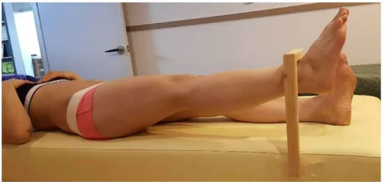
Figure 2. Measurement of ASLR(active straight leg raise).
EO RA
IO
Figure 3. Placements of electromyography, reflective markers and pelvic belt. (EO : external oblique abdominis, RA : rectus abdominis, IO : internal oblique abdominis).
ing the compression sites for the compression with elastic compression belt. Before the trial, the partic- ipants were asked to kick a ball to determine the dominant leg. Dominant legs were right in all participants. Right leg was used for the ASLR, rais- ing the leg for 5 s so that it touched a target bar that was set 20 ㎝ above the ground (Figure 2). The task was performed with and without a pelvic com- pression belt and the order was set randomly and the task was repeated three times. The data for the middle 3 s during the ASLR were analyzed.
The activities of the EO (at the midpoint between the oblique angle of ribs and iliac crest), IO (halfway between the midline and anterior superior iliac spine), and RA (2 ㎝ lateral to the umbilicus) (Cram and
Kasman, 1998) were recorded using electromyography (Delsys, Boston, MA, USA) (Figure 3). The sampling rate was set at 1000 ㎐ and the band width was set at 20∼450 ㎐. Acquired data were calculated as the root mean square and were normalized using a per- cent of maximal voluntary isometric contraction (%MVIC) following Kendall et al. (2005). The pelvic angle was measured using a motion-capture system (Oxford Metrics, Oxford, UK; sampling rate 100 ㎐).
Markers were attached to the left and right anterior superior iliac spine and symphysis pubis (Park et al., 2013) (Figure 2). Right pelvic rotation angle was measured that difference angle between initial posi- tion and final position in horizontal plane during ASLR. And anterior pelvic tilt angle was measured
Parameters Conditions
ASLRa ASLR with PCBb p
Left EOc (%MVICd) 33.54±13.41e 31.39±13.99 .042*
Right EO (%MVIC) 32.50±14.11 28.58±12.31 .031*
Left IOf (%MVIC) 21.30±11.30 19.01±9.40 .039*
Right IO (%MVIC) 41.57±14.03 35.24±14.88 .019*
Left RAg (%MVIC) 17.12±8.63 15.90±8.99 .044*
Right RA (%MVIC) 21.68±12.37 20.21±12.35 .042*
Right pelvic rotation angle (˚) 3.96±1.20 4.57±1.39 .008*
Anterior pelvic tilt angle (˚) 4.09±1.26 4.96±1.24 <.001*
aactive straight leg raise, bpelvic compression belt, cexternal oblique, dmaximal voluntary isometric contraction,
emean±standard deviation, finternal oblique, grectus abdominis, *p<.05.
Table 1. Comparison of muscle activities and pelvic angle during ASLR and ASLR with PCB (N=16)
Figure 4. Change rate (%) of right pelvic rotation angle and anterior pelvic tilt angle between ASLR with and without pelvic compression belt. (ASLR: active straight leg raise).
Figure 5. Change rate (%) of MVIC of abdominal muscle activity between ASLR with and without pelvic compression belt. (EO: external oblique, IO: internal oblique, RA: rectus abdominis, MVIC:
maximal voluntary isometric contraction, ASLR: active straight leg raise).
that difference angle in sagittal plane during ASLR.
Vicon Nexus software (ver. 1.5.2) was used to ana- lyze the kinematic data. The values of the muscle activities and pelvic angles used for analyses were the means of repeated three times tasks.
Statistical analysis
For the statistical analyses, we used SPSS ver.
18.0 (IBM, Armonk, NY, USA). The paired t-test was used to find differences between the ASLR with and without a pelvic compression belt, with sig- nificance accepted at p<.05.
Results
Compared to ASLR without the belt, ASLR with the pelvic compression belt showed significantly less activity of the left EO (p=.042), right EO (p=.031), left IO (p=.039), right IO (p=.019), left RA (p=.044), and right RA (p=.042) and a greater right pelvic ro- tation angle (p=.008) and anterior pelvic tilt angle (p<.001) (Table 1). The change rates of right pelvic rotation angle and anterior pelvic tilt angle during ASLR with and without pelvic compression belt were 17.86% and 24.19% (Figure 4). Between activity of left EO, left IO, left RA, right EO, right IO, right RA during ASLR with and without the pelvic com- pression belt, there were 7.41%, 7.40% 8.26%, 12.39%, 15.15% and 7.17% of the change rates respectively (Figure 5).
Discussion
This study is to figure out the effect of pelvic compression belt on abdominal muscle activity and pelvic movement during active straight leg raise (ASLR). During the ASLR, the load is transferred between the lower limbs and spine via the pelvis.
However, if there is the lumbopelvic instability, peo- ple have limited lumbar and pelvic movement control.
In line with this, the optimal lumbopelvic movements must be adjusted by stabilizing muscle and pelvic neutral (Comerford and Mottram, 2012; Jeon et al., 2016b). With the lack of these stabilizing factors, pelvic compression belt is used to increase external stabilization and improve lumbopelvic movements (Oh et al., 2007; Park et al., 2013).
We found that the activity of both right and left rectus abdomominis (RA), external oblique (EO) and internal oblique (IO) was reduced, while in Park et al.
(2013), their activities were not significantly different between ASLR with and without pelvic compression belt. On the other hand, Hu et al. (2010) and Kim et al. (2013) confirmed that the RA, EO, and IO activ- ities were decreased during movement with a pelvic compression belt, since the belt compresses the front of the abdominal wall and creates force closure that reduces and supports the abdominal muscle activity (Pel et al., 2008; Richardson et al., 2002). By reducing the load to muscles, the pelvic belt can require less muscle activation and resolve muscle problems related to ASLR (Mens et al., 1999).
Regarding to pelvic movement, this study found that pelvic rotation and anterior tilt movement be- came significantly greater durring ASLR with a pel- vic belt. Park et al. (2013) also showed the increased pelvic rotation angle during ASLR with a pelvic belt compared to ASLR without the belt although the dif- ference was not significant and sagittal pelvic move- ment was not included. During the early and mid range of the ASLR, the weight of the lower limb and pelvic flexor activation cause the torque and transfer it to the pelvis, which makes pelvic movement.
Against the torque, abdominal muscles play the role to prevent pelvic anterior tilt and rotation with coun- terbalanced lower limbs during the ASLR (Neumann, 2002; Park et al., 2013). Accordingly, it can be said that the reduced abdominal muscle activities during ASLR with a pelvic compression belt may attribute to the increased pelvic rotation and anterior tilt.
The study by Richardson et al.(2002) and Hu et al. (2010) found that pelvic belt supplements the role
of stabilizing muscles. This passive compression is reported as an effective measure for pelvic instability by making forcing closure (Takasaki et al., 2009).
However, Park et al. (2013) indicated that pelvic compression belt only controls the lumbopelvic mo- tion in initial stages as force closure, but the passive compression does not restrict the pelvic movement, suggesting ASLR with self control as an effective way. Similarly, the results in this study suggest that ASLR with pelvic compression belt would not be valuable to control pelvic anterior tilt and rotation.
While pelvic compression has benefits by managing muscle contribution to stability during ASLR, it could make problems with motor control strategies in the long term (Beales et al., 2010).
This study had some limitations. First, we did not investigate lumbar and pelvic muscles such as the multifidus, erector spinae muscles, and gluteus. Second, we did not consider other methods of external stabi- lization that decrease pelvic rotation, such as having the participant touch the anterior superior iliac spine during the ASLR lightly with both hands. Third, we involved only young women, so we can not be gen- eralized for other populations. Finally, the compression force of the belt was not controlled, although a phys- ical therapist adjusted it based on previous experience.
Further studies are needed to investigate the activ- ities of the pelvic muscles and pelvic tilt and rotation during various performance using other manuever such as having the participant lightly touch the ante- rior superior iliac spine during ASLR with both hands to confirm the effect of pelvic belt.
Conclusion
This study investigated the change of lumbopelvic stabilization during ASLR with and without pelvic compression belt. Abdominal muscle activities were decreased but pelvic tilt and pelvic rotation were in- creased during ASLR with pelvic compression belt compared to ASLR without the belt. These results
indicate that pelvic compression belt would not be useful for controlled ASLR as the application of pel- vic compression belt lead to increased pelvic tilt and rotation.
References
Beales DJ, O'Sullivan PB, Briffa NK. The effects of manual pelvic compression on trunk motor con- trol during an active straight leg raise in chron- ic pelvic girdle pain subjects. Man Ther. 2010;
15(2):190-199. https://doi.org/10.1016/j.math.2009.
10.008
Comerford M, Mottram S. Kinetic Control: The Management of Uncontrolled Movement. Australia.
Churchill Livingstone, 2012:164-182.
Cram JR, Kasman GS. Introduction to Surface Electro- myography. Gaithersburg. Aspen, 1998:343-346.
Hoffman SL, Johnson MB, Zou D, et al. Effect of classification-specific treatment on lumbopelvic motion during hip rotation in people with low back pain. Man Ther, 2011;16(4):344-350.
https://doi.org/10.1016/j.math.2010.12.007
Hu H, Meijer OG, van Dieën JH, et al. Muscle activ- ity during the active straight leg raise (ASLR), and the effects of a pelvic belt on the ASLR and on treadmill walking. J Biomech, 2010;
43(3):532-539. https://doi.org/10.1016/j.jbiomech.
2009.09.035
Jeon IC, Hwang UJ, Jung SH, et al. Comparison of gluteus maximus and hamstring electromyo- graphic activity and lumbopelvic motion during three different prone hip extension exercises in healthy volunteers. Phys Ther Sport, 2016a;22:
35-40. https://doi.org/10.1016/j.ptsp.2016.03.004 Jeon IC, Kwon OY, Weon JH, et al. Comparison of
psoas major muscle thickness measured by so- nography during active straight leg raising in subjects with and without uncontrolled lumbo- pelvic rotation. Man Ther, 2016b;21:165-169.
https://doi.org/10.1016/j.math.2015.07.006
Jung HS, Jeon HS, Oh DW, et al. Effect of the pel- vic compression belt on the hip extensor activa- tion patterns of sacroiliac joint pain patients during one-leg standing: A pilot study. Man Ther, 2013;18(2):143-148.
Kendall FD, McCreary EK, Provance PG, et al.
Muscles Testing and Function With Posture and Pain. 5th ed. Baltimore: Lippincott Williams &
Wilkins, 2005:146-155.
Kim YR, Kim JW, An DH, et al. Effects of a pelvic belt on the EMG activity of the abdominal muscles during a single-leg hold in the hook- lying position on a round foam roll. J Phys Ther Sci, 2013;25(7):793-795. https://doi.org/10.
1589/jpts.25.793
Kisner C, Colby LA, Borstad J. Therapeutic Exercise:
Foundations and techniques. Philadelphia, PA, F.A. Davis Co., 2007:76, 641-643.
Lee DG: The Pelvic Girdle, 3rd ed. Edinburgh.
Churchill Livingstone, 2004:206-209, 328-329.
Linek P, Saulicz E, Wolny T, et al. Intra-rater reli- ability of B-mode ultrasound imaging of the ab- dominal muscles in healthy adolescents during the active straight leg raise test. PM R, 2015;
7(1):53-59. https://doi.org/10.1016/j.pmrj.2014.07.007 Mens J, Inklaar H, Koes BW, et al. A new view on adduction-related groin pain. Clin J Sport Med, 2006;16(1):15-19.
Mens J, Vleeming A, Snijders CJ, et al. The active straight leg raising test and mobility of the pel- vic joints. Eur Spine J. 1999;8(6):468-473.
Neumann DA. Kinesiology of the Musculoskeletal System, 2nd ed. ST. Louis, Mosby, 2002:350-354, 446-447.
Noh KH, Kim JW, Kim GM, et al. The influence of dual pressure biofeedback units on pelvic rota- tion and abdominal muscle activity during the active straight leg raise in women with chronic lower back pain. J Phys Ther Sci, 2014;26(5):
717-719. https://doi.org/10.1589/jpts.26.717 Oh JS, Cynn HS, Won JH, et al. Effects of perform-
ing an abdominal drawing-in maneuver during
prone hip extension exercises on hip and back extensor muscle activity and amount of anterior pelvic tilt. J Orthop Sports Phys Ther, 2007;
37(6):320-324. https://doi.org/10.2519/jospt.2007.
2435
Park KH, Ha SM, Kim SJ, et al. Effects of the pel- vic rotatory control method on abdominal muscle activity and the pelvic rotation during active straight leg raising. Man Ther, 2013;18(3):220- 224. https://doi.org/10.1016/j.math.2012.10.004 Park KN, Cynn HS, Kwon OY, et al. Effects of the
abdominal drawing-in maneuver on muscle ac- tivity, pelvic motions, and knee flexion during active prone knee flexion in patients with lumbar extension rotation syndrome. Arch Phys Med Rehabil, 2011;92(9):1477-1483. https://doi.org/10.
1016/j.apmr.2011.03.020
Pel JJ, Spoor CW, Goossens RH, et al. Biomechanical model study of pelvic belt influence on muscle and ligament forces. J Biomech. 2008;41(9):1878- 1884. https://doi.org/10.1016/j.jbiomech.2008.04.002 Richardson CA, Snijders CJ, Hides JA, et al. The re- lation between the transversus abdominis mus- cles, sacroiliac joint mechanics, and low back pain. Spine (Phila Pa 1976), 2002;27(4):399-405.
https://doi.org/10.1097/00007632-200202150-00015 Sadeghisani M, Sobhani V, Kouchaki E, et al.
Comparison of lumbopelvic and hip movement patterns during passive hip external rotation in two groups of low back pain patients with and without rotational demand activities. Ortop Tra- umatol Rehabil, 2015;17(6):611-618. https://doi.org/
10.5604/15093492.1193032
Sahrmann, SA. Diagnosis and Treatment of Movement Impairment Syndromes, St. Louis, Mosby, 2001:
110-119.
Sawle L, Freeman J, Marsden J, et al. Exploring the effect of pelvic belt configurations upon athletic lumbopelvic pain. Prosthet Orthot Int, 2013;37(2):
124-131. https://doi.org/10.1177/0309364612448806 Scholtes SA, Norton BJ, Lang CE, et al. The effect
of within-session instruction on lumbopelvic
This article was received November 14, 2018, was reviewed November 14, 2018, and was accepted December 24, 2018.
motion during a lower limb movement in people with and people without low back pain. Man Ther, 2010;15(5):496-501. https://doi.org/10.1016/
j.math.2010.05.003
Takasaki H, Iizawa T, Hall T, et al. The influence of increasing sacroiliac joint force closure on the hip and lumbar spine extensor muscle firing pattern.
Man Ther. 2008;14(5):484-489. https://doi.org/10.
1016/j.math.2008.11.003