Ⅰ. 서 론
이 병소는 1987년 처음으로 Padayachee와 Van Wyk
1)에 의해“sialo-odontogenic cyst(타액 치성낭)”로 보고되 었다. 유두상 증식을 보이는 다양한 두께의 중층편평상피로 피복되어 있는 다방성의 형태를 보인다고 하였으며 이는 중 심성 점액표피암(central mucoepidermoid carcinoma)
처음으로 사용되었는데, 그들은 이 병소를 치성 기원의 변 이된 낭으로 보았다. 그 후 1992년 WHO의 치성낭에 대한 분류에서 선양치성낭이라는 용어가 받아들여져 현재까지 주로 사용되고 있다
3).
선양치성낭은 조직형태학적으로 측방치주낭(lateral periodontal cyst), 포상 치성낭(botryoid odontogenic cyst), 중심성 점액표피암(central mucoepidermoid car- 오지수∙김수관∙김학균∙윤정훈*
조선대학교 치과대학 구강악안면외과학교실, *구강병리학교실
선양치성낭의 임상 및 병리조직학적 분석
CLINICAL AND HISTOPATHOLOGIC ANALYSIS OF GLANDULAR ODONTOGENIC CYSTS OF THE JAWS
Ji-Su Oh, Su-Gwan Kim, Hak-Kyun Kim, Jung-Hoon Yoon * Department of Oral and Maxillofacial Surgery & *Oral Pathology,
College of Dentistry, Chosun University
The glandular odontogenic cyst is an uncommon odontogenic cyst as a distinct entity. We reviewed a series of 7 glandular odontogenic cysts of the jaws experienced between 2003 and 2006 at the department of Oral and Maxillofacial surgery, Chosun university.
The study group consisted of 3 females (42.9%) and 4 males (57.1%), with an age range of 31 to 75 years and mean age was 58.6 years. The maxilla was involved in 5 cases (71.4%) and the mandible in 2 cases (28.6%). Three cases involved impacted tooth. Clinically 6 cases showed swelling and tenderness. All the lesion presented well-defined unilocular radiolucent lesion radiographically. Provisional clinical diagno- sis was varied, incisional biopsy was done in 1 case. Histopathologically, those were lined by non-kera- tinized stratified epithelium and thickened epithelial segments (plaques) are seen within the lining epithe- lium. And epithelial lining contains eosinophilic cuboidal type cells, mucous cells and mucin pools in micro- cystic areas are identified.
All cysts were treated by enucleation. All cases are not recurred during follow up period.
Key words: Glandular odontogenic cyst, Sialo-odontogenic cyst, Botryoid odontogenic cyst, Central mucoepidermoid carcinoma
Abstract
다
10). 방사선학적으로 선양치성낭은 이 병소만의 특이한 특 징을 가지고 있지 않다. 대개는 다방성이나 단방성의 방사 선 투과상을 보인다
3,6).
치료는 소파술(curettage)이나 적출술(enucleation)과 같은 보존적인 처치부터 en block resection과 같은 근치적 인 처치도 가능하다
7).
본 연구의 목적은 악골에 발생한 선양 치성낭 7 증례의 임 상 병리학적 특징 및 감별진단에 대해 문헌고찰과 더불어 보고하고자 한다.
Ⅱ. 증례보고
2003년부터 2006년까지 조선대학교 치과병원 구강악안 면외과에 내원하여 조직형태학적으로 선양치성낭(GOC)으 로 진단된 7명의 환자를 대상으로 하였다. 모두 7 증례가 보고되었으며 환자의 성별, 나이, 임상 및 방사선학적 특징, 조직형태학적 특징, 치료법, 추적기간, 재발률에 대해서 조 사하였다. 이 중 여자가 3명, 남자가 4명으로 발생연령은 31세에서 75세까지로, 평균 연령은 58.6세였다. 저혈압을 가지고 있는 1명의 환자를 제외하고 특이할 만한 전신질환 은 없었다. 임상적으로 무통성의 종창을 보이는 환자는 3명
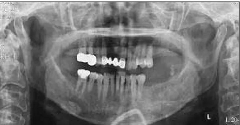
Fig. 1. Panoramic radiograph showing well-defined unilocular radiolucen- cy in the maxilla.
Fig. 2. Well-defined unilocular radiolucent lesion at the anterior maxilla
seen in the panoramic radiograph.
이었으며, 3명은 동통이 있는 종창을 보였고 1명은 종창이 나 동통이 없는 무증상을 보였다. 2명의 환자에서 흡인시 갈색의 장액성 삼출액이 배출되었다. 발생 위치는 5명은 상 악에서 발생하였으며, 이 중 1명은 상악의 전방부에서, 2명 의 환자는 하악에 발생하였다.
방사선학적으로 7명의 환자 모두에서 비교적 경계가 좋은 둥근 모양을 가진 단방성의 방사선 투과상(Figs. 1, 2)을 보였다. 이 중 3명의 환자에서 매복치를 동반하였으며, 2명 의 환자에서 상악에 과잉치를 가지고 있었고 1명의 환자는 하악 제3 대구치를 포함하고 있었다. 상악에 발생한 5명의 환자 중 상악동을 침범한 경우는 없었다.
cyst)로 진단되었으며 1명의 환자는 치성각화낭(odonto- genic keratocyst)로 진단되었으며 1명의 환자에서는 잔존 낭(residual cyst)로 진단되었다. 과잉치와 제3 대구치를 포함하고 있는 2명의 환자는 함치성낭(dentigerous cyst) 으로 추정되었다. 1명의 환자는 수술 전 조직검사가 시행되 었는데 조직형태학적으로 선양치성낭으로 진단되었다 (Figs. 3, 4).
치료는 모든 환자에서 낭적출술(enucleation)을 시행하 였다. 적출된 낭은 육안적으로 회색을 띄는 낭성 조직이었 으며 길이는 1.8cm에서 3.0cm까지로 평균 길이는 2.3cm 였다.
Table 1. Type and Teatment of Glandular Odontogenic Cyst
Patient Age/gender Location Radiological Treatment Follow-up Recurrence
features (months)
1 61/F Lt. Mx. Unilocular Enucleation 4 None containing tooth
2 58/M Lt. Mn. Unilocular Enucleation 4 None containing tooth(#38)
3 31/M Rt. Mn. Unilocular Enucleation 1 None 4 75/M Ant. Mx. Unilocular Enucleation 12 None 5 69/F Rt. Mx. Unilocular Enucleation Lost Not known 6 58/M Rt. Mx. Unilocular Enucleation 8 None 7 58/F Lt. Mx. Unilocular containing tooth Enucleation 1 None
Abbreviations: Lt, left; Rt, right; Mx, maxilla; Mn, mandible; Ant, anterior Fig. 3. The lining epithelium showing thickened epithelial
segments and ciliated columnar epithelium and mucous cells.
Fig. 4. The mucous cells and mucin stained by muci-
carmine are seen.
수술 후 추적기간은 추적이 되지 않는 1명을 제외하고 1 개월에서 12개월까지로 평균 3.3개월이었다. 추적기간 동 안 재발과 관련된 임상적 및 방사선학적 증상은 없었다 (Table 1).
Ⅲ. 고 찰
선양치성낭은 1987년 처음으로 보고된 이후에 현재까지 보고된 예가 많지 않아 임상적 및 치료법 등에 관해 분명히 정립되어 있지 않다
1). 2006년 Sittitavornwong 등
7)이 33 편의 논문에서 보고된 선양치성낭 64예에 대한 연구에서 발생연령은 11세에서 90세까지로 평균 49.4세였으며, 명 확한 성별의 차이는 없었고 80%에서 하악에 발생하였다고 하였다. 상악에 발생한 예 중 84%는 전방부에서 발생하였 다고 하였다. 임상적 증상은 무통성이나 동통을 동반한 종 창이 84%로 대부분을 차지하고 있다고 하였다. 방사선학 적으로 다방성의 방사선 투과상을 나타내는 경우가 52%였 으며 단방성의 경우가 36%로 방사선학적으로 악골에 발생 한 치성낭과 감별할 수 있을만한 특이한 소견은 보이지 않 는다고 하였다
7). 병리조직학적으로 선양치성낭은 다양한 두 께의 중층편평상피로 피복되며, 점액세포와 mucin pool이 상피층에서 많이 관찰되고
8), 상피의 유두상 증식(plaque) 이 여러 부위에서 나타난다고 하였다
2,3,6,7).
Gardner 등
2)은 Padayachee와 Van Wyk 등이 mucous pool과 mucin pool을 타액선 기원으로 이해하기에는 근거 가 부족하다고 하여 이 병소를 타액 치성낭으로 명한 것은 부적절하다고 주장하였다. 그들은 선양 치성낭이라는 용어 를 처음으로 명명하였으며, 다음과 같은 병리조직학적 특징 들을 기술하였다.
1. 다양한 두께의 중층편평상피에 의해 피복되어 있으며, 결합조직에 염증세포의 침윤은 관찰되지 않는다.
2. 상피의 표층은 호산성의 입방세포로 구성되어 있으며, 여러 부위에 불규칙한 상피의 유두상 증식을 포함하고 있다.
3. 상피 내에 mucin pool이 존재하며, 호산성의 입방세포 로 피복되어 있다.
4. mucous cell이 나타날 수 있다.
5. 기저세포는 과염색화되고, 공포화될 수도 있다.
6. epithelial sphere를 관찰할 수 있으며, nuclear polarization은 관찰되지 않는다.
7. 때로 결합조직 하방에서 불규칙한 석회화물질을 관찰 할 수 있다.
저자들도 7예에 대해서 Gardner 등의 병리조직학적 특성 을 근거하여 선양 치성낭으로 진단하였다.
선양치성낭은 다방성의 방사선학적 투과상의 소견을 보이
며, 조직학적으로 epithelial sphere가 존재한다는 점이 포 상 치성낭(botryoid odontogenic cyst)과 유사하다고 하였 다
9). 병소의 상피가 유두상의 증식을 나타내며 낭벽의 내부 로 하방 증식하는 것이 선양 치성낭의 종양 잠재력을 나타 내는 증거라고 Panagiota 등
10)이 언급하였으며 중심성 점 액표피암종(central mucoepidermoid carcinoma)과도 유 사한 점이라고 하였다. 선양 치성낭은 악골에 발생하는 다 른 치성기원의 낭과는 달리 약간 공격적인 성향을 보이는 것으로 보고되고 있으며 이와 관련하여 적출술과 같은 보존 적인 치료법 외에도 en bloc까지도 시행된 보고가 있다. 본 증례에서는 추적조사가 되지 않은 1명을 포함하여 추적기 간이 짧아 재발율에 대한 정확한 평가가 부족하지만 여러 문헌에 보고된 바로는 평균 25%의 재발율을 보이고 있어 8) 선양 치성낭으로 진단된 환자에 대해서는 긴 기간동안의 면밀한 관찰이 요하리라 생각된다.
Ⅳ. 결 론
선양치성낭은 악골에 발생하는 흔하지 않은 치성기원의 낭으로 본 연구에서는 2003년부터 2006년까지 조선대학교 치과병원 구강악안면외과에서 선양치성낭으로 진단된 7예 를 경험하여 이에 대한 임상적, 방사선학적, 조직병리학적 특징을 알아보았다. 남녀의 차이는 없었으며 모두 중장년층 에서 나타났다. 임상적으로는 문헌에서 고찰된 바와는 달리 상악에서 호발하였으며, 7명중 6명의 환자에서 종창을 주 소로 내원하였다. 방사선학적으로는 모두 비교적 경계가 명 확한 단방성의 방사선 투과상을 보였으며 3예에서 제3 대 구치를 비롯한 매복치를 포함하고 있었다. 병리조직학적으 로 비각화성 중층편평상피로 피복되어 있었으며 호산성의 입방세포로 피복된 선 구조나 점액세포도 관찰되었다. 다발 성으로 상피들의 유두상 증식이 관찰되기도 하였다. 치료는 모두 적출술을 시행하였으며 추적기간 중의 재발은 없었다.
참고문헌