조골세포내 PDE4에 의한 PTH 신호의 음성적 조절
박효정·노아롱새미·이정민·임미정# 숙명여자대학교 약학대학
(Received June 28, 2010; Revised October 6, 2010; October 7, 2010)
The Negative Role of PDE4 on PTH-induced Signaling in Osteoblasts
Hyojung Park, A Long Saemi Noh, Jung-Min Lee and Mijung Yim# College of Pharmacy, Sookmyung Women’s University, Seoul 140-742, Korea
Abstract — We explored the role of phosphodiesterase 4 (PDE4) on parathyroid (PTH)-induced signaling in osteoblasts.
PTH was shown to increase the activity of PDE, mainly PDE4, in osteoblasts, which is partly attributable to elevated PDE4B and PDE4D mRNA expression. The use of PDE4 inhibitor strengthened the PTH-induced extracellular signal-reg- ulated kinase (ERK) and p38 MAP kinase (MAPK) activation. Furthermore, the PDE4 inhibitor stimulated PTH-induced receptor activator of nuclear factor-
κB ligand (RANKL) expression in osteoblasts, which in turn increased osteoclast for-
mation. Taken together, these data suggest the negative role of PDE4 on PTH-induced signaling in osteoblasts.Keywords PTH, PDE4, osteoblast, osteoclast
사회가 고령화됨에 따라 노인성 질환도 크게 증가하고 있는 추 세이다. 그 중 골다공증은 뼈 퇴행성 질환으로 골밀도가 감소하 고 그에 대한 질적 변화로 인해 뼈의 강도가 약해져서 골절이 일 어날 가능성이 높은 상태를 의미한다. 골다공증은 노인의 삶의 질을 급격히 저하시키는 문제점을 지님에도 불구하고 발병율이 매우 높아 그에 따른 대책이 시급한 실정이다.
뼈를 구성하는 두가지 세포 중 조골세포는 골을 구성하는 기 질성분을 분비하고 합성하며 골표면 사이의 무기질 이동에 관여 하는 골세포로 중요한 역할을 수행한다. 한편, 파골세포는 골의 기질 성분을 용해시켜 칼슘염과 인산염을 유리시키는 골흡수 분 해를 담당함으로써 골의 농도를 유지시키는 기능을 수행한다. 또 한 조골 및 파골세포는 밀접한 관계를 맺고 있어, 파골세포의 분 화는 조골세포에 의해 엄격하게 조절된다.1,2)즉, 조골세포는 파 골세포 분화인자인 M-CSF(Macrophage Colony-Stimulating Factor, or CSF-1) 및 RANKL(Receptor Activator of Nuclear Factor-kappaB Ligand, OPGL, ODF, or TRANCE)을 통해 파골 세포의 분화를 조절함으로써 체내 골형성과 골파괴의 동적인 평
형을 유지한다.2-5)
부갑상선 호르몬(parathyroid hormone, PTH)은 대표적인 골 대사 조절물질로, 조골세포내 cAMP 농도를 증가시킴으로써 protein kinase A(PKA) 경로를 매개해 RANKL 발현과 파골세 포 분화를 촉진시킨다고 알려져 있다.6,7)
Cyclic AMP(cAMP)는 조골세포내에서 2차 전달자로 작용하 며 세포증식, 분화 및 골형성과 같은 중요 과정을 조절하는 것으 로 알려져 있다. 세포내 cAMP 농도는 매우 정교하게 조절되고 있으며, adenylate cyclase에 의한 합성과 phosphodiesterases (PDEs)에 의한 분해로 항상성을 유지한다.8,9)즉, 세포막 수용체 에 PGE2, PTH, β-adrenergic receptor agonist 등이 결합하면 G 단백질을 매개해 adenylate cyclase가 활성화되고 세포내 cAMP 농도가 증가한다. 한편, cAMP의 유일한 비활성화 기전은 PDEs에 의한 것으로, cAMP는 PDE에 의해 5'-AMP로 분해되 어 신호가 종결된다. 따라서 adenylate cyclase의 활성화 또는 PDE의 억제는 세포내 cAMP의 농도를 증가시킨다. 최근 많은 세포에서 adenylate cyclase의 활성화는 PDE의 활성화와 연계 되어 있으며, 이것이 세포내 cAMP 항상성 조절에 중요한 역할 을 담당함이 밝혀졌다.10,11)그러나 이러한 일련의 보고에도 불구 하고, PDE에 의한 조골세포내 cAMP의 조절은 아직 충분히 밝 혀지지 않았다. 따라서 본 연구자는 조골세포내 PTH에 의한
#본 논문에 관한 문의는 저자에게로
(전화) 02-710-9572 (팩스) 02-710-9871 (E-mail) [email protected]
종설
adenylate cyclase의 활성화가 PDE에 의해 조절되는 기전을 조 사하였기에 보고하는 바이다.
실험방법
마우스 조골세포의 일차 배양
생후 0~1일의 ICR mouse로부터 두개골 피부를 벗긴 후 두 개골을 적출하였다. 이때 후두골 근육 부착부분은 제거하였다.
부착된 근육, 혈구 등을 제거한 후 α-MEM으로 가볍게 세척하 였다. 0.1% collagenase와 0.2% dispase 혼합효소 용액에 넣어 37oC에서 5분간 진탕시킨 후 상등액을 버리고 새로운 효소 용액 을 가하였다. 37oC에서 약 10분간 진탕하여 상등액을 모으는 조 작을 4회 반복하였다. 원심 분리한 후 10% FBS 함유 α-MEM 으로 약 3~5×10⁴세포/100 mm plastic dish가 되도록 접종하였 다. 5% CO2, 37oC에서 3-4일간 배양한 세포를 이후 조골세포로 실험에 사용하였다.
마우스 골수세포의 배양
ICR mouse(6~9주, 수컷)를 경추 탈골한 후 70% 에탄올로 소 독하였다. 경골 부분의 피부를 절개하여 부착 근육을 떼어냈다.
경골 원심부를 절단하고 슬개골을 탈골시켜 경골을 적출하였다.
뼈 양끝을 조금 잘라 한 쪽 끝에 25G의 주사바늘을 꽂고 α-MEM 을 흘려보내 골수세포를 시험관에 모았다. 원심 분리한 후 α- MEM에 현탁하고 2배의 Gey's solution을 가해 적혈구를 제거 했다. 원심 분리한 후 10% FBS가 함유된 α-MEM으로 재현탁 했다.
공배양에 의한 파골세포의 분화유도
초기 배양한 조골세포 5×10³세포/well와 골수세포 1×10⁵ 세포/well을 10% FBS가 함유된 α-MEM으로 PTH(100 nM), rolipram(10 mM), 또는 PTH와 rolipram의 존재하에서 7일간 공 배양했다. 배양이 끝난 세포는 10% formalin으로 10분간 고정한 후 ethanol-aceton(1 : 1)로 1분간 재고정하여 TRAP(tartrate- resistant acid phosphatase) staining을 했다. 3개 이상의 핵을 가 진 TRAP+ 세포를 다핵 파골세포로 판정했다.
PDE 활성 측정
조골세포를 PTH(100 nM) 로 20분간 처리한 후, PDE lysate buffer(20 mM Tris-HCl, pH 8.0, 1 mM EDTA, 0.2 mM EGTA, 50 mM sodium fluoride, 10 mM sodium pyrophosphate, 0.5 µg/ml leupeptin, 0.7µg/ml pepstatin, 4 µg/ml aprotinin, and 2 mM PMSF)에 현탁하고 초음파로 파쇄하였다. 원심분리로 상 등액을 얻은 후 일부를 PDE 활성 buffer에 (20 mM Tris-HCl, pH 8.0, 10 mM MgCl2, 1.25 mM β-mercaptoethanol,0.14 mg/
ml BSA, 1 µM cAMP, and 0.2 µCi [3H] cAMP) 첨가하여 34oC 에서 10분간 반응시켰다. 2분간 끓여 반응을 종결시키고, 5'- nucleotidase를 넣어 37oC에서 10분간 배양하였다. 반응액을 AG1-X2 수지 컬럼에 걸고 빠져나온 [3H]adenosine을 scintillation counting으로 정량했다. PDE4 활성은 동일 실험을 10 mM rolipram 존재하에서 수행하고, rolipram 비반응성 활성을 전체 PDE 활성에서 뺀 값으로 얻었다. 단백질 농도는 BSA를 대조군 으로 Bio-Rad protein reagent(Hercules, CA)를 사용하여 측정 하였다.
Western blot 분석
조골세포를 PTH(100 nM), rolipram(10 mM), 또는 PTH와 rolipram으로 처리하여 일정시간 배양 후, lysate buffer로 용해 하고 원심분리 하였다. 20 µg의 단백질을 SDS-PAGE(poly acrylamide gel electrophoresis)로 변성 분리하고, 이를 80 v에서 1시간 45분 처리하여 PVDF membrane에 이전시켰다. 이를 5%
skim milk가 함유된 PBST 용액으로 blocking하고, 1차 항체로 서 anti-RANKL(1:1000, Santa cruiz), p-p38(1:2000, Cell signaling), p38(1:2000, Cell signaling), p-ERK(1:2000, Cell signaling), ERK(1:2000, Cell signaling) 또는 β-actin(1:4000, Sigma) 항체와 각각 반응시켰다. PBST로 5회 세정하고 HRP (Horseradish peroxidase)가 결합된 2차 항체와 반응시킨 후 ECL Advance(Amersham. CO.)로 발색시켜 분석하였다.
RNA 분리 및 RT-PCR 분석
Total RNA 추출은 easy-Blue(iNtRON Bio technology co,.Ltd, Korea)를 이용하였다. cDNA는 1 mg의 total RNA를 oligodT primer, 10 mM dNTP, 1 unit RNase inhibitor 그리고 4 unit Script reverse transcriptase(Fermentas, Life science)로 42oC에서 60분 처리하여 합성한 후, 70oC에서 10분 가열함으로 써 반응을 중지 시켰다. Polymerase chain reaction(PCR)은 아 래의 프라이머를 사용하여 94 3분간 처리한 후, 94oC 30초, 58oC 45초 72oC 1분의 과정을 32회 또는 28회(GAPDH) 반복하였다.
PCR 결과물은 전기영동으로 확인하였다.: PDE4A, 5'-ggaactcaca- cacctgtcg-3'(forward), 5'-gttcttgtgctaagaggtcc-3'(reverse); PDE4B, 5'-tggaaatcctggctgccat-3'(forward), 5'-tccacagaagctgtgtgct-3' (reverse); PDE4C, 5'-tggtatcagagtaggattcc-3'(forward), 5'-ctctg- tgtaaaccttggctg-3'(reverse); PDE4D, 5'-cggaactcgctctgatgt-3' (forward), 5'-acagaggcgttgtgcttg-3'(reverse); GAPDH, 5'-gaagg- tcggtgtgaacggatttggc-3'(forward), 5'-catgtaggccatgaggtccaccac-3' (reverse).
통계처리
실험결과는 평균+표준편차로 표기하였고, Student's t-test로
분석하여 p 값이 0.05 미만일 때 통계적으로 유의하다고 판단하 였다.
실험결과 및 고찰
PTH 자극에 따른 조골세포내 PDE 활성의 증가
조골세포에 대한 PTH의 처리는 adenylate cyclase를 활성화 시켜 최종적으로 cAMP 농도를 올리는 것으로 알려져있다.7)먼 저 PTH에 의한 adenylate cyclase의 상승이 PDE를 활성화 시 키는지 여부를 조사하였다. 조골세포에서 PTH의 처리는 유의적 으로 PDE를 활성화시켰으며, PDE4 저해제를 이용한 실험에서 PTH에 의해 증가된 PDE 활성의 약 80%가 PDE4에 해당함이 밝혀졌다(Fig. 1A). 이는 PTH가 adenylate cyclase를 자극하여 조골세포내 cAMP의 농도를 일시적으로 상승시키는 한편, 동시 에 PDE4를 활성화시킴으로써 cAMP의 항상성을 유지하는 것으 로 생각된다. 이상의 결과는 조골세포에서 PDE4가 PTH 신호 의 중요한 조절자로 작용할 가능성을 제시하는 것이라 할 수 있다.
PDE4는 4개의 isotype이 존재하며, 조골세포는 그 중 PDE4A, 4B, 4D를 발현함이 보고된 바 있다.12) PTH 처리로 인한 PDE4 isotype의 발현량 변화를 RT-PCR 방법으로 분석하였다(Fig. 1B).
기존의 보고와 일치되게 조골세포는 PDE4A, 4B와 4D를 발현 하였으며, PTH 처리로 PDE4A는 큰 변화 없는 반면, PDE4B와 PDE4D는 그 발현량이 각각 처리후 약 5배, 1.8배로 증가하는 것을 확인할 수 있었다. PDE4C는 PTH 처리로 아무 변화를 보 이지 않았다. 따라서 조골세포내 PTH 자극에 따른 cAMP의 항 상성 유지에는 PDE4B와 4D의 발현 증가에 따른 PDE4 활성화 증강이 관여할 가능성이 제시되었다.
PDE4 저해제에 의한 조골세포내 PTH 매개 신호의 증폭 조골세포내 cAMP는 다양한 신호전달 경로의 활성화에 관여 함이 알려져 있다. 그 중 MAPK인 ERK와 p38의 신호전달경로 가 PDE4에 의한 cAMP의 농도변화에 영향을 받는지 조사하였 다. 먼저 ERK는 PTH 처리뿐 아니라 PDE4 저해제의 처리로도 활성화가 증가하였으며, PTH와 PDE4 저해제의 병용처리가 가 장 강한 활성화를 보였다(Fig. 2A). 이는 PDE4가 PTH 처리뿐 아니라 조골세포의 basal cAMP 농도도 동시에 조절함을 의미하 는 결과이다. PTH와 PDE4 저해제의 병용처리가 ERK 활성화 의 강도를 증강시켰음에도 불구하고 활성화가 크게 지속되지 않 는 것으로 나타났으며, 활성화 시간은 5분에 피크를 보였다(Fig.
2A). 또한 p38의 활성화 역시 PTH, PDE4 저해제 단독처리로
Fig. 1 − The Effects of PTH on PDE in osteoblasts. Calvarial osteoblastic cells were cultured with vehicle or 100 nM PTH in the absence or presence of 10µM rolipram, a PDE4 inhibitor. The PDE acticity (A) or the mRNA level of PDE4 isozymes (B) were measured as described in Materials and methods. In (A), The PDE4 activity was calculated by subtracting the rolipram-insensitive activity from the total PDE activity. Values are the mean±SD of triplicate cultures in a representative experiment. *: p<0.05.
증가하였으며, PTH와 PDE4 저해제의 병용처리로 가장 증가하 였다. 활성화 시간은 PTH처리와 동일하게 5~15분에서 피크를 보였다(Fig. 2B).
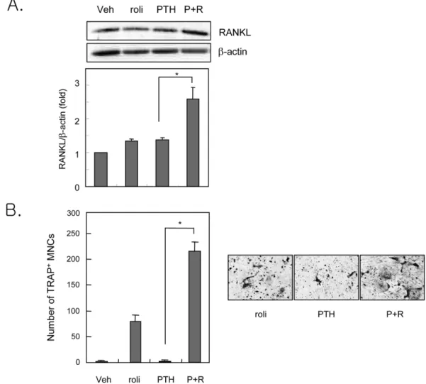
PDE4 저해제에 의한 PTH 반응성 RANKL의 발현 증가 조골세포내 cAMP의 증가는 RANKL의 발현과 연관되어 있으 며, 특히 ERK와 p38에 의한 신호전달경로는 RANKL 발현을 매 개하는 것으로 보고된바 있다.13)따라서 PDE에 의한 조골세포 내 cAMP의 항상성 유지 기전이 RANKL 발현에 미치는 영향을 항 RANKL 항체로 조사하였다. PTH와 PDE4 저해제의 병용투 여는 PTH에 의한 RANKL 단백질 발현을 증강시켰다(Fig. 3A).
조골세포에 의한 RANKL의 발현은 파골세포 형성과 비례적 관 계가 있으므로 공배양계를 사용하여 PDE4에 의한 cAMP의 항 상성 유지가 파골세포 형성에 미치는 영향을 조사하였다. PTH 와 PDE4 저해제의 사용은 단독 처리에 비해 파골세포를 유의하 게 증가시키는 것으로 나타났다(Fig. 3B). 이는 조골세포내 PDE4 에 의한 cAMP의 항상성 유지가 RANKL 발현을 통해 PTH에
의한 파골세포 형성을 조절함을 의미한다. 이상의 결과로 PTH 는 조골세포내 PDE4를 활성화시킴으로써 RANKL에 의한 파골 세포의 과분화를 억제하는 것으로 밝혀졌다. 본 연구를 통해 PTH 에 대한 조골세포내 항상성 유지의 새로운 기전이 제시되었으며, 이를 이용한 골대사 장애 치료제 등의 개발도 가능할 것으로 생 각된다.
결 론
PTH는 조골세포내 PDE의 활성을 증가시켰으며, 이는 대부분 PDE4의 활성증가에 기인하였다. PTH는 4개의 PDE4 isotype 중 PDE4B와 PDE4D mRNA의 발현을 증가시켰으며, 이러한 발 현 증가가 PDE4 활성 증가로 이어진 것으로 추정되었다. PDE4 저해제는 PTH에 의한 하류신호전달 분자인 ERK와 p38의 활성 화를 증가시켰으며, RANKL 단백질 발현의 증강을 통해 파골세 포 분화도 촉진시키는 것으로 나타났다. 이상의 결과는 첫째, PTH에 의한 신호를 PDE4가 음성적으로 조절하며, 둘째, PTH Fig. 2 − The effects of PDE4 inhibitor on PTH-induced signaling pathways in osteoblasts. Calvarial osteoblastic cells were treated with veh or 100 nM PTH in the absence or presence of 10µM rolipram. The expression of p-ERK (A) or p-p38 (B) was determined by western blotting assay. Values are the mean±SD of triplicate cultures in a representative experiment. Veh: vehicle, P: PTH, R: rolipram, *:
p<0.05.
는 조골세포내 PDE4를 활성화시킴으로써 RANKL에 의한 파골 세포의 과분화를 억제함을 시사하는 것이라 할 수 있다.
감사의 말씀
본 연구는 숙명여자대학교 2009학년도 교내연구비 지원에 의 해 수행되었으므로 이에 감사드립니다.
참고문헌
1) Takahashi, N., Akatsu, T., Udagawa, N., Sasaki, T., Yamaguchi, A., Moseley, J. M., Martin, T. J. and Suda, T. : Osteoblastic cells are involved in osteoclast formation. Endocrinology 123, 2600 (1988).
Fig. 3 − The effects of PDE4 inhibitor on PTH-induced osteoclast formation. A, Calvarial osteoblastic cells were treated with veh or 100 nM PTH in the absence or presence of 10µM rolipram. The expression of RANKL was determined by western blotting assay. B, Calvarial osteoblastic cells were cocultured with bone marrow cells in the absence or presence of 100 nM PTH and/or 10µM rolipram for 7 days. Cells were then fixed and stained for TRAP. TRAP-positive (+) multinucleated cells (MNCs) were counted. Values are the mean±SD of triplicate cultures in a representative experiment. Veh: vehicle, P: PTH, R: rolipram, *: p<0.05.
2) Suda, T., Takahashi, N., Udagawa, N., Jimi, E., Gillespie, M. T.
and Martin, T. J. : Modulation of osteoclast differentiation and function by the new members of the tumor necrosis factor receptor and ligand families. Endocr. Rev. 20, 345 (1999).
3) Wong, B. R., Rho, J., Arron, J., Robinson, E., Orlinick, J., Chao, M., Kalachikov, S., Cayani, E., Bartlett, F. S. 3rd, Frankel, W. N., Lee, S. Y. and Choi, Y. : TRANCE is a novel ligand of the tumor necrosis factor receptor family that activates c-Jun N- terminal kinase in T cells. J. Biol. Chem. 272, 25190 (1997).
4) Yasuda, H., Shima, N., Nakagawa, N., Yamaguchi, K., Kinosaki, M., Mochizuki, S., Tomoyasu, A., Yano, K., Goto, M., Murakami, A., Tsuda, E., Morinaga, T., Higashio, K., Udagawa, N., Takahashi, N. and Suda, T. : Osteoclast differentiation factor is a ligand for osteoprotegerin/osteoclastogenesis- inhibitory factor and is identical to TRANCE/RANKL. Proc.
Natl. Acad. Sci. USA 95, 3597 (1998).
5) Lacey, D. L., Timms, E., Tan, H. L., Kelley, M. J., Dunstan, C. R., Burgess, T., Elliott, R., Colombero, A., Elliott, G., Scully, S., Hsu, H., Sullivan, J., Hawkins, N., Davy, E., Capparelli, C., Eli, A., Qian, Y. X., Kaufman, S., Sarosi, I., Shalhoub, V., Senaldi, G., Guo, J., Delaney, J. and Boyle, W. J. : Osteoprotegerin ligand is a cytokine that regulates osteoclast differentiation and activation. Cell 93, 165 (1998).
6) Kondo, H., Guo, J. and Bringhurst, F. R. : Cyclic adenosine monophosphate/protein kinase A mediates parathyroid hormone/parathyroid hormone-related protein receptor regulation of osteoclastogenesis and expression of RANKL and osteoprotegerin mRNAs by marrow stromal cells. J. Bone.
Miner. Res. 17, 1667 (2002).
7) Datta, N. S. and Abou-Samra A. B. : PTH and PTHrP signaling in osteoblasts. Celllular Signalling 21, 1245 (2009).
8) Conti, M. and Beavo, J. : Biochemistry and physiology of cyclic nucleotide phosphodiesterases: Essential components in cyclic nucleotide signaling. Annu. Rev. Biochem. 76, 481 (2007).
9) Essayan, D. M. : Cyclic nucleotide phosphodiesterases. J.
Allergy Clin. Immunol. 108, 671 (2001).
10) Bruss, M. D., Richter, W., Horner, K., Jin, S. L. and Conti, M. : Critical role of PDE4D in beta2-adrenoceptor-dependent cAMP signaling in mouse embryonic fibroblasts. J. Biol. Chem.
283, 22430 (2008).
11) Xin, W., Tran, T. M., Richter, W., Clark, R. B. and Rich, T. C. : Roles of GRK and PDE4 activities in the regulation of beta2 adrenergic signaling. J. Gen. Physiol. 131, 349 (2008).
12) Wakabayashi, S., Tsutsumimoto, T., Kawasaki, S., Kinoshita, T., Horiuchi, H. and Takaoka, K. : Involvement of phospho- diesterase isozymes in osteoblastic differentiation. J. Bone.
Miner. Res. 17, 249 (2002).
13) Takami, M., Cho, E. S., Lee, S. Y., Kamijo, R. and Yim, M. : Phosphodiesterase inhibitors stimulate osteoclast formation via TRANCE/RANKL expression in osteoblasts: possible involvement of ERK and p38 MAPK pathways. FEBS Lett. 31, 832 (2005).