Copyrightⓒ 2008, The Korean Academy of Oral Biology
149
Journal of Oral Biology
Porphyromonas Gingivalis Lipopolysaccharide Increases Monocyte Adhesion to Microvascular Endothelium by Induction of Adhesion Molecules
Su-Ryun Kim1*, Hyun-Joo Park1*, Soo-Kyung Bae2, Ji-Hyun Park1, Hyo-Sun Kim1, Tae Hyeon Koo3 and Moon- Kyoung Bae1*
1 School of Dentistry, 2 School of Medicine, Pusan National University, Pusan 602-739,
3 Korea Food & Drug Administration, Seoul, 122-704, South Korea
(Received August 4, 2008 ; Revised October 7, 2008 ; Accepted October 20, 2008)
Porphyromonas gingivalis, a major periodontal pathogen, has been implicated in the initiation and progression of periodontal disease. Endothelial dysfunction (Editor note:
Aberrant and dysfunction are somewhat redundant. The authors may want to choose one or the other.) contributes to chronic periodontal inflammation. Using cDNA-represen- tational difference analysis, we found that P.gingivalis lipopolysaccharide differentially induces a number of genes in human microvascular endothelial cells. Among these upregulated genes, we focused on intercellular adhesion molecule-1 (VCAM-1), which is crucial for leukocyte recruitment during vascular inflammation. P. gingivalis LPS significantly increased the expression of vascular cell adhesion molecule-1 (VCAM-1) as well as ICAM-1.
Promoter assays revealed that the transcription of these cell adhesion molecules was mainly regulated by nuclear factor- κB (NF-κB) in endothelial cells. Furthermore, P. gingivalis LPS significantly increased leukocyte adhesiveness to microvascular endothelial cells and to aortic endothelium.
Taken together, our results demonstrate that P. gingivalis LPS activates microvascular endothelial cells through NF- κB-dependent expression of cell adhesion molecules.
Keywords : cDNA representational difference analysis;
Cell adhesion molecules; human microvascular endothelial cells; Porphyromonas gingivalis lipopolysaccharide
INTRODUCTION
Porphyromonas gingivalis, black-pigmented, gram- negative anaerobe, has been recognized as a major pathogen in periodontal diseases (Wang and Ohura, 2002). The P.
gingivalis possesses a number of virulence factors such as peptidoglycan, fimbriae, gingipain, and lipopolysaccharide (LPS) which are considered as etiological agents in the initiation and progression of chronic periodontitis. P.
gingivalis LPS has been shown to stimulate the production of proinflammatory mediators within inflamed periodontal tissue (Bainbridge and Darveau, 2001).
A key event in inflammatory processes is the recruitment and transmigration of leukocytes across the vascular endothelium of microvasculature within inflamed tissues (Rao et al., 2007; Ley et al., 2007). This process is predomi- nantly mediated by endothelial cell adhesion molecules (ECAMs), such as vascular cell adhesion molecule-1 (VCAM-1) and intercellular adhesion molecule-1 (ICAM-1), which engage circulating leukocytes and adhere them to vascular endothelial cells lining the vessel wall (Muller, 2003).
In this study, we have used the technique of cDNA representational difference danalysis to detect genes differentially expressed in P. gingivalis LPS-stimulated microvascular endothelial cells. This strategy enables us to identify a number of molecules that may be involved in endothelial dysfunction in P. gingivalis LPS-mediated inflammatory response. Of differentially expressed genes, a transcript encoding ICAM-1 was further characterized. We found that P. gingivalis LPS significantly enhances the expression of ICAM-1 and VCAM-1 via NF-κB activation,
*Corresponding author: Moon-Kyoung Bae, Department of Oral Physiology, School of Dentistry, Pusan National University, Pusan 602-739, South Korea, Tel.: +82-51-240-7974; Fax.: +82-51-245- 8388; E-mail: [email protected]
*Denotes co-first authors
which increases leukocyte adhesion to endothelial cells.
These findings indicated that P. gingivalis LPS exerts pro- inflammatory effects on microvascular endothelial cells by promoting leukocyte-endothelial cell interaction.
MATERIALS AND METHODS
Reagents
Mouse monoclonal anti-ICAM-1 and anti-VCAM-1 antibodies were obtained from Santa Cruz Biotechnology (Santa Cruz, CA, USA). Human α-tubulin antibody was purchased from Biogenex (San Ramon, CA, USA). Calcein- AM was obtained from Molecular Probes (Eugene, OR, USA). P. gingivalis LPS was purchased from Invivogen (Carlsbad, CA, USA).
Cell culture
Human microvascular endothelial cells (HMECs) were obtained from the CDC (Atlanta, GA, USA). These cells were maintained in MCDB (Gibco, Gaithersburg, MD, USA) supplemented with 10 % FBS (Invitrogen), 1 % antibiotics (Hyclone, Logan, UT, USA) 1µg/ml hydro- cortisone (Sigma, St. Louis, MO, USA) and 10 ng/ml hEGF (Upstate Biotechnology, Lake Placid, NY, USA) at 37oC under a humidified 5 % CO2 incubator. U937 monocytic cells were grown in RPMI-1640 (Invitrogen) with 10 % FBS (Invitrogen) and 1 % antibiotics (Hyclone).
cDNA-representational difference analysis (RDA) cDNA-RDA was performed according to GeneFisher PCR subtraction system (TAKARA, Shuzo, Tokyo, Japan) on total RNA isolated from HMECs after 8 h of P. gingivalis LPS treatment. Briefly, cDNAs were prepared by reverse transcription of mRNA, isolated by Micro-Fast Track kit, using an oligo(dT) primer and Superscript II reverse transcriptase (Invitrogen). cDNAs were digested with DpnII and ligated to the adaptors. Amplicons were made by PCR amplification of the adaptor-ligated DpnII cDNA fragments.
Driver DNA was prepared by digesting amplicons with DpnII, and tester DNA was prepared by gel purification of digested amplicons followed by ligation to other adaptors.
The first subtractive hybridization was performed using a tester-driver ratio of 1:100 to obtain difference product 1 (DP1). Subtractive hybridization was repeated using tester- driver ratios of 1:800 for the second round to obtain difference product 2 (DP2). The DP2 products were ligated into pGEM-T vector, and then all constructs were seque- nced by automatic DNA sequencing analysis.
RT-PCR
Total RNA was isolated from HMECs with a TRIzol reagent kit (Invitogen). cDNA synthesis was performed on 3µg of total RNA with a reverse transcription kit (Promega, Madison, WI, USA). The oligonucleotide primers for PCR
were designed as follows: β-actin, 5’-GACTACCTCAT- GAAGATC-3’ and 5’-GATCCACATCTGCTGGAA-3’;
ICAM-1, 5’-CGATGACCATCTACAGCTTTCCG-G-3’ and 5’-GCTGCTACCACAGTGATGATGACAA-3’; VCAM-1, 5’-GATACAACCGTCTTGGTCAGCCC-3’ and 5’-CAGT- TGAAGGATGCGGGAGTA-TATG-3’.
Transient transfection and reporter gene analysis Cells were plated on 24-well plates and transfected with the luciferase construct and pCMV-β-gal using Lipo- fectamine plus reagents (Invitrogen). Cell extracts were prepared 48 h after transfection, and then analyzed with the β-galactosidase enzyme assay for luciferase activity using an assay kit (Promega) and a luminometer (Turner Biosystems, Sunnyvale, CA, USA). Each extract was assayed at least three times and relative luciferase activity was calculated as RLU/β-galactosidase. ICAM-1 and VCAM-1 luciferase reporter constructs with full-length (ICAM-1: -1350 to +45 bp, VCAM-1: -1716 to +119 bp) and truncated forms (ICAM-1: -485 to +45 bp, VCAM-1: - 213 to +119 bp) were used as previously reported (Kim et al., 2008). These luciferase vectors were kindly provided by Dr. Young-Geun Kwon (Yonsei University, Korea) In vitro monocyte adhesion assay
HMECs were plated on 24 well plates at 5 × 10⁴ cells/
well and incubated with or without P. gingivalis LPS for 16 h. U937 cells were then added (5 × 10⁵ cells/ml) to the confluent monolayers of HMECs and incubated for 30 min.
Non-adherent monocytes were removed by washing twice with PBS. Adherent cells were fixed and washed twice with PBS. Binding of monocytes to the endothelial cells were stained with Diff-Quick (Sysmex, Kobe, Japan), and the adherent cells were counted in 3 separate fields in each well by microscopy (Nikon, Tokyo, Japan). The average number of adherent monocytes was calculated in the three fields for each set of four wells.
Ex vivo monocyte adhesion assay
Male Sprague-Dawley rats (SD, 6 weeks of age) were obtained from Samtako (Seoul, Korea). The aortas were opened longitudinally, and incubated with P. gingivalis LPS for 16 h. The aortas were then incubated for 30 min with 1 × 10⁶ fluorescence-labeled monocytes. After incubation, unbound monocytes were rinsed away; the adherent monocytes were counted in 3 consistent fields using fluorescence microscopy (Nikon).
RESULTS
Identification of ICAM-1 gene in P. gingivalis LPS- stimulated microvascular endothelial cells
To identify genes whose expression was altered in response to P. gingivalis LPS in endothelial cells, we
performed cDNA representational difference analysis (RDA) using cDNA isolated from untreated and microvascular endothelial cells treated with P. gingivalis LPS (10µg/ml). This concentration showed no cytotoxic effect on microvascular endothelial cells (data not shown).
After two rounds of subtractive hybridization/PCR, the DP2 products (Fig. 1A) were subcloned into pGEM-T vectors and sequenced. Among clones, some clones were identified as ICAM-1 (Fig. 1B).
P. gingivalis LPS regulates the expression of ICAM-1 and VCAM-1 in microvascular endothelial cells
Cell adhesion molecules, such as ICAM-1 and VCAM-1, on endothelial cells mediate leukocyte adhesion to the endothelium during inflammation (Rao et al., 2007).
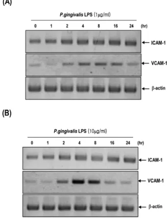
Therefore, we investigated whether P. gingivalis LPS could induce the expression of ICAM-1 and VCAM-1 on human microvascular endothelial cells. Treatment of HMECs with P. gingivalis LPS (10 µg/ml) or P. gingivalis LPS even at low concentration (1µg/ml) significantly increased the mRNA expression of ICAM-1 and VCAM-1 in various time points (Fig. 2A and 2B).
P. gingivalis LPS induces the promoter activity of ICAM-1 and VCAM-1 genes through NF-κB
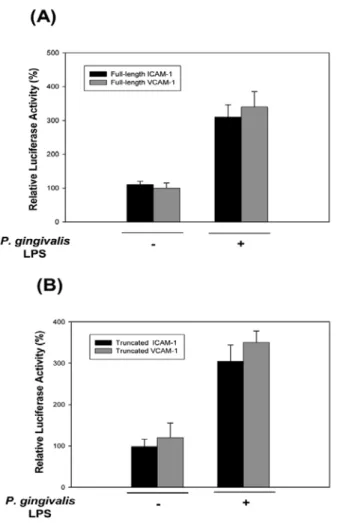
To examine whether the induction of ICAM-1 and VCAM-1 mRNA levels by P. gingivalis LPS is mediated by transcriptional activation of these genes, we carried out promoter analysis with luciferase reporter constructs. The full-length promoter regions of both cell adhesion molecules have transcription factor binding sites, such as NF-κB, TRE, and GATA for the 1.3kb ICAM-1 promoter and NF-κB, TRE and GATA for the 1.8kb VCAM-1 promoter. These luciferase reporter constructs were transfected into HMECs and then incubated with P.
gingivalis LPS, respectively. P. gingivalis LPS significantly increased luciferase reporter activities in full-length constructs-transfected HMECs (Fig. 3A).
Both ICAM-1 and VCAM-1 promoters contain NF-κB binding sites which are located in ~ 200 bp (ICAM-1), and 65 and 75 bp (VCAM-1) upstream of the transcription start site, respectively. These sites are critical for the induction of ICAM-1 and VCAM-1 mRNA transcription (Kim et al., 2008). The treatment of P. gingivalis LPS markedly stimulated promoter activities in these truncated ICAM-1
Fig. 1. Representational difference analysis of gene expression between untreated and P. gingivalis LPS-stimulated HMECs. A, DP2 was run on a 1.5 % agarose gel and stained with ethidium bro- mide. Visible smearing bands were excised from the gel and sub- cloned into the pGEM-T vectors. M, DNA marker; DP2, difference product 2. B, Comparison of nucleotide sequence of identified clone with human ICAM-1 gene.
Fig. 2. P. gingivalis LPS increases the expression of ICAM-1 and VCAM-1 mRNAs in HMECs. HMECs were incubated with or without P. gingivalis LPS (1µg/ml) (A) or (10 µg/ml) (B) for indi- cated times. Total RNAs were isolated and then analyzed by RT- PCR using specific primers for human ICAM-1 and VCAM-1. β- actin served as an internal control.
and VCAM-1 promoters (Fig. 3B). Reporter activities of truncated ICAM-1 and VCAM-1 promoters are similar to those of full-length ICAM-1 and VCAM-1 promoters.
These results indicated that NF-κB binding sites are important for P. gingivalis LPS-induced transcriptional activation of ICAM-1 and VCAM-1 genes.
P. gingivalis LPS induces adhesion of leukocytes to endothelial cells in vitro and ex vivo
Based on uperegulation of ICAM-1 and VCAM-1 by P.
gingivalis LPS, we next investigated whether P. gingivalis LPS regulates the adhesion of leukocytes to endothelium.
As shown in Fig. 4A, incubation of P. gingivalis LPS with HMECs significantly increased the adhesion of U937
monocytic cells to HMECs. To test whether P. gingivalis LPS enhances monocyte binding to the ex vivo aortic endothelium, we performed an ex vivo adhesion assay using fluorescence-labeled monocytes and an aorta isolated from Fig. 3. P. gingivalis LPS induces the transcriptional activities of
ICAM-1 and VCAM-1 genes through NF-κB. A, HMECs were transiently transfected with full-length ICAM-1 and VCAM-1 luciferase vectors that contain ICAM-1 (1.2kb) or VCAM-1 (1.8kb) promoter regions, respectively and then treated with or without P. gingivalis LPS (10µg/ml) for 12 h. B, HMECs were transfected with truncated ICAM-1 and VCAM-1 promoters that contain NF-κB binding site located ~200 bp (ICAM-1) and 65 and 75 bp (VCAM-1) upstream of the transcription start site, respec- tively and incubated with or without P. gingivalis LPS (10µg/ml).
Data is the mean± SE of triplicate experiments relative to the luciferase light units in untreated cells (set at 100 %).
Fig. 4. P. gingivalis LPS increases the adhesion of leukocytes to endothelial cells in vitro and ex vivo. A, HMECs were exposed with or without P. gingivalis LPS (10µg/ml) for 16 h. U937 monocytes were added to HMECs, and numbers of adherent cells were counted. Arrow indicates the adhesion of U937 monocytes to HMECs. B, Rat aorta was isolated, and the incubated with or with- out P. gingivalis LPS (10µg/ml) for 16 h. Fluorescence-stained monocytes were identified and counted by fluorescence micros- copy at 40 × magnification. Data is the mean ± SE relative to adhesion of untreated cells (set at 100 %) in triplicate experiments.
an SD rat. The numbers of fluorescence-labeled adherent monocytes on the surface of the aortic endothelium were markedly increased by treatment of P. gingvialis LPS (Fig.
4B).
DISCUSSION
In this study, we performed cDNA-RDA to identify genes differentially expressed under the treatment of P. gingivalis LPS in HMECs. We identified ICAM-1 gene, one of the endothelial cell adhesion molecules, which is up-regulated in P. gingivalis LPS-stimulated HMECs. We also showed that P. gingivalis LPS induces ICAM-1 and VCAM-1 expression via NF-κB activation in HMECs, leading to adhesion of leukocytes to endothelial cells in vitro and ex vivo.
Periodontal disease is a typical chronic inflammatory disorder which results from infection with periodontal pathogens (Socransky and Haffajee, 1992). The LPS secreted by P. gingivalis, a major periodontal pathogen, may be able to activate various nonimmune cell types such as gingival epithelial cells, periodontal ligament cells, and microvascular endothelial cells in periodontal tissues (Bainbridge and Darveau, 2001). Especially, microvascular endothelial cells lining the microvasculature may play a key role in inflammatory reactions through the recruitment of immune cells into periodontal tissues (Walter et al., 2004;
Mao et al., 2004). In the progress of periodontal diseases, endothelial dysregulation in microvasculature has been associated with the pathogenesis of chronic periodontitis and gingivitis (DeCarlo et al., 2008; Zoellner et al., 2002).
Our work demonstrated the effect of P. gingivalis LPS on microvascular endothelial cells, particularly the ability to regulate leukocyte adhesion. Blockage of endotheial dysregulation may be able to contribute to protecting against periodontogen-mediated vascular inflammation.
Thus, development of anti-vascular inflammatory agents will be beneficial for treatment of periodontal diseases.
P. gingivalis LPS engages with toll-like receptor 4 (TLR4) or TLR2 on cell surface of different cell types, and activates intracellular second messenger such as a number of kinases and transcriptional factors (Wang et al., 2000; Harokopakis and Hajishengallis, 2005). It has been reported that P.
gingivalis LPS-dependent NF-κB activation is mediated by TLR4 in HMECs (Coats et al., 2003). Taken these facts into consideration, P. gingivalis LPS may stimulate leukocyte adhesion to microvascular endothelial cells through TLR4- dependent activation of microvascular endothelial cells. Of course, further investigations are required to elucidate whether TLR4 participates in the P. gingivalis LPS-induced microvascular endothelial dysfunction.
In summary, our results demonstrated the use of cDNA- RDA as an efficient molecular screening method for differentially regulated genes in P. gingivalis LPS-treated
microvascular endothelial cells; We focused that P.
gingivalis LPS exerts pro-inflammatory effects on microvascular endothelial cells by induction of ICAM-1 and VCAM-1 expression via the NF-κB activation. Our findings suggest a pathological role of P. gingivalis LPS in microvascular endothelial dysfunction. We also identified a number of up-regulated genes having known or unknown functions. Further studies on these genes will be useful to provide a global gene expression profile and their novel functions during P. gingivalis LPS-mediated microvascular inflammation.
ACKOWEDGEMENTS
This work was supported for two years by Pusan National University Research Grant (to M-K Bae).
REFERENCES
Bainbridge BW, Darveau RP. Porphyromonas gingivalis lipopolysaccharide: An unusual pattern recognition receptor ligand for the innate host defense system. Acta Odontol Scand. 2001;59:131-8.
Coats SR, Reife RA, Bainbridge BW, Pham TT, Darveau RP.
Porphyromonas gingivalis lipopolysaccharide antagonizes Escherichia coli lipopolysaccharide at toll-like receptor 4 in human endothelial cells. Infect Immun. 2003;71:6799-807.
DeCarlo AA, Cohen JA, Aguado A, Glenn B. Isolation and characterization of human gingival microvascular endothelial cells. J Periodontal Res. 2008;43:246-54.
Harokopakis E, Hajishengallis G. Integrin activation by bacterial fimbriae through a pathway involving CD14, toll- like receptor 2, and phosphatidylinositol-3-kinase. Eur J Immunol. 2005;35:1201-10.
Kim SR, Bae YH, Bae SK, Choi KS, Yoon KH, Koo TH, Jang HO, Yun I, Kim KW, Kwon YG, Yoo MA, Bae MK. Visfatin enhances ICAM-1 and VCAM-1 expression through ROS- dependent NF-kappaB activation in endothelial cells.
Biochim Biophys Acta. 2008;1783:886-95.
Ley K, Laudanna C, Cybulsky MI, Nourshargh S. Getting to the site of inflammation: The leukocyte adhesion cascade updated. Nat Rev Immunol. 2007;7: 678-89.
Mao S, Maeno N, Matayoshi S, Yoshiie K, Fujimura T, Oda H.
The induction of intercellular adhesion molecule-1 on human umbilical vein endothelial cells by a heat-stable component of Porphyromonas gingivalis. Curr Microbiol.
2004;48:108-12.
Muller WA. Leukocyte-endothelial-cell interactions in leukocyte transmigration and the inflammatory response.
Trends Immunol. 2003;24:327-34.
Rao RM, Yang L, Garcia-Cardena G, Luscinskas FW.
Endothelial-dependent mechanisms of leukocyte recruitment to the vascular wall. Circ Res. 2007;101:234-47.
Socransky SS, Haffajee AD. The bacterial etiology of destructive periodontal disease: Current concepts. J
Periodontol. 1992;63:322-31.
Walter C, Zahlten J, Schmeck B, Schaudinn C, Hippenstiel S, Frisch E, Hocke AC, Pischon N, Kuramitsu HK, Bernimoulin JP, Suttorp N, Krüll M. Porphyromonas gingivalis strain-dependent activation of human endothelial cells. Infect Immun. 2004;72:5910-18.
Wang PL, Azuma Y, Shinohara M, Ohura K. Toll-like receptor 4-mediated signal pathway induced by Porphyromonas gingivalis lipopolysaccharide in human gingival fibroblasts.
Biochem Biophys Res Commun. 2000;273:1161-7.
Wang PL, Ohura K. Porphyromonas gingivalis lipopolysaccharide signaling in gingival fibroblasts-CD14 and toll-like receptors. Crit Rev Oral Biol Med. 2002;13:
132-42.
Zoellner H, Chapple CC, Hunter N. Microvasculature in gingivitis and chronic periodontitis: Disruption of vascular networks with protracted inflammation. Microsc. Res. Tech.
2002;56:15-31.