• 검색 결과가 없습니다.

Underlying Hemato-oncologic, Gastrointestinal-nutritional Diseases, or Prematurity May Be Risk Factors for Hypovitaminosi D in Children

N/A
N/A
Protected

Academic year: 2021

Share "Underlying Hemato-oncologic, Gastrointestinal-nutritional Diseases, or Prematurity May Be Risk Factors for Hypovitaminosi D in Children"

Copied!
9
0
0

로드 중.... (전체 텍스트 보기)

전체 글

(1)

© Copyright

Keimyung University School of Medicine 2015

Purpose: An adequate vitamin D level is important for normal growth, cancer prevention, controlling hormones, and immune regulation.

However, no study has investigated vitamin D status in patients with serious illnesses, such as malignancies, malabsorption diseases, or prematurity in Korean children. Thus, we analyzed the results of 25-hydroxyvitamin D

3

(25-D) levels in children in a single tertiary medical center. Methods: Children who had their vitamin D level evaluated at Keimyung University Dongsan Medical Center from January 2004 to December 2014 were included. We reviewed the medical records and laboratory test results. Results: Ninety-three children (male:female = 48:45) who had their vitamin D levels measured were included. The most common reason for measuring vitamin D level was hypocalcemia with or without neurological signs.

Among the subjects, 61 children had hypovitaminosis D (25-D < 30 ng/mL). A positive correlation was detected between 25-D and total calcium levels ( p = 0.001). Negative correlations were found between 25-D and immunoreactive parathyroid hormone (iPTH) levels ( p = 0.002) and between 25-D and alkaline phosphatase (ALP) levels ( p = 0.021). Twenty-three subjects (37.7%) had a critical underlying condition, including a hemato-oncologic issue (18.0%), gastrointestinal or nutritional problem (11.5%), and prematurity or low birth weight (8.2%). Conclusion: The levels of total calcium, ALP, and iPTH were correlated with 25-D level. An underlying malignancy, malabsorption issues, or prematurity may be risk factors for hypovitaminosis D in children.

Key Words: Cancer, Children, Malabsorption, Prematurity, Vitamin D

Received: September 14, 2015 Revised: October 5, 2015 Accepted: October 13, 2015

Corresponding Author: Heung Sik Kim, M.D., Department of Pediatrics,

Keimyung University School of Medicine, 56 Dalseong-ro, Jung-gu, Daegu 41931, Korea Tel: +82-53-250-7516

E-mail: [email protected]

• The authors report no conflict of interest in this work.

Department of Pediatrics, Keimyung University School of Medicine, Daegu, Korea Byung Wook Jo, M.D., Nani Jung, M.D., Ye Jee Shim, M.D., Jin Hyeok Choi, M.D.,

Heung Sik Kim, M.D.

Underlying Hemato-oncologic, Gastrointestinal-nutritional Diseases, or Prematurity May Be Risk Factors for Hypovitaminosis

D in Children

(2)

Low levels of vitamin D are associated with rickets and osteomalacia. More recently, vitamin D has been recently associated with normal growth and puberty, cancer prevention, control of hormones, and immune regulation [1,2]. Several studies have investigated vitamin D deficiency in breastfed infants and concomitant rickets or neurological symptoms [3-7]. Other studies have been conducted about vitamin D status in healthy Korean children and adolescents [8-11]. However, no study has evaluated vitamin D status in children who have a serious illness, including hemato- oncologic diseases, gastrointestinal-nutritional problems, or prematurity. Thus, in this study, we retrospectively reviewed the results of vitamin D status in children in a single tertiary medical center in Korea.

Materials and Methods

Subjects

Children who had their vitamin D level measured at Keimyung University Dongsan Medical Center from January 2004 to December 2014 were included in this study. We reviewed their medical records for laboratory test results, including total c a l c i u m, i o n i z e d c a l c i u m, p h o s p h o r u s, 25-hydroxyvitamin D

3

(25-D), 1,25-hydroxyvitamin D

3

(1,25-D), immunoreactive parathyroid hormone (iPTH), alkaline phosphatase (ALP) and mean platelet volume (MPV). The presence of underlying disease and why the vitamin D test was performed were reviewed. Body mass index (BMI) was also calculated.

Statistical analysis

Statistical analyses were performed using SPSS

standard deviations. Analysis of variance (ANOVA) analysis with the Bonferroni method was used to compare variables between groups. Linear-by- linear association with the chi-square test was used to compare ratios between the groups. Pearson’s correlation analysis was used to define the correlations between patient variables. Receiver- operator characteristic (ROC) curves were used to calculate the area under curve (AUC) and define cutoff values. An absolute correlation coefficient rho value > 0.3 and a p-value < 0.05 were considered significant. BMI was calculated as weight (kg)/height squared (m2).

Ethics statement

This study was approved by the institutional review board of Keimyung University Dongsan Medical Center (Approval No., 2015-06-061-001), which waived the requirement for informed consent.

Results

Subject characteristics and significant variables

A total of 93 children (male:female = 48:45)

who had their vitamin D level determined were

i n c l u d e d. A m o n g t h e m, 61 c h i l d r e n h a d

hypovitaminosis D; 13 patients had a vitamin D

deficiency (25-D < 10 ng/mL) and 48 patients had

a vitamin D insufficiency (10 ng/mL ≤ 25-D < 30

ng/mL). Thirty-two children had an adequate

vitamin D level (25-D ≥ 30 ng/mL). Significant

differences in total calcium ( p = 0.001), ALP ( p =

0.015), and iPTH were observed ( p < 0.001)

between the three groups. No differences were

found for the sex ratio, 1,25-D, ionized calcium,

phosphorus, MPV, or BMI between the three

(3)

Table 1. Subject characteristics and laboratory results for those who received vitamin D testing Hypovitaminosis D Normal p-value Deficiency Insufficiency 25-D < 10 ng/mL 10 ng/mL ≤ 25-D < 30 ng/mL 25-D ≥ 30 ng/mL N 13 48 32 . Age (yr) 1.9 ± 3.4 5.2 ± 6.7 3.1 ± 3.3 0.076 Sex (M : F) 8 : 5 26 : 22 14 : 18 0.278 25-D (ng/mL) 7.2 ± 2.1 18.3 ± 4.9 54.8 ± 21.9 . 1,25-D (pg/mL) 44.4 ± 26.8 70.0 ± 47.4 63.9 ± 42.6 0.224 Total calcium (mg/dL) 8.0 ± 1.8 9.1 ± 1.5 9.8 ± 1.2 0.001* Ionized calcium (mEq/L) 2.0 ± 0.4 2.2 ± 0.4 2.3 ± 0.3 0.331 Phosphorus (mg/dL) 4.7 ± 2.3 5.1 ± 2.0 5.3 ± 0.9 0.626 ALP (IU/L) 1531 ± 1 174 976.3 ± 846.4 718.4 ± 625.4 0.015* MPV (fL) 8.4 ± 2.0 7.7 ± 1.0 7.5 ± 0.8 0.077 iPTH (pg/mL) 134.6 ± 103.0 64.9 ± 87.8 23.4 ± 16.9 < 0.001* BMI (kg/m

2

) 18.0 ± 3.6 18.7 ± 4.7 18.6 ± 4.4 0.900 25 -D: 2 5- hy dr ox yv ita m in D3 , 1 ,2 5- D: 1 ,2 5- h yd ro xy vi ta m in D3 , AL P: a lk al in e ph osp ha ta se , MPV : m ea n pl at el et v ol um e, iPT H: im m un or ea ct iv e parathyroid hormone, BMI: body mass index.

(4)

results, and respective p -values are described in Table 1.

Reasons for measuring vitamin D level

The most common reason that vitamin D level measured was hypocalcemia (n = 32). Eleven patients had symptomatic neurological signs related to hypocalcemia (seven convulsion, three tetany, and one spasms). The second reason was suspicion of rickets (n = 28), followed by bone problems, including a fracture or osteopenia detected on radiography (n = 11). The fourth reason was a hemato-oncologic work-up during chemotherapy or at the end of therapy (n = 10) (three had hypocalcemia or osteopaenia). The detailed reasons why the pediatrician measured vitamin D level are shown in Figure 1.

variables

Negative correlations were detected between 25-D level and iPTH (rho = -0.315 and p = 0.002) and between 25-D level and ALP (rho = -0.240 and p = 0.021). A positive correlation was found between 25-D and total calcium levels (rho = 0.341 and p = 0.001). No associations were found between 25-D level and other variables, including phosphorus (rho = 0.086 and p = 0.415), 1,25-D (rho = -0.086 and p = 0.534), ionized calcium (rho

= 0.185 and p = 0.208), MPV (rho = -0.094 and p = 0.368), or BMI (rho = -0.009 and p = 0.938). These results correspond to the ANOVA analysis shown in Table 1. The details of the correlations and statistical differences between the three groups detected by ANOVA with the Bonferroni method are shown in Figure 2.

The highest AUC was detected for total calcium

Fig. 1. Reasons for measuring vitamin D level in children in a single tertiary medical

center.

(5)

Fig. 2. Correlations between vitamin D level and other significant variables. 25-D: 25-hydroxyvitamin D

3

, ALP: Alkaline phosphatase, iPTH: immunoreactive parathyroid hormone.

p = p =

p =

p =

p =

p =

p = p =

p =

(6)

specificity 72.5% when the cut-off value was 9.2 mg/dL in case of dividing the children into groups

AUC was highest for iPTH (AUC, 0.749) in those grouped by 25-D < 30 ng/mL and 25-D ≥ 30

Table 2. Cut-off values of variables in the patients with hypovitaminosis using a receiver-operator characteristic curve analysis

25-D (ng/mL) Variables AUC Cut-off Sensitivity (%) Specificity (%)

10 Total calcium 0.751 9.2 mg/dL 76.9 72.5

ALP 0.674 1068 IU/L 61.5 70.0

iPTH 0.741 66.2 pg/mL 69.2 82.5

30 Total calcium 0.692 9.8 mg/dL 63.9 65.6

ALP 0.623 717 IU/L 60.7 62.5

iPTH 0.749 23.5 pg/mL 72.1 62.5

25-D: 25-hydroxyvitamin D

3

, AUC: area under curve, ALP: alkaline phosphatase, iPTH: immunoreactive parathyroid hormone.

Table 3. Vitamin D status in children with underlying hemato-oncologic diseases

No. Sex Age (yr) Diagnosis 25-D (ng/mL) Time point of vitamin D test

1 F 2.3 BT 9.2 End therapy

2 M 12.7 WT 9.9 Several years after therapy

3 F 13.0 NHL 10.9 End therapy

4 M 15.4 BT 16.0 Several years after therapy

5 F 21.3 AA 17.7 During follow up

6 M 17.3 CML 18.9 During therapy

7 M 15.4 ALL 19.3 During therapy

8 F 11.8 LCH 20.7 During therapy

9 M 15.8 ALL 20.9 During therapy

10 F 13.0 ALL 25.7 During therapy

11 M 4.6 ALL 29.9 During therapy

12 F 8.5 ALL 44.7 During therapy

13 M 5.0 NBL 77.1 Several years after therapy

14 M 8.0 HLH 84.6 Several years after therapy

25-D: 25-hydroxyvitamin D

3

, BT: brain tumor, WT: Wilms’ tumor, NHL: non-Hodgkin’s lymphoma, AA: aplastic

anemia, CML: chronic myeloid leukemia, ALL: acute lymphoblastic leukemia, LCH: Langerhans cell

histiocytosis, NBL: neuroblastoma, HLH: hemophagocytic lymphohistiocytosis.

(7)

ng/mL, with sensitivity of 72.1% and specificity of 62.5% when the cut-off value was 23.5 pg/mL. The respective AUC, cut-off values, sensitivities, and specificities are shown in Table 2.

U n d e r l y i n g d i s e a s e s i n c h i l d r e n w i t h hypovitaminosis D

Fourteen patients who had a hemato-oncologic disease had their vitamin D levels checked. Among them, two were vitamin D deficient and nine were

insufficient. The 25-D levels of the subjects and the time points of the test are shown in Table 3.

Patients 1 and 7 were also treated with the bisphosphonate derivatives, pamidronate and alendronate for osteopenia. Seven children had gastrointestinal or nutritional problems and were tested for vitamin D. All had hypovitaminosis D.

The vitamin D levels and disease profiles are shown in Table 4. Nine subjects were premature or low birth weight and had their vitamin D levels

Table 4. Vitamin D status in children with underlying gastrointestinal and nutritional problems

No. Sex Age (yr) Diagnosis 25-D (ng/mL)

1 M 0.9 Microvillous inclusion disease 4.6

2 M 1.8 Food allergy 11.5

3 M 0.0 Eosinophilic gastroenterocolitis 11.8

4 F 1.1 History of necrotizing enterocolitis 11.9

5 F 0.0 History of duodenal atresia, after operation 15.1

6 F 1.6 Diet restriction due to atopic dermatitis and food allergy 15.3

7 M 5.4 Non erosive reflux disease, quadriparesis 22.8

25-D: 25-hydroxyvitamin D

3

.

Table 5. Vitamin D status in children who were premature or low birth weight

No. Sex Age (yr) Diagnosis 25-D (ng/mL)

1 M 0.4 Prematurity (IUP 35 + 5 wks, 2,520 gm) 5.3

2 M 0.1 LBW, Twin (IUP 37 + 4 wks, 2,200 gm) 6.9

3 F 0.6 Prematurity, LBW (IUP 35 + 2 wks, 2,000 gm) 12.1

4 M 1.0 Prematurity, LBW (IUP 34 + 5 wks, 2,140 gm) 22.0

5 M 0.3 LBW (IUP 38 wks, 2,360 gm ) 26.0

6 F 0.2 Prematurity, LBW (IUP 30 + 3 wks, 1,380 gm) 30.2

7 F 0.3 Prematurity, LBW, Twin (IUP 34 + 3 wks, 1,960 gm) 35.6

8 M 0.0 Prematurity, VLBW (IUP 31 wks, 1,410 gm) 36.8

9 M 1.0 Prematurity (IUP 36 wks, 2,760 gm) 99.9

25-D: 25-hydroxyvitamin D

3

, IUP: intrauterine period, LBW: low birth weight, VLBW: very low birth weight.

(8)

D. The vitamin D levels, gestational ages, and birth weights are shown in Table 5.

Among the 61 patients with hypovitaminosis, 23 subjects (37.7%) had an underlying serious illness, including hemato-oncologic issues (11cases, 18.0%), gastrointestinal or nutritional problems (7cases, 11.5%), and prematurity or low birth weight (5cases, 8.2%).

Discussion

This is the first vitamin D status survey in children including underlying serious illnesses in Korea. The negative correlation we found between iPTH and vitamin D was also reported previously [9]. We analyzed the AUC of the ROC curves for total calcium, ALP, and iPTH. Our results will help pediatricians determine when to evaluate about vitamin D in a practical clinical setting because hypovitaminosis D has no specific symptoms or signs. For example, if total calcium is < 9.2 mg/dL and ALP is > 1068 IU/L, the pediatrician can check 25-D suspecting deficiency.

Negative correlations have been consistently reported between 25-D level and obesity [9,12-14].

Although mechanisms are not fully understood, sedentary life sytle can affect low expose of sun.

and increased adipocyte uptake of Vitamin D can make low circulating vitamin D. However, we found not difference in BMI between the three g r o u p s (T a b l e 1). P a t i e n t s u n d e r g o i n g chemotherapy for a hemato-oncologic disease under or those with gastrointestinal/nutritional problems often lose weight. Thus, we cannot generalize the negative correlation between vitamin D level and BMI in children with an underlying disease.

Low vitamin D level is strongly associated with

level of proinflammatory cytokines. This may lead to high MPV level. And high MPV increases risk of cardiac disease. However, we found no correlation.

This may have occurred because of the smaller number of subjects and the different vitamin D deficiency cut-offs used in the two studies (25-D level 20 ng/dL in previous study vs. 30 ng/mL in this study).

Some limitations of this study should be discussed. First, not all hemato-oncologic patients in our center from 2004 to 2014 were included in the vitamin D evaluation. We suggest that vitamin D status should be checked at a cancer diagnosis and then regularly until the end of chemotherapy c o n s i d e r i n g t h e r e l a t i o n s h i p b e t w e e n carcinogenesis and vitamin D [1,16,17]. Second, we could not investigate the correlation between iron and vitamin D. Iron status was not routinely checked with vitamin D. Iron status is generally correlated with vitamin D level in breastfed infants [5,6]. However, the opposite result in hemato- oncologic patients may have occurred because of repeated transfusions.

In conclusion, vitamin D status and other correlated variables were somewhat different between children with an underlying disease and healthy individuals. Total calcium was positively correlated with 25-D level. ALP and iPTH were negatively correlated with 25-D level. Underlying hemato-oncologic, gastrointestinal-nutritional diseases, or prematurity can be risk factors for hypovitaminosis D in children.

References

1. Kim SY. The pleiomorphic actions of vitamin D and its

importance for children. Ann Pediatr Endocrinol Metab

2013;18:45-54.

(9)

2. Shin YH, Shin HJ, Lee YJ. Vitamin D status and childhood health. Korean J Pediatr 2013;56:417-23.

3. Song JY, Shin YL, Yoo HW. Clinical characteristics of symptomatic hypocalcemic infants. J Korean Soc Pediatr Endocrinol 2002;7:95-104.

4. Kim JW, Han HS. Clinical characteristics of nutritional rickets in Korean infants. J Korean Soc Pediatr Endocrinol 2010;15:51-7.

5. Yoon JW, Kim SW, Yoo EG, Kim MK. Prevalence and risk factors for vitamin D deficiency in children with iron deficiency anemia. Korean J Pediatr 2012;55:206- 11.

6. Jin HJ, Lee JH, Kim MK. The prevalence of vitamin D deficiency in iron-deficient and normal children under the age of 24 months. Blood Res 2013;48:40-5.

7. Choi YJ, Kim MK, Jeong SJ. Vitamin D deficiency in infants aged 1 to 6 months. Korean J Pediatr 2013;56:205-10.

8. Han SW, Kang HR, Kim HG, Kim JH, Uhm JH, Seo JY. Subclinical vitamin D insufficiency in Korean school-aged children. Pediatr Gastroenterol Hepatol Nutr 2013;16:254-60.

9. Chung IH, Kim HJ, Chung S, Yoo EG. Vitamin D deficiency in Korean children: prevalence, risk factors, and the relationship with parathyroid hormone levels.

Ann Pediatr Endocrinol Metab 2014;19:86-90.

10. Jung SS, Kim MS, Lee DY. Serum vitamin D status in children and adolescence with diabetes according to season and age. Ann Pediatr Endocrinol Metab

2014;19:13-9.

11. Lee DY, Kwon AR, Ahn JM, Kim YJ, Chae HW, Kim DH, et al. Relationship between serum 25-hydroxy- vitamin D concentration and risks of metabolic syndrome in children and adolescents from Korean National Health and Nutrition Examination survey 2008-2010. Ann Pediatr Endocrinol Metab 2015;20:46-52.

12. Bell NH, Epstein S, Greene A, Shary J, Oexmann MJ, Shaw S. Evidence for alteration of the vitamin D-endocrine system in obese subjects. J Clin Invest 1985;76:370-3.

13. Liel Y, Ulmer E, Shary J, Hollis BW, Bell NH. Low circulating vitamin D in obesity. Calcif Tissue Int 1988;43:199-201.

14. Jung CH, Mok JO. Vitamin D and obesity. Korean J Obes 2014;23:236-41.

15. Cumhur Cure M, Cure E, Yuce S, Yazici T, Karakoyun I, Efe H. Mean platelet volume and vitamin D level.

Ann Lab Med 2014;34:98-103.

16. Gandini S, Boniol M, Haukka J, Byrnes G, Cox B, Sneyd MJ, et al. Meta-analysis of observational studies of serum 25-hydroxyvitamin D levels and colorectal, breast and prostate cancer and colorectal adenoma. Int J Cancer 2011;128:1414-24.

17. Wu X, Zhou T, Cao N, Ni J, Wang X. Role of vitamin

D metabolism and activity on carcinogenesis. Oncol

Res 2015;22:129-37.

수치

Table 1. Subject characteristics and laboratory results for those who received vitamin D testing Hypovitaminosis DNormalp-value DeficiencyInsufficiency 25-D &lt; 10 ng/mL10 ng/mL ≤ 25-D &lt; 30 ng/mL25-D ≥ 30 ng/mL N 134832
Fig. 1. Reasons for measuring vitamin D level in children in a single tertiary medical  center.
Fig. 2. Correlations between vitamin D level and other significant variables. 25-D: 25-hydroxyvitamin D 3 ,  ALP: Alkaline phosphatase, iPTH: immunoreactive parathyroid hormone.
Table 3. Vitamin D status in children with underlying hemato-oncologic diseases
+2

참조

관련 문서

Vitamin D status affects serum parathyroid hormone concentrations during winter in female adolescents: associations with forearm bone mineral density.. Budak N, Cicek B,

Conclusion: Although serum vitamin D levels were severely low in terminally ill cancer patients, we found no association between severe vitamin D deficiency

Sahota et al.[4] previously reported that among patients with vitamin D deficiency, those with low serum PTH levels had significantly higher BMD than those with high serum

Odds ratios (95% confidence intervals) for the non-linear relationship between serum 25-hydroxy vitamin D (25(OH)D) concentration and prevalence of (A) anemia and (B) iron

Adult population-based studies have noted that low vitamin D levels were independently related to NAFLD [8-11] and a study of Korean adults showed that vitamin D deficiency

[19] classified serum 25(OH)D levels of ≤20 ng/mL as vitamin D deficiency based on data obtained from 6011 participants of the Korean National Health and Nutrition Examination

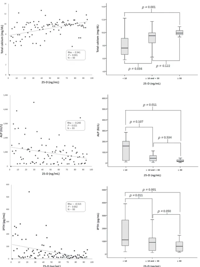
Most of the Ca in the body is stored in the bone, and serum Ca levels are regulated mainly by parathyroid hormone (PTH), vitamin D, and calcitonin via bony uptake of Ca

Serum vitamin D concentrations and associated severity of acute lower respiratory tract infections in Japanese hospitalized children.. Vitamin D deficiency and acute lower respiratory