Genetic Variation of Genes Involved in Dihydrotestosterone Metabolism and the Risk of Prostate Cancer
Sunita R. Setlur
1, Chen X. Chen
2, Ruhella R. Hossain
2, Jung Sook Ha
1, Vanessa E. Van Doren
2, Birgit Stenzel
5, Eberhard Steiner
5, Derek Oldridge
2, Naoki Kitabayashi
2, Samprit Banerjee
3, Jin Yun Chen
1, Georg Schäfer
5, Wolfgang Horninger
5, Charles Lee
1, Mark A. Rubin
2, Helmut Klocker
5, and Francesca Demichelis
2,4Abstract
Purpose: Dihydrotestosterone (DHT) is an important factor in prostate cancer (PCA) genesis and disease progression. Given PCA's strong genetic component, we evaluated the possibility that variation in genes in- volved in DHT metabolism influence PCA risk.
Experimental Design: We investigated copy number variants (CNV) and single nucleotide polymorphisms (SNP). We explored associations between CNV of uridine diphospho-glucuronosyltransferase (UGT) genes from the 2B subclass, given their prostate specificity and/or involvement in steroid metabolism and PCA risk. We also investigated associations between SNPs in genes (HSD3B1, SRD5A1/2, and AKR1C2) involved in the conversion of testosterone to DHT, and in DHT metabolism and PCA risk. The population consisted of 426 men (205 controls and 221 cases) who underwent prostate-specific antigen screening as part of a PCA early detection program in Tyrol, Austria.
Results: No association between CNV in UGT2B17 and UGT2B28 and PCA risk was identified. Men car- rying the AA genotype at SNP rs6428830 (HSD3B1) had an odds ratio (OR) of 2.0 [95% confidence intervals (95% CI), 1.1-4.1] compared with men with GG, and men with AG or GG versus AA in rs1691053 (SRD5A1) had an OR of 1.8 (95% CI, 1.04-3.13). Individuals carrying both risk alleles had an OR of 3.1 (95% CI, 1.4-6.7) when compared with men carrying neither (P = 0.005). Controls with the AA genotype on rs7594951 (SRD5A2) tended toward higher serum DHT levels (P = 0.03).
Conclusions: This is the first study to implicate the 5α-reductase isoform 1 (SRD5A1) and PCA risk, sup- porting the rationale of blocking enzymatic activity of both isoforms of 5α-reductase for PCA chemopreven- tion. Cancer Epidemiol Biomarkers Prev; 19(1); 229 –39. ©2010 AACR.
Introduction
Dihydrotestosterone (DHT), the most potent male hor- mone, has long been considered an important factor in prostate cancer (PCA) disease progression through direct activation of the androgen receptor (1). Testosterone (T) is converted to DHT by 5α-reductase and is then glucuro- nidatized by members of the uridine diphospho-glucur- onosyltransferase (UGT) family for excretion. The
REduction by DUtasteride of prostate Cancer Events (REDUCE) trial (2) recently reported that dutasteride, an inhibitor of 5α-reductase types I and II, reduced PCA risk in men at higher levels of risk of the disease (American Urological Association in Chicago, IL, April 27, 2009). This trial consisted of a total of 1,516 patients with PCA, with 659 in the dutasteride arm and 857 in the placebo arm. The REDUCE trial showed that dutasteride significantly lowered the risk of all biopsy-detectable PCA by 23% (P < 0.0001) over 4 years. These findings are consistent with the results of the Prostate Cancer Pre- vention Trial, which also showed a significant reduction in the 7-year period prevalence of biopsy-detectable PCA with finasteride, a type II 5α-reductase–selective inhibitor (3). Thus, two large clinical studies show that decreasing production of DHT using 5α-reductase inhibitors can effec- tively decrease the incidence of clinically localized PCA.
Interindividual levels of DHT, and possibly the re- sponse to inhibitors of DHT, may be influenced by germ line polymorphisms. The extent of polymorphisms has been catalogued by the HapMap consortium, which cate- gorized single nucleotide polymorphisms (SNP) in sev- eral widely disparate populations (4). A recent discovery
& Prevention
Authors' Affiliations:
1Department of Pathology, Brigham and Women's Hospital and Harvard Medical School, Boston, Massachusetts;
2
Department of Pathology and Laboratory Medicine,
3Department of Public Health, and
4Institute for Computational Biomedicine, Weill Cornell Medical College, New York, New York; and
5Department of Urology, Innsbruck Medical University, Innsbruck, Austria
Note: Supplementary data for this article are available at Cancer Epidemiology Biomarkers and Prevention Online (http://cebp.aacrjournals.
org/).
Corresponding Author: Francesca Demichelis, Institute for Computa- tional Biomedicine, Weill Cornell Medical College, 1305 York Avenue, Y 1307 (or Box 140), New York, NY 10065. Phone: 646-962-5616; Fax: 215- 440-9354. E-mail: [email protected]
doi: 10.1158/1055-9965.EPI-09-1018
©2010 American Association for Cancer Research.
shows that common polymorphisms exist not only as SNPs (i.e., single base pair alterations) but also as larger genomic regions of DNA gain and loss called copy number variation (CNV; refs. 5, 6). Mounting evidence suggests that these two types of polymorphisms predis- pose individuals to risk of disease (7). PCA is a common disease that may be highly influenced by genetic variation.
PCA has the strongest hereditary component of com- mon cancers, as illustrated by a study of monozygotic twins, which suggested that 42% of the incident PCA risk is genetically linked (8). More recent studies using geno- mewide analyses have identified risk loci comprising SNPs on chromosome 8q24 and 17p to be associated with PCA (9-14). How such polymorphisms affect the growth of PCA or the regulation of androgens, however, is inad- equately addressed in the abovementioned genomewide studies. An alternative approach to genomewide studies has been to focus on the role of polymorphisms involving genes regulating the steroid hormone pathway as a risk factor for developing PCA. Two recent examples studied SNPs related to 5α-reductase type II (SRD5A2; ref. 15) and 3β-hydroxysteroid dehydrogenase 1 (HSD3B1;
ref. 16). The role of the second type of germ line polymor- phism, the CNVs, has been examined by four studies that analyzed the copy number state of UGT2B17 (UDP glu- curonosyltransferase 2 family, polypeptide B17) for risk of PCA (17-20). This gene maps to chromosome 4 and plays a central role in the catabolism of T and DHT.
The studies reported conflicting results regarding the as- sociation of genomic copy number alterations of this gene with PCA risk. Park et al. (20) and Karypidis et al.
(18) showed that the deletion polymorphism of this gene
results in greater risk of PCA in Caucasian patients. Stud- ies by Gallagher et al. (17) and Olsson et al. (19) did not find any association between the deletion polymorphism and risk for PCA. This candidate CNV involving a UGT locus is intriguing due to their prostate-specific role in DHT catabolism and the important role of that gene in prostate gland maintenance and growth. However, the extent to which CNV in genes involved in the metabo- lism of T or DHT may predispose a man to a higher risk of having PCA is still unresolved.
Using a short sequence oligonucleotide array platform (Genome-Wide Affymetrix 6.0 SNP), we were able to test genetic variation in seven genes involved in the metabo- lism and catabolism of T and DHT. In particular, we ex- plored for associations between UGT genes that mediate glucuronidation and clearance of several compounds in- cluding steroid hormones, bile acids, bilirubin, xenobio- tics, and drugs (21). We focused on UGT2B17, UGT2B15, UGT2B7, and UGT2B28, UGTs from the UGT2B protein subclass, because of their prostate specificity and/or in- volvement in steroid metabolism. Specifically, UGT2B17 mediates the glucuronidation and subsequent clearance of DHT in the basal cells whereas UGT2B15 mediates the clearance of T and DHT in the luminal epithelial cells.
UGT2B17, but not UGT2B15, is downregulated by DHT.
The mechanism of action of the UGTs in the prostate is shown in Fig. 1. Stromal cells also play a role in steroid metabolism in hormone-sensitive tissues. We also inves- tigated the association between SNPs in genes directly re- lated to the conversion of T in DHT, and in DHT catabolism and risk of PCA. Specifically, we investigated HSD3B1 (1p12), the two steroid-5α-reductase genes, SRD5A1 (5p15.31) and SRD5A2 (2p23.1), which catalyze the conversion of T into the more potent androgen DHT, and the aldo-keto-reductase family 1 member C2, AKR1C2 (10p15.1).
One significant limitation to recent genetic studies in the field of PCA has been the use of cases from surgical cohorts and controls from a disparate control population.
In most of these studies, controls are most frequently de- fined as having no reported PCA but may have no re- corded prostate-specific antigen (PSA) level, and in none of the studies have the controls undergone system- atic prostate needle biopsy evaluation to confirm their disease-free status. To overcome these limitations, we identified cases and controls from the same population of men screened for elevated serum PSA as part of a re- gional PCA early detection trial (22-24).
All men on trial underwent intense PSA screening.
Abnormal results (age-adjusted PSA levels as low as 1.25 ng/mL) led to prostate needle biopsy evaluation.
Controls with negative prostate needle biopsies contin- ued to be followed with regular PSA evaluations. The current study therefore is the first to use a PSA-screened clinical trial population to explore the risk of PCA based on genetic variation of genes involved in DHT metabo- lism and their effect on DHT and T serum levels, both in the form of CNVs and SNPs.
Translational Relevance
Two recent clinical trials showed a reduction of
prostate cancer diagnosis after treatment with dihy-
drotestosterone (DHT) inhibitors implicating the im-
portant role DHT metabolism plays in prostate
cancer development. Our study evaluates the poten-
tial role of genetic germ line variation in genes along
the DHT pathway on the risk of prostate cancer. Al-
though we did not observe associations between copy
number polymorphisms for UGT2B17 or UGT2B28
and risk of prostate cancer, we did identify significant
associations between single nucleotide polymorph-
isms located in HSD3B1 and SRD5A1 with increased
risk of prostate cancer. Individuals carrying both risk
alleles were three times more likely to have prostate
cancer compared with men carrying neither. This is
the first study to implicate the 5α-reductase isoform
1 (SRD5A1) in prostate cancer risk, supporting the ra-
tionale of blocking the enzymatic activity of both iso-
forms of 5α-reductase for the chemoprevention of
prostate cancer.
Materials and Methods
Cohort Description
The blood DNA samples were obtained from the Tyrol early PCA detection program, Innsbruck, Austria (Table 1). This cohort was comprised of men between 41 and 75 y of age who had undergone PSA screening since 1993. A serum PSA of >1.25 ng/mL was used as the lowest cutoff for cancer detection by biopsy. The PSA cutoff was age-adjusted (24-29). Controls were de- fined as men with normal PSA levels for 3 y following an increase in PSA and a negative biopsy. The mean follow-up time without cancer diagnosis of the control in- dividuals was 92 ± 59 mo. In this study, we used DNA
from prospectively collected peripheral blood lympho- cyte cells from men who underwent biopsy for elevated age-adjusted PSA levels (n = 426; 205 control subjects and 221 case patients). Here, we considered PSA levels at the time of initial enrollment in the study.
Serum Androgen Hormone Measurements
Serum samples stored at −80°C were thawed at 4°C and vortexed. Serum levels of androgen hormones T and DHT were determined using commercial diagnostic assays. T was measured by RIA (testosterone Coute-A- Count RIA, 27466 TKTT1; Siemens Diagnostics) and DHT by ELISA (Diachrome 5α DHT ELISA, DB52021;
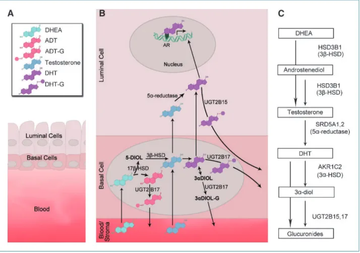
Figure 1.
Summary of DHT metabolism focusing on the genes evaluated in this study. A. The schematic representation of the prostate gland depicts the three major compartments involved in DHT metabolism. The luminal epithelial cells are present in both the normal benign state and prostate cancer, and basal cells (secondary layer present) are present in benign glands and partially present in the precursor cancer lesion, prostatic intraepithelial neoplasia. The stroma/blood interface (referred to as blood) allows for the inflow and outflow of DHT and other metabolites from and to the rest of the body, respectively.
Stromal cells are important in steroid metabolism in hormone-sensitive tissues. B. The function of the UGT enzymes in the prostate compartments is
depicted here, demonstrating how the UGT2B17 and UGT2B15 enzymes work in concert to facilitate the clearance of androgens and their by-products
in the prostate. The androgens produced by testis and adrenals are conjugated in the basal cells by UGT2B17. The substrates for this enzyme include
DHEA, testosterone, ADT, DHT, and 3 α-diol. UGT2B17 has high affinity for testosterone and DHT. UGT2B15 functions in the luminal cells to facilitate
the clearance of DHT and testosterone. DHT is depicted as a potent activator of androgen receptor in the luminal cells responsible for maintenance
and growth in the normal state. C. The conversion of testosterone to DHT and subsequent breakdown is depicted in a schematic pathway. Right, the
genes evaluated in this study, which encode enzymes that play a key role in this pathway (DHT, dihydrotestosterone; ADT, androsterone; 3α-diol,
3 α-androstanediol; 17β-diol, androstenediol; G, glucuronide; UGT, uridine diphospho-glucuronosyltransferases; DHEA, dehydroepiandrosterone; HSD,
hydroxysteroid dehydrogenase).
Diachrome), respectively, according to the recommended procedures.
Sample DNA Preparation
Isolation of genomic DNA from blood samples was car- ried out in a high-throughput fashion using the QIAamp 96 DNA Blood kit (Qiagen). Ficoll-purified peripheral blood mononuclear cells were manually resuspended in 400 μL of chilled phosphate buffer saline, allowed to equilibrate to room temperature, and split into two aliquots. The aliquots were added to 20 μL of Qiagen protease in the company-provided collection microtubes to facilitate nuclear and cellular lysis. The resulting lysates were processed according to the guidelines of the manu- facturer and finally resuspended in 100 μL of nuclease-free distilled water. DNA quality and quantity were evaluated by electrophoresis and NanoDrop spectrophotometry (NanoDrop; Thermo Scientific), respectively.
CNV and SNP Genotype Evaluation
Data were generated using Affymetrix Genome-Wide Human SNP Array 6.0. Briefly, genomic DNA was pro- cessed for the Affymetrix 6.0 whole genome platform
(Affymetrix, Inc.) according to the protocols of the manufacturer. The DNA was digested with the enzymes NspI and StyI (New England Biolabs) and ligated to the respective adapters (Affymetrix) using T4 DNA ligase (New England Biolabs). The ligated DNA was amplified (Clontech), purified using magnetic beads (Agencourt), labeled, fragmented, and hybridized to the arrays. Fol- lowing hybridization, the arrays were washed and stained with streptavidin-phycoerythrin (Invitrogen Corporation) and scanned. Data processing was done using Affymetrix Power Tools. SNP genotype calls were generated using the Birdseed (v2) algorithm (30). All samples included in this study passed strict data quality control measures.
In addition, taking advantage of array-based genotype information, we applied an inter-sample similarity test to check for relatedness called the SNP panel identifica- tion assay (31) and tested for population stratification (32). Copy number data was evaluated by measuring the median of the log 2 values of the ratios between the normalized hybridization intensity of each target sample and a reference model built using a subset of the all-male control subjects and unrelated Caucasian HapMap female samples. For highly polymorphic loci (i.e., UGT2B17 and UGT2B28), the copy number offset caused by the presence of the CNV signal in the refer- ence model was computationally corrected.
SNP Selection
For each gene directly related to the conversion of T to DHT and in DHT catabolism, we investigated the haplo- block structure considering 10 kb flanks using Haplo- view (33) with default parameters. We selected one or more available SNPs for each block based on minor allele frequency as evaluated in the control group (intervals:
0.03-0.2, 0.21-0.3, and 0.31-0.5). Only SNPs with at most 3% of data missing were considered. This procedure led to the selection of 15 SNPs, specifically two for HSD3B1 (rs6428830 and rs10754400), seven for SRD5A1 (rs566202, rs4702379, rs248803, rs3797177, rs8192249, rs30434, and rs1691053), three for SRD5A2 (rs4952197, rs7594951, and rs806645), and three for AKR1C2 (rs11252866, rs11252867, and rs11816204).
Quantitative PCR for UGT2B17
The relative standard curve quantitative PCR (qPCR) method was used to investigate UGT2B17 copy numbers.
DNA from HapMap NA07022 was used for the standard curve with all assays being set up in triplicates. Thermal cycling and quantification were carried out using 7900HT Sequence Detection System from ABI with an annealing temperature of 62°C. Rho guanine nucleotide exchange factor 11 (ARHGEF11; forward, 5′-TCTCTGCTCCC- TCACTCTCAA-3′; reverse, 5′-TGTGCCTCTTCCATC- GTTCT-3′) was used as the endogenous control gene because it was copy number invariant across the samples. The UGT2B17 primer pair (forward, 5′-CAGT‐
TGCCTGTAGCCACATTT-3′; reverse, 5′-TGGTGGAT- GAAGAAGACTTTTG-3′) was designed to specifically Table 1. Study cohort demographics
Controls
( n = 205) Cases
( n = 221) P
Age (y)
Mean 58.9 61.7 0.0001
Median 59 62.0
Range 41-76 43-77
PSA (ng/mL)
≤4 126 87
4.1-10.0 54 99
10.1-20.0 22 23
>20.0 3 12 <0.0001
Free PSA (%)
Mean 17.5 16.6 0.12
Median 16. 15.1
Range 5.1-58.6 5.2-59.6
T (ng/mL)
Mean 4.4 4.5 NS
Median 4.3 4.5
Range 1.5-9.0 1.8-10.5
DHT (ng/mL)
Mean 0.77 0.71 NS
Median 0.58 0.54
Range 0.16-5.8 0.2-4.1
DHT/T
Mean 0.19 0.17 NS
Median 0.14 0.14
Range 0.02-1.26 0.04-1.1
Abbreviation: NS, not significant.
amplify UGT2B17. This primer set was checked for specificity using the BLAST, BLAT, and the Eland-based algorithms outlined below.
Statistical Analysis
χ
2test and Wilcoxon test were applied for categorical and continuous variables, respectively, to assess the sta- tistical significance of differences observed in the control and case groups. Two-sided P ≤ 0.05 values were con- sidered statistically significant. For each polymorphic location considered in the study, we applied binary lo- gistic regression to evaluate the risk for PCA. Each re- gression was also considered with age as covariate (age adjustment). We report odds ratios (OR) and 95%
confidence intervals (CI). Logistic regression P values are unadjusted. With respect to the potential increased risk for PCA in the presence of UGT2B17 deletion previ- ously reported by other authors, we estimated that we were powered (β = 80%; α = 0.05) to detect an OR of 1.9 or above and 1.6 or above for homozygous deletion and any deletion, respectively (proportions equal to 0.13 and 0.40 for deletions were considered, respectively).
Analysis was done using R 2.9.0 (ref. 34; accessed April 23, 2008).
Primer Analysis of Published Studies
The specificity of the primers we designed for this study and those designed by other groups (17-20) was evaluated by implementing an Eland-like
6alignment al- gorithm based on the short-read alignment indexing strategy for finding perfect and 1- to 2-bp mismatch hits of short sequences. Briefly, every n-mer sequence was broken into four nonoverlapping tiles. If a pair of tiles was found to have a perfect match with a paired 6-mer tile index of the genome, an ungapped extension was done to determine if the subalignment generated a hit of interest. By design, the algorithm detects perfect match,
single base pair mismatch, and two–base pair mismatch hits with perfect sensitivity and specificity. The hits that generated amplicons of ≤2 kb were reported.
Results
Association of Age, PSA, Free PSA, DHT and T Levels with Cases and Controls
The cases and controls included in the study were se- lected to be age-matched. The cases were, on average, 2 years older than the controls (P = 0.001; Table 1). Initial PSA levels ranged between 0.4 and 29.2 ng/mL and be- tween 1.25 and 469 ng/mL in controls and cases, respec- tively (P < 0.0001). No association was observed for serum-free PSA, DHT, T, and DHT over T ratios with respect to end point (case/control). We evaluated the behavior of PSA, free PSA, and DHT/T with respect to age in the control group and observed a moderate increase of PSA and free PSA with age (P < 0.001 for linear trends) and no trend for DHT/T (P = 0.1). We also observed that individuals with high DHT levels tend to have low PSA and vice versa. We evaluated in- dividual ethnicity by population stratification analysis (32), which revealed that all individuals reflect Cauca- sian ancestry.
Incidence of Germ Line CNVs and SNP Genotypes in a Study Cohort
A total of 426 cases and controls were used to evaluate the germ line copy number frequencies for the genes of interest. Table 2 shows the breakdown of the copy num- ber analysis results of the two genes exhibiting CNVs, UGT2B17 and UGT2B28. Due to poor coverage, we were not able to assess UGT2B15 copy number state in our co- hort. The observed frequencies of the homozygous and hemizygous deletion in UGT2B17 (9.8% and 44.9%, re- spectively) and UGT2B28 (3.4% and 22.0%, respectively) in controls are consistent with previous reports (35).
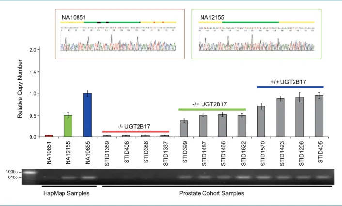
qPCR confirmed the copy number of UGT2B17 deter- mined by the Affymetrix SNP array platform (Fig. 2).
The PCR amplicons were sequenced to confirm the Table 2. Copy number polymorphism at UGT2B17 and UGT2B28 in controls and cases and PCA risk association results
Gene CN Controls,
n (%) Cases,
n (%) Crude OR (95% CI) P Age-adjusted
OR (95% CI)
P
UGT2B17 +/+ 93 (45.4) 11 (50.2) 1 Reference Reference
+/ − 92 (44.9) 87 (39.4) 0.79 (0.5-1.2) 0.26 0.77 (0.51-1.16) 0.21
−/− 20 (9.8) 23 (10.4) 0.96 (0.5-1.9) 0.91 0.88 (0.45-1.73) 0.71
UGT2B28 +/+ 153 (74.6) 164 (74.2) Reference Reference
+/ − 45 (22.0) 53 (24) 1.10 (0.70-1.73) 0.68 1.14 (0.72-1.81) 0.58
−/− 7 (3.4) 4 (1.8) 0.53 (0.15-1.86) 0.32 0.50 (0.14-1.79) 0.29
Abbreviation: CN, copy number ( −/−, homozygous deletion; +/−, hemizygous deletion; +/+, wild-type).
6
A.J. Cox, unpublished data.
specificity of the products amplified. The samples that had either one or two copies of the gene showed the spe- cific product (Fig. 2, right, inset). A nonspecific product was observed in the samples that were homozygous for deletion. Sequencing identified this as UGT2B15, which shares 95% nucleotide homology with UGT2B17 (Fig. 2, left, inset). We did not detect any copy number alterations in the UGT2B7 gene, or in SRD5A1, SRD5A2, HSD3B1, and AKR1C2. HSD3B1 was reported by two independent studies as copy number polymorphic in <1% of Cauca- sian individuals (36, 37).
We identified 15 SNPs on HSD3B1 (2), SRD5A1 (7), SRD5A2 (3), and AKR1C2 (3) based on linkage disequilib- rium, minor allele frequency, and platform design con- straints. Supplementary Table S1 summarizes the characteristics of the selected SNPs in terms of location, minor allele frequency in Caucasian HapMap indivi- duals, study controls and cases, and Hardy-Weinberg equilibrium. It also reports P values for allele and geno- type frequency differences based on Fisher exact test.
Supplementary Fig. S1 depicts allele scatter plots for each SNP and shows good cluster separation for low genotype frequencies.
Clinical Parameters and Genetic Polymorphism To assess the influence of genetic variants on PSA, DHT, and T serum levels, we considered individuals from the study control group. We observed higher levels of DHT and DHT/T ratio in the presence of minor allele homozygous genotype for one intronic SNP on SRD5A2, rs7594951 (nonparametric test, P = 0.028 and 0.046 for DHT and DHT over T, respectively). The minor allele fre- quency for rs7594951 in controls was 0.14.
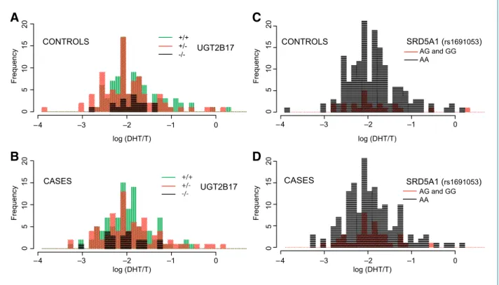
Association of Polymorphisms and PCA Risk The individuals with germ line deletion polymorph- isms in UGT2B17 and UGT2B28 were examined for con- ferring risk to PCA. We did not find any association between the copy number states of either gene and risk of PCA (Table 2), nor any association between copy num- ber and serum levels of DHT. Figure 3A and B shows the copy number counts in cases and controls in the light of DHT over T levels for cases and controls. No association between copy number state and risk for PCA was de- tected even when considering gene-gene interaction (data not shown) nor in combination with SNPs.
Figure 2.
Confirmation of the copy number state of UGT2B17 by qPCR. Three HapMap samples and 12 samples from the Tyrol PSA Screening Cohort were selected based on known copy number states (HapMap, far left) and array-based copy number data. Array-based copy number calls are color-coded:
UGT2B17 −/− (red), UGT2B17+/− (green), and UGT2B17+/+ (blue). The qPCR results agree with the array-based copy number calls. The PCR amplicons were sequenced to confirm the specificity of the amplified products (top, insets). The samples that had either one or two copies of the gene showed the specific product (top right, inset). A nonspecific product was observed in the samples that were homozygous for deletion (−/−; top left, inset).
Sequencing identified this as UGT2B15. Primer sequences (yellow bars), amplicons (green bars), nucleotides specific to UGT2B15 (black bars), and
nucleotides in which the mismatches between UGT2B17 and UGT2B15 may have occurred, leading to the UGT2B15 product (orange bars).
When assessing the association between SNPs and risk for PCA (Table 3), we observed that men with a minor allele homozygous genotype in rs6428830 (HSD3B1) had an age-adjusted OR of 2.0 (95% CI, 1.1-4.1) as com- pared with men with GG. The rs6428830 SNP is 2.5 kb apart from the locus previously reported by Park et al.
(ref. 16; rs1047303) as associated with increased risk of PCA in Caucasian men with a family history of PCA. In- dividuals with AG or GG versus AA in rs1691053 (SRD5A1) have an OR of 1.8 (95% CI, 1.04-3.13). Figure 3C and D shows genotype counts in cases and controls in the light of DHT over T levels for cases and controls. Interest- ingly, rs1691053 risk allele shows an OR of 2.2 (95% CI, 1.2-4.0) for early onset risk. We then evaluated the effect of carrying both risk alleles at rs6428830 and at rs1691053, and estimated an OR of 3.1 (95% CI, 1.4-6.7) when compared with carrying neither of these risk alleles (P = 0.005).
Discussion
Our study examined the association of polymorphisms in UGT2B genes (UGT2B7, UGT2B17, and UGT2B28) and in genes involved in the metabolism of T and DHT with the risk of PCA. We used a well-defined cohort for the
study in which the cases and controls were intensively screened for PSA and the controls had a negative prostate biopsy and a mean follow-up without cancer diagnosis for 7.6 years. We were unable to detect any association between the deletion polymorphism of UGT2B genes examined and the risk of PCA. We also investigated if polymorphisms in these genes would result in differen- tial levels of serum hormones.
The association between UGT2B17 deletion and PCA risk was previously investigated leading to conflicting re- sults (17-20). One primary difference between our study and the previous studies is the use of whole genome ar- ray platform for the assessment of copy number states and independent verification by qPCR on a subset of samples (three HapMap samples sequenced at the UGT2B17 locus were included as a gold standard;
ref. 38), whereas the other studies used qPCR as the only platform for genotyping. Because UGT2B17 shares 95%
sequence homology with the UGT2B15 gene, designing specific primers for PCR is necessary for accuracy. Of in- terest, the genomic region encompassing UGT2B17 and UGT2B15 (4q13.2) is a complex genomic region whose reference sequence was recently reinterpreted (38).
Hence, we tested the specificity of our primers and the primers used in other studies by applying an alignment
Figure 3.