Analysis of the Expression Patterns of Thymosin β4, Vascular Endothelial Growth Factor, and Hypoxia-Inducible Factor-1α in Various Tumors Using Tissue Microarray
Bo-Young Lee
1, Seung-Hyun Lee
2, Byung-Kwon Ahn
2, Mee Sun Ock
1and Hee-Jae Cha
1,3*
1
Department of Parasitology and Gnenetics,
2Department of Surgery,
3Institute for Medical Science, Kosin University College of Medicine, Busan 602-703, Korea
Received December 23, 2010 /Accepted December 30, 2010
Thymosin β4 (TB-4) has been reported to play a key role in tumor growth, metastasis and angiogenesis.
In addition, TB-4 induced the expression of vascular endothelial growth factor (VEGF) and stabilized the hypoxia-inducible factor (HIF)-1α in melanoma cells. Although the importance of thymosin β4 in angiogenesis and metastasis has been proven, there are few studies that show the expression patterns of TB-4, VEGF and HIF-1α. This study was conducted to analyze the relationship among these proteins in various tumors. Using tissue microarray analysis, we investigated the expression patterns of TB-4, VEGF and HIF-1α in various tumors to identify the expression patterns and relationships of these pro- teins in certain tumors. TB-4 was highly expressed in osteosarcoma, colon adenocarcinoma, esophageal squamous cell carcinoma, kidney and urinary bladder transitional carcinoma, lung cancer, and liver cancer. HIF-1α was highly expressed in nasal cavity inverted papilloma, lung cancer, and esophageal squamous cell carcinoma. The expression patterns of TB-4 and HIF-1α were almost similar and co-localized. VEGF expression was high in the blood vessels in tumors, but usually not high in the tumors themselves. VEGF was moderately expressed in stomach cancer, liver angiosarcoma, gall bladder adenocarcinoma, and uterus endometrial adenocarcinoma. The expression patterns of VEGF shows sim- ilarities in certain tumors including stomach cancer, osteosarcoma, liposarcoma, lung cancer, liver cancer, gall bladder adenocarcinoma, esophageal squamous cell carcinoma, stomach cancer, colorectal carcinoma and renal cell carcinoma. These results suggest that the expression patterns of TB-4, HIF-1α and VEGF were co-localized and related to tumorigenesis and angiogenesis of certain tumors.
Key words : Thymosin β4, VEGF, HIF-1α, tissue microarray, various tumors
*Corresponding author
*Tel:+82-51-990-6428, Fax:+82-51-990-3081
*E-mail : [email protected]
서 론
사이모신 베타 4(thymosin β4)는 43 아미노산으로 구성된 4.9 kDa의 작은 크기의 단백질이다. 사이모신 베타는 베타 1부 터 베타 15의 15개의 아형들이 보고되어 있으며 이 외에도 알파와 감마그룹의 여러 아형들이 존재하며 각 아형 별로 기 능이 다른 것으로 보고되고 있다 [12]. 사이모신 베타 4는 actin 과 결합하며 주로 F-actin을 G-actin으로 분리하는 actin depo- lymerization을 증가시키는 펩티드로서 actin cytoskeleton의 구조를 변경하는데 관여하여 직접적으로 세포의 이동을 조절 한다 [18]. 처음 사이모신 베타 4가 발견될 때는 갑상선에서 분 비되는 펩티드로써 면역반응이나 염증반응에도 관여하는 cy- tokine이나 성장인자일 것으로 알려졌으나 최근 여러 가지 연 구를 통해 혈관신생 (angiogenesis)을 증가시키고[3,13] 상처 치유 (wound healing) [14,15]를 촉진하며 모발 성장(hair growth) 등을 증진시킨다고 보고되었다[16,17]. 그 외에도 최
근 사이모신 베타 4는 integrin-linked kinase와 결합하여 sur- vival kinase Akt의 인산화(phosphorylation)를 증가시켜 심근 세포의 이동과 생존을 증가시킨다는 보고가 있어 여러 세포의 이동 및 생리작용에 사이모신 베타 4가 중요한 역할을 담당하 고 있음이 밝혀졌다 [1].
사이모신 베타 4는 암의 성장 및 전이현상에도 중요한 기능 을 하는 것이 보고 되고 있다 . 사이모신 베타4는 쥐의 악성 섬유육종세포의 이동과 전이를 촉진시키며 [9] B16 쥐 흑색종 세포의 성장 , 전이 및 혈관형성을 촉진시킨다고 보고 되었다 [2]. 사이모신 베타4를 과 발현시킨 경우 SW480 대장암 세포의 침윤 및 전이가 촉진되었으며 [19] 사이모신 베타4가 in- tegrin-linked kinase를 통해 대장암에서 상피세포의 간질세포 로의 변이 (epithelial-mesenchymal transition)를 촉진시키는 것으로 보고되었다 [4]. 유방암에서도 사이모신 베타 4의 발현 이 보고되었으며 [10,11] 소세포성 폐암 등에서도 사이모신 베 타 4가 높이 발현되는 것이 보고되었다[5].
최근 본 연구그룹에서는 사이모신 베타 4가 혈관내피증식인
자 (Vascular endothelial growth factor, VEGF)의 발현을 증가
시키는 것을 보고하였으며 이러한 VEGF의 발현증가가 혈관
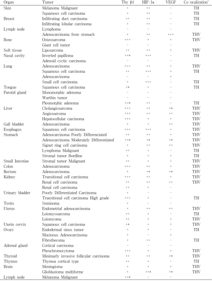
Table 1. Expression of thymosin β4 (Thy β4), HIF-1α and VEGF in various human tumors
Organ Tumor Thy β4 HIF-1α VEGF Co-ocalization
1Skin Melanoma Malignant + ++ - TH
Squamous cell carcinoma + ++ - TH
Breast Infiltrating duct carcinoma ++ ++ - TH
Infiltrating lobular carcinoma + ++ - TH
Lymph node Lymphoma - - -
Adenocarcinoma from stomach + ++ +++ THV
Bone Osteosarcoma +++ + + THV
Giant cell tumor - - -
Soft tissue Liposarcoma ++ ++ + THV
Nasal cavity Inverted papilloma ++* +++ - TH
Adenoid cyctic carcinoma - - -
Lung Adenocarcinoma +++ ++ + THV
Squamous cell carcinoma ++ +++ + TH
Adenocarcinoma - - +
Small cell carcinoma + +++ - TH
Tongue Squamous cell carcinoma +* + - TH
Parotid gland Monomorphic adenoma - - -
Warthin tumor - - -
Pleomorphic adenoma ++* ++ - TH
Liver Cholangiosarcoma +++ ++ +* THV
Angiosarcoma +++ ++ ++ THV
Hepatocellular carcinoma +++ + + THV
Gall bladder Adenocarcinoma + + ++ THV
Esophagus Squamous cell carcinoma +++ +++ + THV
Stomach Adenocarcinoma Poorly Differenciated ++ ++ + THV
Adenocarcinoma Moderately Differentiated ++* +* +* THV
Signet ring cell carcinoma + ++ ++ THV
Lymphoma Malignant ++ + - TH
Stromal tumor Bordline + + + TH
Small Intestine Stromal tumor Malignant ++ + + THV
Colon Adenocarcinoma +++ ++ + THV
Rectum Adenocarcinoma + +* +* THV
Kidney Transitional cell carcinoma +++ ++ + THV
Renal cell carcinoma ++ ++ ++ THV
Renal cell carcinoma ++ + -
Urinary bladder Poorly Differentiated Carcinoma + - -
Transitional cell carcinoma High grade +++ + - TH
Testis Seminoma + - -
Uterus Endometrial adenocarcinoma + ++ ++ THV
Leiomyosarcoma ++ + - TH
Leiomyoma ++ + + THV
Uterin cervix Squamous cell carcinoma +* + + THV
Ovary Endodermal sinus tumor + + - TH
Mucinous Adenocarcinoma + - +
Fibrothecoma + ++ - TH
Adrenal gland Cortical carcinoma - - -
Pheochromocytoma +++ + + THV
Thyroid Minimarly invasive follicular carcinoma ++ ++ +* THV
Thymus Thymoa cortical type ++ + - TH
Brain Meningioma + + + THV
Glioblastoma multiforme + ++* +* THV
Lymph node Melanoma Malignant ++* - -
+: weakly positive, ++: moderate, +++: marked, *: Focal positive
1: Co-localazation of Thy β4 and HIF-1α (TH), Thyβ4, VEGF and HIF-1α (TVH)
Fig. 2. Immunohistochemical detection of HIF-1α in various human tumors. Original magnification ×200. Tissue microarray slides were immunostained with mouse monoclonal antibody to HIF-1α (1:100) and stained with DAB.
Fig. 3. Immunohistochemical detection of VEGF in various human tumors. Original magnification ×200. Tissue microarray slides were immunostained with rabbit polyclonal antibody to VEGF (1:2,000) and stained with permanent red.
신생을 촉진시키며 암세포의 성장과 전이를 촉진시키는 것을 규명하였다 [2]. 이후 사이모신 베타 4에 의한 VEGF의 발현증 가가 VEGF 발현에 직접적인 역할을 하는 hypoxia-inducible factor (HIF)-1α의 단백질 안정성을 사이모신 베타 4가 올려 줌으로 인해 상대적으로 증가한 HIF-1α가 VEGF 발현을 촉진 시키는 것을 규명하였다 [6].
사이모신 베타 4는 혈관형성을 촉진하며 암의 성장 및 전이 에 중요한 역할을 담당하는 단백질이다 . 이와 더불어 사이모 신 베타 4의 기능과 관련된 VEGF 및 HIF-1α는 혈관신생에 중요한 역할을 하는 단백질이며 암의 전이 및 성장과 밀접한 관련성을 지닌다 . 따라서 본 연구에서는 tissue micro-array를 사용하여 여러 가지 암 조직에서 사이모신 베타 4, VEGF 및
HIF-1α의 발현 양상을 조사하고 이들 각각의 단백질의 발현 부위 및 발현양상이 서로 연관되었는지를 조사하였다 .
재료 및 방법
Tissue microarray
Tissue microarray는 Super Bio Chip (Super Biochips Laboratories, Seoul, Korea)사로부터 구입하여 사용하였다.
나이와 성별을 제외한 조직을 제공한 환자의 정보는 가지고 있지 않다 .
면역조직화학검사(Immunohistochemistry)
면역조직화학검사를 위해 tissue microarray 슬라이드를 탈
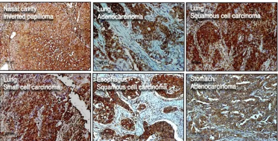
Fig. 1. Immunohistochemical detection of thymosin β4 in various human tumors. Original magnification ×200. Tissue microarray slides were immunostained with rabbit polyclonal antibody to thymosin β4 (1:2,000) and stained with DAB.
파라핀화와 수화를 거친 다음 citrate buffer (0.01 M, pH 6.0) 상에서 전자레인지로 최고출력으로 (700 W 이상) 5분간 두 번 antigen retrieval을 실시하였다. PBS하에서 슬라이드를 충분히 식힌 후 3% hydrogen peroxide 용액에서 5분간 반응하여 endog- enous peroxidase를 제거하였다. 이후 PBS로 5분간 3번 씻어 준 다음 PBS에 희석된 rabbit polyclonal thymosin β4 (1:2,000 dilu- tion; ALPCO Diagnostics, Windham, NH, USA) 혹은 rabbit polyclonal VEGF (1:2,000 dilution, Abcam Inc., Cambridge, MA, USA), mouse monoclonal HIF-1α (1:100 dilution; Novus Biologicals, Littleton, CO, USA) 항체로 4℃에서 overnight 배양 하였다 . 다음날 PBS로 5분씩 3번 씻어 준 후 2차 항체를 처리하여 1시간 동안 반응한다. PBS로 5분 동안 3번 씻어 준 후 Dako’s EnVision Kit (DAKO, Carpinteria, CA, USA)를 사용하여 thy- mosin β4와 HIF-1α의 경우 DAB 발색반응을 VEGF의 경우 per- manent red의 발색반응을 유도하였다. 슬라이드는 이후 대조 염 색을 Meyer's hematoxylin (DAKO, Carpinteria, CA, USA)로 10 초간 염색 후 탈수반응을 거친 다음 Permount (Fishier Scientific, Pittsburgh, PA, USA)로 고정하였다.
정량적 분석(Semiquantitative assessment)
단백질 발현양상의 정도를 측정하기 위해 면역화학염색의 정도를 4가지로 나누어 판단하였다. 발현이 되지 않고 염색이 되지 않은 경우를 negative (-), 약하게 발현되는 경우를 weak
(+), 중간 정도의 발현양상을 보이는 경우를 moderate (++), 강한 발현양상을 보이는 경우를 strong (+++)으로 분류하였 다 . 발현 양상이 국소적인 경우는 focal positive (*)로 표시하 였다 .
결 과
Tissue microarray 슬라이드 상의 50종류, 59가지의 인간 암 조직을 대상으로 사이모신 베타 4, VEGF 및 HIF-1α에 대한 면역조직화학검사를 실시하였으며 자세한 조직 별 발현양상 및 co-localization 여부는 Table 1에 요약되어 있다.
사이모신 베타 4는 골육중(osteosarcoma), 대장 선암(colon adenocarcinoma), 식도 편평세포암(esophageal squamous cell carcinoma), 신장 및 방광의 이행세포암(kidney and uri- nary bladder transitional cell carcinoma), 각 종류의 폐암 (lung cancer) 및 간암(liver cancer)에서 많이 발현되는 것을 관찰 할 수 있었다 (Fig. 1).
HIF-1α은 비강 역위성 유두종(nasal cavity inverted papil- loma), 각종 폐암 및 식도 편평세포암(esophageal squamous cell carcinoma)에서 강한 발현을 보였으며 대체로 발현되는 양상이나 위치가 사이모신 베타 4와 일치하는 것으로 관찰되 었다 (Fig. 2).
VEGF는 암 조직에서보다 암 조직에 분포된 혈관내피에
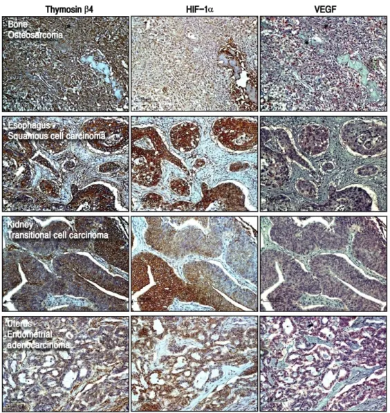
Fig. 4. Immunohistochemical detection of co-localized thymosin β4, HIF-1α and VEGF in various human tumors. Original magnifica- tion ×200. Tissue microarray slides were immunostained with rabbit polyclonal antibody to thymosin β4 (1:2,000), mouse monoclonal antibody to HIF-1α (1:100) and rabbit polyclonal antibody to VEGF (1:2,000). Then slides were stained with DAB (for thymosin β4 and HIF-1α) and permanent red (for VEGF).
서 강하게 발현되는 양상을 나타내었으며 암세포에서는 사 이모신 베타 4나 HIF-1α에 비해 강하게 발현되지 않았다.
위암 (stomach cancer), 간 혈관육종(liver angiosarcoma), 담 낭 선암 (gall bladder adenocarcinoma)과 자궁 내막 선암 (uterus endometrial adenocarcinoma)에서 적당(moderate) 수준의 VEGF 발현이 관찰되었다(Fig. 3). VEGF의 발현 양 상 및 위치는 위암 (stomach cancer), 골육종(osteosarcoma), 지방종 (liposarcoma), 폐암(lung cancer), 간암(liver cancer), 담낭 선암 (gall bladder adenocarcinoma), 식도 편평세포암 (esophageal squamous cell carcinoma), 대장 및 직장암, 신세포암 (renal carcinoma)을 포함하는 특정 암에서 사이 모신 베타 4 및 HIF-1α 과 일치하는 것으로 관찰되었다 (Fig. 4).
고 찰
사이모신 베타 4는 섬유육종, 흑색종, 대장암, 유방암 및 폐암의 성장과 전이를 촉진시킨다는 보고를 통해 암의 성장 과 전이에 관여하는 것이 알려 졌다 [2,4,5,9,10,11,19]. 하지만 아직까지 사이모신 베타 4의 발현양상을 실제 암 조직에서 연구한 결과는 미비한 실정이며 대부분의 연구들이 세포주 를 대상으로 이루어지고 있다 . 본 연구에서는 사이모신 베타 4의 발현양상을 여러 암 조직을 대상으로 분석하였으며 관련 단백질인 VEGF 및 HIF-1α를 동일한 조직에서 함께 관찰하 여 사이모신 베타 4의 발현양상 및 관련 단백질과의 연계여 부를 조사하였다 .
사이모신 베타 4의 여러 가지 기능 중 혈관신생작용은 암의
증식 및 전이와 밀접한 관계를 가지고 있다 . 실제 흑생종의 성장 및 전이 촉진이 사이모신 베타 4의 혈관신생 촉진 작용에 의해 일어나고 이러한 혈관신생의 촉진이 사이모신 베타 4가 혈관신생에 중요한 역할을 하는 VEGF의 발현을 촉진시킴으 로 가능하다고 보고되어 사이모신 베타 4는 혈관신생과 암의 성장 및 전이에 중요한 역할을 하는 단백질인 것이 보고되었 다 [2]. 최근 본 연구진은 사이모신베타 4가 VEGF의 발현 유도 에 중요한 역할을 전사조절인자인 HIF-1α를 통해 VEGF의 발 현양상을 증가시킨다는 것을 보고하였다 [6]. HIF-1α 단백질은 일반적인 상태에서는 잘 분해되어 안정성이 떨어지지만 저 산소의 상태에서는 단백질의 안정화가 증가하여 VEGF의 전 사조절부위에 결합하여 VEGF의 발현을 증가시키는 것으로 알려져 있다 [7,8]. 하지만 사이모신 베타 4는 저 산소 현상이 아닌 일반적인 유산소 상태에서 HIF-1α 단백질의 안정화를 높여 VEGF의 발현을 유도하고[6] 이러한 현상은 기존의 암 조직 내의 저 산소현상이 혈관신생을 유도한다는 기존의 학설 과 외에 저 산소 현상이 일어나지 않아도 암조직 내에서 혈관 신생이 일어날 수 있음에 대한 새로운 이론을 제시하였다 .
따라서 본 연구에서는 암의 성장 및 전이 현상 , 혈관신생에 관여하는 사이모신 베타 4의 발현 양상을 이와 관련된 단백질 인 HIF-1α 및 VEGF의 발현 양상과 함께 관찰하여 상호 비교 함으로 인해 특정 암에서의 발현양상을 조사하였으며 각 단백 질의 발현 양상과 위치를 조사하여 이러한 세 단백질의 상호 관련성 및 상호작용을 조사하였다 . 연구 결과에 의하면 거의 대부분의 조직에서 사이모신 베타 4와 HIF-1α의 발현 양상은 동일하고 발현 위치가 일치하는 것으로 밝혀 졌으며 VEGF의 경우도 특정 암에서 사이모신 베타 4 및 HIF-1α와 동일한 발현 양상을 보이는 것으로 관찰되었다 . 이러한 양상을 통해 사이 모신 베타 4, VEGF 및 HIF-1α은 함께 상호작용하며 이러한 단백질을 통한 혈관신생 및 암의 성장 활동이 특정 암에서 중요한 역할을 담당함을 알 수 있었다 . 또한 본 연구결과를 통한 자료가 암의 성장 및 전이 그리고 암 조직내의 혈관 신생 에서의 사이모신 베타 4, VEGF 및 HIF-1α의 역할을 연구하는 좋은 자료가 될 수 있을 것이다 .
감사의 글
이 논문은 2010년도 정부(교육과학기술부)의 재원으로 한 국연구재단의 지원을 받아 수행된 기초연구사업임 (No. 2010- 0017017).
References