Soo-Hyun Lim1, Kwang Seok Ahn2, Sung-Hoon Kim2& Hyeung-Jin Jang1
1Department of Biochemistry, College of Oriental Medicine, Kyung Hee University, Seoul, Korea
2Department of Oriental Pathology, College of Oriental Medicine, Kyung Hee University, Seoul, Korea
Correspondence and requests for materials should be addressed to H. J. Jang ([email protected])
Accepted 10 November 2009
Abstract
A major source of paeoniflorin (PF) which was from the Paeonia lactiflora root, has been used as a herbal medicine in East Asia for its antiallergic, antiinflamma-tory, and immunoregulatory effects. However, only few details are known about the mechanism of apop-tosis induced by this compound. The present study was undertaken to further elucidate the molecular mechanism of apoptosis and the changes of gene expression elicited by PF using DNA microarrays and computational gene-expression analysis tools in hu-man leukemia U937 cells. A comparative global tran-scription analysis between treatment with PF and anisomycin (AM) that induces apoptosis in U937 cells revealed that c-Jun-NH2-kinase (JNK) pathway related genes were less expressed in PF-treated cells. Eluci-dation of the mechanisms by which PF conducts its anti-cancer activities through comparative analysis of the gene expression is necessary to provide a solid foundation for its use as a promising agent in preven-tion and treatment strategies.
Keywords:Paeoniflorin, Anisomysin, Comparative genom-ics, U937, Apoptosis
The root of Paeonia lactiflora pall (also called
Paeo-niae alba), a major source of paeoniflorin (PF) has
been used as a herbal medicine in East Asia for its
anti-allergic, anti-inflammatory, and
immuno-regulato-ry effects. PF has also been shown to exert anticancer
and anti-proliferative activities in cancer cells
1(e.g
human leukemia Jurkat cells, human gastric carcinoma
cells). In this study, this apoptosis was mediated
thro-ugh the reduction of mitochondrial membrane
poten-tial, activation of caspase, and fragmentation of DNA.
Additionally, PF induced the phosphorylation of three
mitogen-activated protein (MAP) family kinases,
extra-cellular signal-regulated kinase (ERK), c-Jun
amino-terminal kinase (JNK), and p38 MAP kinase.
But recent studies have shown the contradictory
results that cell viability was not affected after PF
treat-ment in human leukemia U937 cells
2. Apoptosis is
thought to be an important response to most of the
chemotherapeutic agents in leukemia cells and other
kinds of cells. Although apoptosis was not detected in
this experiment, it was observed that PF can change
the apoptotic pathway related gene expression. The
recent experiment indicated that the apoptotic
mitocho-ndrial caspase pathway is significant for cell death
induced by anisomycin (AM) in human lymphoma
U937 cells
3. AM, which is purified from Streptomyces
griseolus, was first published as an antibiotic for
pro-tozoa by Sobin and Tanner
4. AM is also known as a
potent apoptosis inducer through the activation of JNK.
It has been reported that JNK activation stimulates
apoptosis
5-7.
In this AM experiment, like the PA experiment, gene
expression was also analyzed using a GeneChip
®sys-tem with Human Genome U133A Array which was
spotted with 22,283 probe sets. And AM also changed
the apoptotic pathway related gene expression.
Tran-scriptome analyses of human cells treated with these
therapeutic agents have only recently emerged, and no
comparative analysis between different therapeutic
agents has been made. So elucidation of the
mechani-sms by which PF conducts its anti-cancer activities
through comparative analysis of the gene expression
with AM is necessary to provide a solid foundation for
its use as a potential agent in prevention and treatment
strategies.
Transcriptome Changes in Response to AM
and PA
Gene-expression profiling of U937 cells exposed to
1
μM AM for 0 to 6 h was performed by GeneChip
®oligonucleotide expression arrays. Of the 22,283 probe
Gene Expression Profiling Reveals that Paeoniflorin
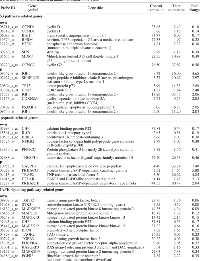
Table 1. List of significantly up- or down regulated genes based on comparison between experimental (paeoniflorin (PF)-treated for 3 h) and control (non-treated) in human leukemia U937 cells.
Probe ID Gene Gene title Control Treat Fold
symbol expression expression change
p53 pathway-related genes down
208711_s_at CCND1 cyclin D1 25.65 2.49 0.10
208712_at CCND1 cyclin D1 8.66 1.18 0.14
206083_at BAI1 brain-specific angiogenesis inhibitor 1 34.77 6.03 0.17
219370_at RPRM reprimo, TP53 dependent G2 arrest mediator candidate 22.33 4.97 0.22
222176_at PTEN phosphatase and tensin homolog 7.81 2.32 0.30
(mutated in multiple advanced cancers 1)
209260_at SFN stratifin 2.89 1.12 0.39
205655_at MDM4 Mdm4, transformed 3T3 cell double minute 4, 22.35 10.99 0.49
p53 binding protein (mouse)
202770_s_at CCNG2 cyclin G2 76.50 37.87 0.50
up
209542_x_at IGF1 insulin-like growth factor 1 (somatomedin C) 3.44 16.00 4.65
202627_s_at SERPINE1 serpin peptidase inhibitor, clade E (nexin, plasminogen 3.57 10.62 2.97 activator inhibitor type 1), member 1
220804_s_at TP73 tumor protein p73 3.99 11.53 2.89
203904_x_at CD82 CD82 molecule 31.27 77.64 2.48
211577_s_at IGF1 insulin-like growth factor 1 (somatomedin C) 17.26 35.47 2.06
211156_at CDKN2A cyclin-dependent kinase inhibitor 2A 4.74 9.73 2.05
(melanoma, p16, inhibits CDK4)
220402_at P53AIP1 p53-regulated apoptosis-inducing protein 1 3.06 6.27 2.05
209540_at IGF1 insulin-like growth factor 1 (somatomedin C) 5.50 11.20 2.04
Apoptosis-related genes down
207993_s_at CHP calcium binding protein P22 37.81 6.53 0.17
215561_s_at IL1R1 interleukin 1 receptor, type I 2.65 0.51 0.19
210538_s_at BIRC3 baculoviral IAP repeat-containing 3 6.66 2.03 0.30
211524_at NFKB2 nuclear factor of kappa light polypeptide gene enhancer 2.79 1.07 0.38
in B-cells 2 (p49/p100)
213950_s_at PPP3CC Protein phosphatase 3 (formerly 2B), catalytic subunit, 3.82 1.56 0.41
gamma isoform
202688_at TNFSF10 tumor necrosis factor (ligand) superfamily, member 10 57.40 26.58 0.46
up
210955_at CASP10 caspase 10, apoptosis-related cysteine peptidase 4.49 33.24 7.40
207228_at PRKACG protein kinase, cAMP-dependent, catalytic, gamma 2.52 14.64 5.80
204413_at TRAF2 TNF receptor-associated factor 2 5.50 26.62 4.84
214618_at CFLAR CASP8 and FADD-like apoptosis regulator 1.14 2.43 2.13
212559_at PRKAR1B protein kinase, cAMP-dependent, regulatory, type I, beta 44.33 90.49 2.04
MAPK signaling pathway-related genes down
209908_s_at TGFB2 transforming growth factor, beta 2 22.75 1.36 0.06
211078_s_at STK3 serine/threonine kinase 3 (STE20 homolog, yeast) 7.55 0.59 0.08
216139_s_at MAPK8IP3 mitogen-activated protein kinase 8 interacting protein 3 49.35 4.34 0.09
216435_at MAP2K5 Mitogen-activated protein kinase kinase 5 10.78 1.32 0.12
206249_at MAP3K13 mitogen-activated protein kinase kinase kinase 13 14.42 2.15 0.15
207993_s_at CHP calcium binding protein P22 37.81 6.53 0.17
205447_s_at MAP3K12 mitogen-activated protein kinase kinase kinase 12 37.22 6.64 0.18
206382_s_at BDNF brain-derived neurotrophic factor 7.62 1.65 0.22
204878_s_at TAOK2 TAO kinase 2 26.35 6.07 0.23
209747_at TGFB3 transforming growth factor, beta 3 20.22 6.02 0.30
215305_at PDGFRA platelet-derived growth factor receptor, alpha polypeptide 9.60 3.05 0.32
205801_s_at RASGRP3 RAS guanyl releasing protein 3 (calcium and DAG-regulated) 3.54 1.16 0.33 213177_at MAPK8IP3 mitogen-activated protein kinase 8 interacting protein 3 22.03 7.38 0.34
204380_s_at FGFR3 fibroblast growth factor receptor 3 7.87 2.72 0.35
206178_at PLA2G5 phospholipase A2, group V 3.59 1.24 0.35 205699_at MAP2K6 mitogen-activated protein kinase kinase 6 11.57 4.07 0.35 214786_at MAP3K1 mitogen-activated protein kinase kinase kinase 1 34.64 12.20 0.35 214571_at FGF3 fibroblast growth factor 3 (murine mammary tumor virus 4.53 1.62 0.36
integration site (v-int-2) oncogene homolog)
210059_s_at MAPK13 mitogen-activated protein kinase 13 6.78 2.57 0.38 211524_at NFKB2 nuclear factor of kappa light polypeptide gene enhancer 2.79 1.07 0.38
in B-cells 2 (p49/p100)
206103_at RAC3 ras-related C3 botulinum toxin substrate 3 5.76 2.22 0.39 (rho family, small GTP binding protein Rac3)
209189_at FOS v-fos FBJ murine osteosarcoma viral oncogene homolog 185.89 72.01 0.39
201465_s_at JUN jun oncogene 53.59 21.36 0.40
211371_at MAP2K5 mitogen-activated protein kinase kinase 5 15.30 6.14 0.40 209951_s_at MAP2K7 mitogen-activated protein kinase kinase 7 17.83 7.18 0.40 208893_s_at DUSP6 dual specificity phosphatase 6 72.41 29.29 0.40 213950_s_at PPP3CC Protein phosphatase 3 (formerly 2B), catalytic subunit, 3.82 1.56 0.41
gamma isoform
210477_x_at MAPK8 mitogen-activated protein kinase 8 19.80 8.46 0.43 222164_at FGFR1 fibroblast growth factor receptor 1 33.50 14.43 0.43
(fms-related tyrosine kinase 2, Pfeiffer syndrome)
205463_s_at PDGFA platelet-derived growth factor alpha polypeptide 18.54 8.15 0.44 201041_s_at DUSP1 dual specificity phosphatase 1 94.48 43.20 0.46 203930_s_at MAPT microtubule-associated protein tau 13.81 6.38 0.46 215498_s_at MAP2K3 mitogen-activated protein kinase kinase 3 94.64 44.52 0.47 207822_at FGFR1 fibroblast growth factor receptor 1 22.92 10.80 0.47
(fms-related tyrosine kinase 2, Pfeiffer syndrome)
219714_s_at CACNA2D3 calcium channel, voltage-dependent, alpha 2/delta 3 subunit 47.61 22.77 0.48 212912_at RPS6KA2 ribosomal protein S6 kinase, 90 kDa, polypeptide 2 70.07 33.73 0.48 214367_at RASGRP2 RAS guanyl releasing protein 2 (calcium and DAG-regulated) 8.33 4.09 0.49 215992_s_at RAPGEF2 Rap guanine nucleotide exchange factor (GEF) 2 2.98 1.47 0.49 215050_x_at MAPKAPK2 mitogen-activated protein kinase-activated protein kinase 2 52.30 26.09 0.50 up
214368_at RASGRP2 RAS guanyl releasing protein 2 (calcium and DAG-regulated) 2.86 58.46 20.43 211485_s_at FGF18 fibroblast growth factor 18 1.77 29.62 16.69 205590_at RASGRP1 RAS guanyl releasing protein 1 (calcium and DAG-regulated) 0.97 13.88 14.28 203131_at PDGFRA platelet-derived growth factor receptor, alpha polypeptide 1.74 12.53 7.20 217515_s_at CACNA1S calcium channel, voltage-dependent, L type, alpha 1S subunit 4.26 28.83 6.76 203649_s_at PLA2G2A phospholipase A2, group IIA (platelets, synovial fluid) 3.66 23.29 6.37 221310_at FGF14 fibroblast growth factor 14 2.28 14.12 6.20 207228_at PRKACG protein kinase, cAMP-dependent, catalytic, gamma 2.52 14.64 5.80 205558_at TRAF6 TNF receptor-associated factor 6 9.45 53.48 5.66 204421_s_at FGF2 fibroblast growth factor 2 (basic) 2.97 16.17 5.44 204413_at TRAF2 TNF receptor-associated factor 2 5.50 26.62 4.84
215195_at PRKCA protein kinase C, alpha 1.06 5.08 4.78
201743_at CD14 CD14 molecule 1.49 6.98 4.68
204200_s_at PDGFB platelet-derived growth factor beta polypeptide 3.36 14.14 4.21 (simian sarcoma viral (v-sis) oncogene homolog)
208432_s_at CACNA1E calcium channel, voltage-dependent, R type, alpha 1E subunit 2.11 8.65 4.09 210226_at NR4A1 nuclear receptor subfamily 4, group A, member 1 9.21 36.44 3.96 211143_x_at NR4A1 nuclear receptor subfamily 4, group A, member 1 3.18 12.45 3.92 220780_at PLA2G3 phospholipase A2, group III 2.88 10.49 3.64 207222_at PLA2G10 phospholipase A2, group X 4.78 17.34 3.63 206311_s_at PLA2G1B phospholipase A2, group IB (pancreas) 0.95 3.33 3.52 210675_s_at PTPRR protein tyrosine phosphatase, receptor type, R 1.61 5.59 3.47 211499_s_at MAPK11 mitogen-activated protein kinase 11 12.70 42.89 3.38 207050_at CACNA2D1 calcium channel, voltage-dependent, alpha 2/delta subunit 1 2.55 8.22 3.22 62987_r_at CACNG4 calcium channel, voltage-dependent, gamma subunit 4 28.76 90.44 3.14 211533_at PDGFRA platelet-derived growth factor receptor, alpha polypeptide 1.16 3.43 2.97 211372_s_at IL1R2 interleukin 1 receptor, type II 1.02 3.02 2.95 214284_s_at FGF18 Fibroblast growth factor 18 3.66 9.89 2.70 Table 1. Continued.
Probe ID Gene Gene title Control Treat Fold
sets analyzed and identified for exposure periods of 0,
1, 2, 3 and 6 h, respectively
3. To identify genes
res-ponsive to PF treatment in U937 cells, they carried
out global-scale DNA microarray analysis of cells
cul-tured at 0, 1, and 3 h after PF treatment (160 mg/mL,
30 min)
2. These results indicated that the number of
expressed probe sets was approximately constant
among the different samples. Complete lists of probe
sets from all samples are available on the Gene
Expression Omnibus (http://www.ncbi.nlm.nih.gov/
geo/query/acc.cgi?acc=GSE8229 and http://www.ncbi.
nlm.nih.gov/geo/query/ acc.cgi?acc=GSE8228).
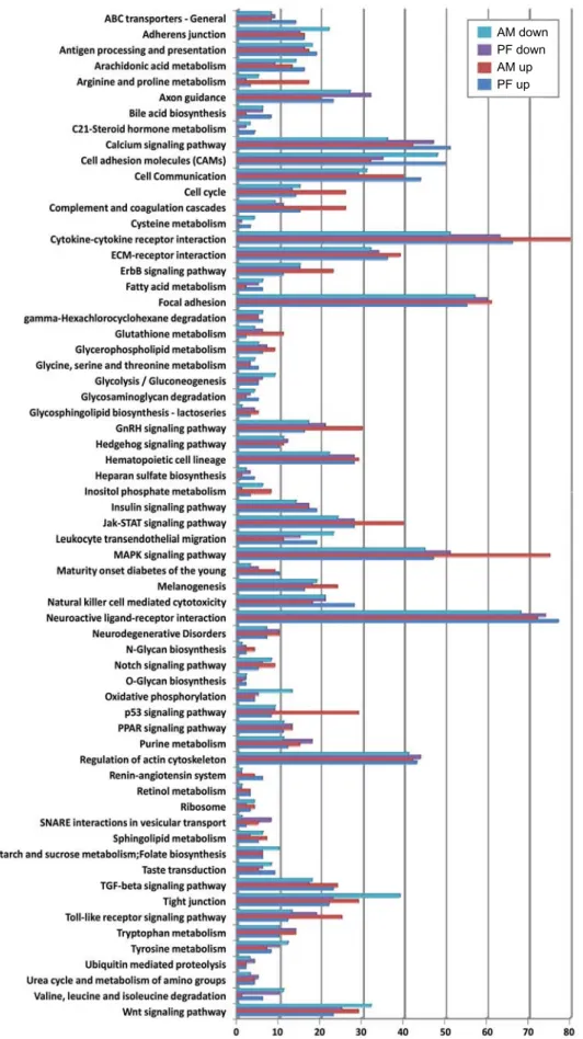
Functional Classifications Analysis
A comparative global transcription analysis between
treatment with PF and AM that induces apoptosis in
U937 cells revealed that functional classifications of
the responding genes are provided in Figure 1 (the
hypothetical, unclassified, unknown is omitted).
Func-tional classes are taken from Gene Ontology category
in GenPlex v3.0 software.
Metabolic Pathway Analysis
The pathways from the Kyoto Encyclopaedia of
Genes and Genomes (KEGG)
8were downloaded and
imported in to the GenPlex v3.0 software and visually
inspected for changes based on the 2,159 genes from
the t-test analysis. These metabolic pathways were
compiled into tables to organize the data based on
metabolic pathways. Striking features were revealed
by inspection. First, the p53 dependent pathway genes
were compiled and organized in to Table 1. All genes
were significantly upregulated and downregulated.
Second, the apoptosis pathway was organized in to
Table 2. Third, MAPK-related genes were compiled
and organized in to supplementary Table 1.
Discussion
As expected, paeoniflorin (PF) treatment triggered
the expression of genes involved in antiinflammatory,
antiimmuno-regulatory effects, anticancer and
antipro-liferative activities. PF has been proposed and shown
to elicit similar responses to anisomycin (AM) through
MAPK signaling pathway and apoptosis which were
presumed to account for the major anticancer
9-11and
apoptosis
1,12,13(Figure 1). Hence, it has been
speculat-ed that PF functions by similar mechanisms as other
antiinflamation
14-16. The p53 pathway senses a variety
of stress signals which will reduce the fidelity of cell
growth and division, and responds by initiating cell
cycle arrest, senescence, or apoptosis. Correspondingly,
p53 related genes using cyclin D1 (CCND1)
17, p53
binding protein (MDM4)
18, and phosphatase and tensin
homolog (PTEN)
19,20were all downregulated. Cyclin
D1 gene CCND1 was strongly repressed (0.1-fold). The
mRNA level of MDM4 (Murine Double Minute 4)
decreased under PF (0.49-fold). MDM4 shares
signifi-cant structural homology with MDM2 (Murine Double
Minute 2) and interacts and regulates transcriptional
activity of the tumor suppressor p53. In tumors with
wild-type p53, there is often overexpression of MDM2
or MDM4 leading to functional inactivation of p53
18.
This study explores two p53-regulated gene products,
PTEN (0.3-fold) and IGF-1 (4.65-fold), each of which
negatively regulates the IGF-1-AKT-mTOR pathways
after PF.
Exposure to PF, however, induced a variety of genes
involved in apoptosis including p53 dependent and
p53 independent
21. Most notably, Caspase-10
activa-tion, in turn, depended on caspase-8, which cleaved
caspase-10 directly
22. The caspase-8 homologous
cellu-lar FLICE-like inhibitory protein (cFLIP) can also be
recruited to the DISC. cFLIP acts as an anti-apoptotic
regulator by interfering with activation of caspases-8
and -10 at the DISC
23. Caspase-10 were among the
most heavily upregulated 6.4-fold (Table 1). CASP8
and FADD-like apoptosis regulator gene (CFLAR) was
upregulated by 2.13-fold.
Signaling from transforming growth factor beta
(TGF
β stimulates the MAPK pathway
24. The MAPK
201983_s_at EGFR epidermal growth factor receptor (erythroblastic leukemia 2.88 7.30 2.53 viral (v-erb-b) oncogene homolog, avian)
212647_at RRAS related RAS viral (r-ras) oncogene homolog 20.00 48.72 2.44 215365_at CACNB2 calcium channel, voltage-dependent, beta 2 subunit 1.31 3.10 2.37
206706_at NTF3 neurotrophin 3 6.10 14.03 2.30
208449_s_at FGF8 fibroblast growth factor 8 (androgen-induced) 0.64 1.40 2.18 202340_x_at NR4A1 nuclear receptor subfamily 4, group A, member 1 10.04 21.58 2.15 215688_at RASGRF1 Ras protein-specific guanine nucleotide-releasing factor 1 12.05 24.16 2.01 Table 1. Continued.
Probe ID Gene Gene title Control Treat Fold
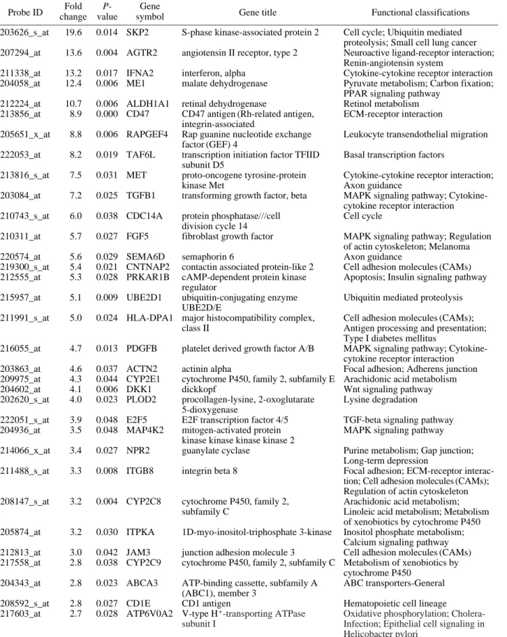
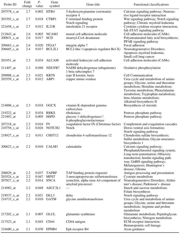
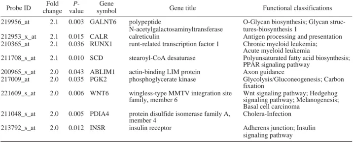
Table 2. List of significantly Up-regulated genes based on comparison between experimental (paeoniflorin (PF)-treated for 3 h) and experimental (anisomycin (AM)-treated for 3 h) in human leukemia U937 cells.
Probe ID changeFold valueP- symbolGene Gene title Functional classifications 203626_s_at 19.6 0.014 SKP2 S-phase kinase-associated protein 2 Cell cycle; Ubiquitin mediated
proteolysis; Small cell lung cancer 207294_at 13.6 0.004 AGTR2 angiotensin II receptor, type 2 Neuroactive ligand-receptor interaction;
Renin-angiotensin system
211338_at 13.2 0.017 IFNA2 interferon, alpha Cytokine-cytokine receptor interaction 204058_at 12.4 0.006 ME1 malate dehydrogenase Pyruvate metabolism; Carbon fixation;
PPAR signaling pathway 212224_at 10.7 0.006 ALDH1A1 retinal dehydrogenase Retinol metabolism 213856_at 8.9 0.000 CD47 CD47 antigen (Rh-related antigen, ECM-receptor interaction
integrin-associated
205651_x_at 8.8 0.006 RAPGEF4 Rap guanine nucleotide exchange Leukocyte transendothelial migration factor (GEF) 4
222053_at 8.2 0.019 TAF6L transcription initiation factor TFIID Basal transcription factors subunit D5
213816_s_at 7.5 0.031 MET proto-oncogene tyrosine-protein Cytokine-cytokine receptor interaction;
kinase Met Axon guidance
203084_at 7.2 0.025 TGFB1 transforming growth factor, beta MAPK signaling pathway; Cytokine-cytokine receptor interaction 210743_s_at 6.0 0.038 CDC14A protein phosphatase///cell Cell cycle
division cycle 14
210311_at 5.7 0.027 FGF5 fibroblast growth factor MAPK signaling pathway; Regulation of actin cytoskeleton; Melanoma 220574_at 5.6 0.029 SEMA6D semaphorin 6 Axon guidance
219300_s_at 5.4 0.021 CNTNAP2 contactin associated protein-like 2 Cell adhesion molecules (CAMs) 212555_at 5.3 0.028 PRKAR1B cAMP-dependent protein kinase Apoptosis; Insulin signaling pathway
regulator
215957_at 5.1 0.009 UBE2D1 ubiquitin-conjugating enzyme Ubiquitin mediated proteolysis UBE2D/E
211991_s_at 5.0 0.024 HLA-DPA1 major histocompatibility complex, Cell adhesion molecules (CAMs); class II Antigen processing and presentation;
Type I diabetes mellitus
216055_at 4.7 0.013 PDGFB platelet derived growth factor A/B MAPK signaling pathway; Cytokine-cytokine receptor interaction 203863_at 4.6 0.037 ACTN2 actinin alpha Focal adhesion; Adherens junction 209975_at 4.3 0.044 CYP2E1 cytochrome P450, family 2, subfamily E Arachidonic acid metabolism 204602_at 4.1 0.006 DKK1 dickkopf Wnt signaling pathway 202620_s_at 4.0 0.023 PLOD2 procollagen-lysine, 2-oxoglutarate Lysine degradation
5-dioxygenase
222051_s_at 3.9 0.048 E2F5 E2F transcription factor 4/5 TGF-beta signaling pathway 204936_at 3.5 0.048 MAP4K2 mitogen-activated protein MAPK signaling pathway
kinase kinase kinase kinase 2
214066_x_at 3.4 0.027 NPR2 guanylate cyclase Purine metabolism; Gap junction; Long-term depression
211488_s_at 3.3 0.008 ITGB8 integrin beta 8 Focal adhesion; ECM-receptor interac- tion; Cell adhesion molecules (CAMs); Regulation of actin cytoskeleton 208147_s_at 3.2 0.004 CYP2C8 cytochrome P450, family 2, Arachidonic acid metabolism;
subfamily C Linoleic acid metabolism; Metabolism of xenobiotics by cytochrome P450 205874_at 3.2 0.030 ITPKA 1D-myo-inositol-triphosphate 3-kinase Inositol phosphate metabolism;
Calcium signaling pathway 212813_at 3.0 0.042 JAM3 junction adhesion molecule 3 Cell adhesion molecules (CAMs) 217558_at 2.8 0.038 CYP2C9 cytochrome P450, family 2, subfamily C Metabolism of xenobiotics by
cytochrome P450 204343_at 2.8 0.023 ABCA3 ATP-binding cassette, subfamily A ABC transporters-General
(ABC1), member 3
208592_s_at 2.8 0.027 CD1E CD1 antigen Hematopoietic cell lineage 217603_at 2.7 0.028 ATP6V0A2 V-type H++
-transporting ATPase Oxidative phosphorylation;
Cholera-subunit I Infection; Epithelial cell signaling in
207578_s_at 2.7 0.002 HTR4 5-hydroxytryptamine (serotonin) Calcium signaling pathway; Neuroac-receptor 4 tive ligand-receptor interaction
203392_s_at 2.7 0.018 CTBP1 C-terminal binding protein Wnt signaling pathway; Notch signaling Notch signaling pathway; Chronic myeloid leukemia 221658_s_at 2.7 0.012 IL21R interleukin 21 receptor Cytokine-cytokine receptor interaction;
Jak-STAT signaling pathway 212843_at 2.6 0.003 NCAM1 neural cell adhesion molecule Cell adhesion molecules (CAMs) 200831_s_at 2.6 0.017 SCD stearoyl-CoA desaturase Polyunsaturated fatty acid biosynthesis;
PPAR signaling pathway 209663_s_at 2.6 0.020 ITGA7 integrin alpha 7 Focal adhesion
206665_s_at 2.4 0.017 BCL2L1 BCL2-like 1 (apoptosis regulator Bcl-X) Neurodegenerative Disorders; Apoptosis myeloid leukemia; Small cell lung cancer
201951_at 2.3 0.034 ALCAM activated leukocyte cell adhesion Cell adhesion molecules (CAMs) molecule
211407_at 2.3 0.040 NDUFB7 NADH dehydrogenase (ubiquinone) Oxidative phosphorylation 1 beta subcomplex 7
209008_x_at 2.3 0.021 KRT8 type II keratin, basic Cell Communication
203559_s_at 2.3 0.012 ABP1 copper amine oxidase Urea cycle and metabolism of amino groups; Glycine, serine and threonine metabolism; Histidine metabolism; Tyrosine metabolism; Phenylalanine metabolism; Tryptophan metabolism; beta-Alanine metabolism;
Alkaloid biosynthesis II 214006_s_at 2.3 0.018 GGCX vitamin K-dependent gamma- Biosynthesis of steroids
carboxylase
219222_at 2.3 0.034 RBKS ribokinase Pentose phosphate pathway 221892_at 2.3 0.005 H6PD glucose 1-dehydrogenase/// Pentose phosphate pathway
6-phosphogluconolactonase
207218_at 2.3 0.016 F9 coagulation factor IX (Christmas factor) Complement and coagulation cascades 210756_s_at 2.2 0.026 NOTCH2 Notch Dorso-ventral axis formation;
Notch signaling pathway 218927_s_at 2.2 0.011 CHST12 chondroitin 4-sulfotransferase 12 Chondroitin sulfate biosynthesis;
Sulfur metabolism; Glycan structures-biosynthesis 1
200623_s_at 2.2 0.010 CALM3 calmodulin Calcium signaling pathway; Phosphatidylinositol signaling system; Long-term potentiation; Olfactory transduction; Insulin signaling path- way; GnRH signaling pathway; Melanogenesis; Huntington’s disease; Glioma
208829_at 2.2 0.037 TAPBP TAP binding protein (tapasin) Antigen processing and presentation 203524_s_at 2.2 0.047 MPST 3-mercaptopyruvate sulfurtransferase Cysteine metabolism
207827_x_at 2.2 0.014 SNCA synuclein, alpha (non A4 component of Neurodegenerative Disorders; Alzhei-amyloid precursor) mer’s disease; Parkinson’s disease 214982_at 2.2 0.048 ASCC3L1 --- Starch and sucrose metabolism;
Folate biosynthesis 219537_x_at 2.2 0.021 DLL3 delta Notch signaling pathway
216733_s_at 2.2 0.010 GATM glycine amidinotransferase Urea cycle and metabolism of amino groups; Glycine, serine and threonine metabolism; Arginine and proline metabolism
217202_s_at 2.1 0.007 GLUL glutamine synthetase Glutamate metabolism; Peptidoglycan biosynthesis; Nitrogen metabolism 217523_at 2.1 0.045 CD44 CD44 antigen ECM-receptor interaction;
Hematopoietic cell lineage 216680_s_at 2.1 0.030 EPHB4 Eph receptor B4 Axon guidance
Table 2. Continued.
pathways are deeply involved in signaling for various
immune responses including apoptosis. MAPKs are
serine/threonine kinases (STK), which include the
ex-tracellular signal-related kinases (ERKs), p38 kinases,
and c-Jun N-terminal kinases (JNKs)
25. Activation of
MAPK pathway often occurs in response to growth
factor stimulation of receptor tyrosine kinases, which
are coupled to the activation of Ras G-proteins, such
Shc and Grb2, and quinine nucleotide exchange
fac-tors such as SOS
26,27. In this study, we detected the
downregulation of TGFB, STK3, MAPK8IP3, NFKB2,
and FOS in Pf-treated human leukemia U937 cells
(Table 1).
Fibroblast growth factor (FGF) signals play
funda-mental roles in development and tumorigenesis.
Thy-roid cancer is an example of a tumor with
nonoverlap-ping genetic mutations that up-regulate
mitogen-acti-vated protein kinase (MAPK). Here, we show that FGF
receptors (FGF18, 16.69-fold; FGF14, 6.2-fold; FGF2,
5.44-fold), which are expressed mainly in U937 cells,
propagates MAPK activation and promotes tumor
pro-gression. These data unmask an epigenetically
con-trolled FGFR signal that imposes precisely on the
intra-genically modified BRAF/MAPK pathway to
modu-late human leukemia U937 cells. Interestingly, our
data showed that genes associated with the cell cycle,
cytokine-cytokine receptor interaction, Jak-STAT
sig-naling pathway, MAPK sigsig-naling pathway, and p53
signaling pathway were highly upregulated in
AM-treated than PF-AM-treated U937 cells (Figure 1 and Table
3). The genes encoding enzymes responsible for the
F-box and leucine-rich repeat protein 1 (SKP2),
angio-tensin II receptor, type 2, interferon, alpha, Rap
gua-nine nucleotide exchange factor (GEF) 4, TGF
β, and
FGF exhibited the highly upregulated genes after
PF-exposure than AM-treated. This Study also revealed
that PF induced several IGF, caspase-10, Ras, RASGRP
and FGF genes, which are essential for MAPK
acti-vation and promotes tumor progression.
Taken together, these results indicate that PF may
have potential efficacy for the treatment of anticancer
and induction of apoptosis. However, it may possible
for proliferation and differentiation by activating FGF
signaling and MAPK pathway. A comparative global
transcription analysis between treatment with PF and
AM that induces apoptosis in U937 cell revealed that
JNK pathway related genes were less expressed in
PF-treated cells. Elucidation of the mechanisms by
which PF conducts its anticancer activities through
comparative analysis of the gene expression is
neces-sary to provide a solid foundation for its use as an agent
in prevention and treatment strategies.
Materials & Methods
Data Analysis
The MAS5 algorithm was used to evaluate the
expr-ession signals generated by the Affymetrix Human
Genome U133A Array. Global scaling normalization
was then performed and the normalized data were
log-transformed with base 2. Next, fold change was
applied to select the differentially expressed genes
(DEGs) using a fold change threshold of 2.0-fold and
a P
⁄0.05 to indicate significance. Each probe set used
in the Affymetrix GeneChip produces a detection call,
with P (present call) indicating good quality, M
(mar-ginal call) indicating intermediate quality and A
(abs-219956_at 2.1 0.003 GALNT6 polypeptide O-Glycan biosynthesis; Glycan struc-
N-acetylgalactosaminyltransferase tures-biosynthesis 1
212953_x_at 2.1 0.015 CALR calreticulin Antigen processing and presentation
210365_at 2.1 0.036 RUNX1 runt-related transcription factor 1 Chronic myeloid leukemia; Acute myeloid leukemia
211708_s_at 2.1 0.010 SCD stearoyl-CoA desaturase Polyunsaturated fatty acid biosynthesis;
PPAR signaling pathway
200965_s_at 2.0 0.043 ABLIM1 actin-binding LIM protein Axon guidance
217009_at 2.0 0.035 PGK2 phosphoglycerate kinase Glycolysis/Gluconeogenesis; Carbon
fixation
221609_s_at 2.0 0.006 WNT6 wingless-type MMTV integration site Wnt signaling pathway; Hedgehog
family, member 6 signaling pathway; Melanogenesis;
Basal cell carcinoma 211048_s_at 2.0 0.005 PDIA4 protein disulfide isomerase family A, Cholera-Infection
member 4
213792_s_at 2.0 0.012 INSR insulin receptor Adherens junction; Insulin
signaling pathway
Table 2. Continued.
Figure 1. Gene Ontology classifications of genes based on comparison of gene ex-pression between experimen-tal (paeoniflorin (PF)-treated and anisomycin (AM)-treated) and control (non-treated) in human leukemia U937 cells. AM down
PF down AM up PF up
ent call) indicating relatively low reliability. Therefore,
probe sets that resulted in A calls in the compared
groups were removed to filter false positives. The
2.0-fold DEGs were clustered using the GenPlex
TMv3.0
software (ISTECH Inc., Korea) using hierarchical
clus-tering with Pearson correlation as a similarity measure
and complete linkage as the linkage method. In
addi-tion, gene ontology significance analysis was
conduct-ed to investigate the functional relationships among
the 2.0-fold DEGs using high-throughput GoMiner.
The 2.0-fold DEGs were then mapped to relevant
path-ways using GenPlex
TMv3.0 software (ISTECH Inc.,
Korea). The pathway resources were provided by the
KEGG database.
Data analysis was also performed with the
Gene-Spring GX v. X (Agilent Technologies). The following
parameters were employed for GCOS expression
analy-sis:
α
1=
=0.04,
α
2=
=0.06, and
τ=
=0.015; target signal was
scaled to 150. Genes that received “absent” calls from
50% or more of the replicates in GeneSpring were not
used for the analysis. Finally, gene expression changes
with statistical significance were identified by an upper
1-tailed t-test (P cutoff value, 0.05). “Fold-change” was
calculated as the ratio between the signal averages of
Table 3. List of significantly Down-regulated genes based on comparison between experimental (paeoniflorin (PF)-treated for 3 h) and experimental (anisomycin (AM)-treated for 3 h) in human leukemia U937 cells.Probe ID changeFold valueP- symbolGene Gene title Functional classifications 211449_at 0.2 0.008 MSH6 DNA mismatch repair protein MSH6 Colorectal cancer
208116_s_at 0.2 0.002 MAN1A1 mannosyl-oligosaccharide N-Glycan biosynthesis alpha-1,2-mannosidase
211496_s_at 0.2 0.004 PDC phosducin Olfactory transduction
212759_s_at 0.2 0.008 TCF7L2 transcription factor 7-like 2 Wnt signaling pathway; Acute myeloid leukemia
206751_s_at 0.3 0.038 PCYT1B choline-phosphate cytidylyltransferase Aminophosphonate metabolism 202669_s_at 0.3 0.004 EFNB2 ephrin-B Axon guidance
203710_at 0.3 0.022 ITPR1 inositol 1,4,5-triphosphate receptor, Calcium signaling pathway type 1
214378_at 0.3 0.019 TFPI tissue factor pathway inhibitor Complement and coagulation cascades 213055_at 0.3 0.007 CD47 CD47 antigen (Rh-related antigen, ECM-receptor interaction
integrin-associated
204062_s_at 0.3 0.032 ULK2 unc51-like kinase Regulation of autophagy; mTOR signaling pathway
201334_s_at 0.4 0.000 ARHGEF12 Rho guanine nucleotide exchange factor Axon guidance; Regulation of actin
(GEF) 12 cytoskeleton
202861_at 0.4 0.038 PER1 period circadian protein Circadian rhythm
209050_s_at 0.4 0.048 RALGDS ral guanine nucleotide dissociation Colorectal cancer; Pancreatic cancer stimulator
212067_s_at 0.4 0.043 C1R complement component 1, Complement and coagulation cascades r subcomponent
201963_at 0.4 0.038 ACSL1 long-chain fatty-acid-CoA ligase Fatty acid metabolism 201602_s_at 0.4 0.013 PPP1R12A protein phosphatase 1, regulatory Focal adhesion
(inhibitor) subunit 12
203544_s_at 0.4 0.019 STAM signal transducing adaptor molecule Jak-STAT signaling pathway 204635_at 0.4 0.006 RPS6KA5 mitogen-, stress activated protein kinases MAPK signaling pathway; Bladder
cancer
205105_at 0.4 0.000 MAN2A1 alpha-mannosidase II N-Glycan biosynthesis
202981_x_at 0.4 0.006 SIAH1 ubiquitin ligase SIAH1 p53 signaling pathway; Wnt signaling pathway
206769_at 0.4 0.014 TMSB4Y thymosin, beta 4 Regulation of actin cytoskeleton 217028_at 0.5 0.039 CXCR4 chemokine (C-X-C motif) receptor 4 Cytokine-cytokine receptor interaction 210512_s_at 0.5 0.046 VEGFA vascular endothelial growth factor A/B, Cytokine-cytokine receptor interaction
PGF
212249_at 0.5 0.013 PIK3R1 phosphoinositide-3-kinase, regulatory ErbB signaling pathway subunit
205448_s_at 0.5 0.010 MAP3K12 mitogen-activated protein kinase kinase MAPK signaling pathway kinase 12
203096_s_at 0.5 0.010 RAPGEF2 Rap guanine nucleotide exchange factor MAPK signaling pathway (GEF) 2
204633_s_at 0.5 0.031 RPS6KA5 mitogen-, stress activated protein kinases MAPK signaling pathway; Bladder cancer
four untreated and four treated cultures. That is, results
are from biological quadruplicate experiments. Genes
with a 2.0-fold or more induction or repression were
used in this analysis.
Acknowledgements
This work was supported by the Korea Science and
Engineering Foundation (KOSEF) grant funded by
the Korea government (MEST) (No. 2009-0063466).
References
1. Tsuboi, H. et al. Paeoniflorin induces apoptosis of lymphocytes through a redox-linked mechanism. J Cell Biochem 93:162-172 (2004).
2. Salunga, T. L. et al. Identification of genes responsive to paeoniflorin, a heat shock protein-inducing com-pound, in human leukemia U937 cells. Int J Hyper-thermia 23:529-537 (2007).
3. Hori, T. et al. Molecular mechanism of apoptosis and gene expressions in human lymphoma U937 cells treat-ed with anisomycin. Chem Biol Interact 172:125-140 (2008).
4. Jiménez A, V. D. Anisomycin and related antibiotics (ed. E, H. F.) Springer-Verlag, New York (1979). 5. Xia, Z., Dickens, M., Raingeaud, J., Davis, R. J. &
Greenberg, M. E. Opposing effects of ERK and JNK-p38 MAP kinases on apoptosis. Science 270:1326-1331 (1995).
6. Zanke, B. W. et al. The stress-activated protein kinase pathway mediates cell death following injury induced by cis-platinum, UV irradiation or heat. Curr Biol 6: 606-613 (1996).
7. Park, J. et al. Activation of c-Jun N-terminal kinase antagonizes an anti-apoptotic action of Bcl-2. J Biol Chem 272:16725-16728 (1997).
8. Ogata, H. et al. KEGG: Kyoto Encyclopedia of Genes and Genomes. Nucleic Acids Res 27, 29-34 (1999). 9. Washida, K., Itoh, Y., Iwashita, T. & Nomoto, K.
And-rogen modulators from the roots of Paeonia lactiflora (paeoniae radix) grown and processed in nara prefec-ture, Japan. Chem Pharm Bull (Tokyo) 57:971-974 (2009).
10. Wu, H., Li, W., Wang, T., Shu, Y. & Liu, P. Paeoni-florin suppress NF-kappaB activation through modu-lation of I kappaB alpha and enhances 5-fluorouracil-induced apoptosis in human gastric carcinoma cells. Biomed Pharmacother 62:659-666 (2008).
11. Hung, J. Y., Yang, C. J., Tsai, Y. M., Huang, H. W. & Huang, M. S. Antiproliferative activity of paeoniflo-rin is through cell cycle arrest and the Fas/Fas ligand-mediated apoptotic pathway in human non-small cell lung cancer A549 cells. Clin Exp Pharmacol Physiol
35:141-147 (2008).
12. Zhong, S. Z., Ge, Q. H., Li, Q., Qu, R. & Ma, S. P. Peoniflorin attentuates Abeta ((1-42))-mediated neu-rotoxicity by regulating calcium homeostasis and ameliorating oxidative stress in hippocampus of rats. J Neurol Sci 280:71-78 (2009).
13. Nizamutdinova, I. T. et al. Paeonol and paeoniflorin, the main active principles of Paeonia albiflora, pro-tect the heart from myocardial ischemia/reperfusion injury in rats. Planta Med 74:14-18 (2008).
14. Jiang, B., Qiao, J., Yang, Y. & Lu, Y. Inhibitory ef-fect of paeoniflorin on the inflammatory vicious cy-cle between adipocytes and macrophages. J Cell Bio-chem (2009).
15. Zhang, L. L. et al. Paeoniflorin suppresses inflamma-tory mediator production and regulates G protein-coupled signaling in fibroblast-like synoviocytes of collagen induced arthritic rats. Inflamm Res 57:388-395 (2008).
16. Huang, H. et al. A genome-wide microarray analysis reveals anti-inflammatory target genes of paeonol in macrophages. Inflamm Res 57:189-198 (2008). 17. Guo, Z., Wang, J., Yang, J., Wu, N. & Zhang, Y. The
Role of p53 and p65 in heat shock-induced inhibi-tion of cyclin D1. Biochim Biophys Acta 1789:758-762 (2009).
18. Kulkarni, D. et al. A polymorphic variant in human MDM4 associates with accelerated age of onset of estrogen receptor negative breast cancer. Carcinoge-nesis 30:1910-1915 (2009).
19. Regina, S. et al. Increased tissue factor expression is associated with reduced survival in non-small cell lung cancer and with mutations of TP53 and PTEN. Clin Chem 55:1834-1842 (2009).
20. Feng, Z. et al. The regulation of AMPK beta1, TSC2, and PTEN expression by p53: stress, cell and tissue specificity, and the role of these gene products in modulating the IGF-1-AKT-mTOR pathways. Can-cer Res 67:3043-3053 (2007).
21. Konstantakou, E. G. et al. Human bladder cancer cells undergo cisplatin-induced apoptosis that is asso-ciated with p53-dependent and p53-independent res-ponses. Int J Oncol 35:401-416 (2009).
22. Chen, H., Xia, Y., Fang, D., Hawke, D. & Lu, Z. Cas-pase-10-mediated heat shock protein 90 beta cleavage promotes UVB irradiation-induced cell apoptosis. Mol Cell Biol 29:3657-3664 (2009).
23. Walczak, H. & Haas, T. L. Biochemical analysis of the native TRAIL death-inducing signaling complex. Methods Mol Biol 414:221-239 (2008).
24. Lamar, J. M., Iyer, V. & DiPersio, C. M. Integrin al-pha3beta1 potentiates TGFbeta-mediated induction of MMP-9 in immortalized keratinocytes. J Invest Dermatol 128:575-586 (2008).
25. Sohn, K. H. et al. Genome wide expression profile of Asiasarum sieboldi in LPS-stimulated BV-2 Micro-glial Cells. Molecular & Cellular Toxicology 4:205-210 (2008).
/2 activity in cells expressing neuronal nitric-oxide synthase. J Biol Chem 279:3933-3940 (2004). 27. Sim, S. et al. NADPH oxidase-derived reactive
oxy-gen species-mediated activation of ERK1/2 is required for apoptosis of human neutrophils induced by Enta-moeba histolytica. J Immunol 174:4279-4288 (2005).