Effects of Beech-wood Creosote on Intestinal Microflora in Rat
Jeong A Kim1, Da Yoon Yu1, In Sung Kim1, Chul Young Lee1, Dong Kee Jeong2, Sang Suk Lee3, In Soon Choi4 and Kwang Keun Cho1*
1Department of Animal Resources Technology, Gyeongnam National University of Science and Technology, Jinju 52725, Korea
2Faculty of Biotechnology, Jeju National University, Jeju 63243, Korea
3Department of Animal Science and Technology, Sunchon National University, Sunchon 57922, Korea
4Department of Biological Science, Silla University, Busan 46958, Korea Received March 6, 2018 /Revised June 27, 2018 /Accepted July 24, 2018
Diarrhea is one of the main disorders which cause the highest level mortality of the post-weaning economic animal. Beech-wood creosote has been used as a traditional anti-diarrheic medicament for a long time. The present study was conducted to investigate the effects of dietary supplementation of Beech-wood on growth performance and intestinal microbiota in rats. Twelve 4-week-old rats were randomly assigned to one of four dietary groups and fed a basal diet supplemented with none (CON), 0.5% apramycin (ANTI), 0.4% creosote (Creo 0.4), or 0.8% creosote (Creo 0.8) for 4 weeks following 1 week of adaptation period to the respective diet. Average daily gain was not influenced by the diet- ary treatment whereas average daily feed intake was greatest for the Creo 0.8 group. In the intestinal microbiota at the level of the phylum, the percentage of Firmicutes bacteria decreased but Bacteroidetes increased in the Creo 0.8 group vs. Control, which resulted in a decreased F/B ratio for the former (p<0.05). Moreover, the percentage of Lachnospiraceae was greater at the level of the family for the Creo 0.8 group than for Control, but the percentages of Turicibacter and Clostridium disporicum were less in the former (p<0.01) at the genus and species levels, respectively. Collectively, the present results in- dicate that dietary supplementation of creosote increases the feed intake and also influence the in- testinal microbiota in rats.
Key words : Beech-wood, creosote, gut microbiota, rat
*Corresponding author
*Tel : +82-55-751-3286, Fax : +82-55-751-3689
*E-mail : [email protected]
This is an Open-Access article distributed under the terms of the Creative Commons Attribution Non-Commercial License (http://creativecommons.org/licenses/by-nc/3.0) which permits unrestricted non-commercial use, distribution, and reproduction in any medium, provided the original work is properly cited.
Journal of Life Science 2018 Vol. 28. No. 7. 849~856 DOI : https://doi.org/10.5352/JLS.2018.28.7.849
서 론
항생제는 가축의 성장과 질병 치료에 중요한 역할을 한다 [41]. 그러나 최근 항생제 내성 문제로 인해 사료 첨가 항생제 가 전면 금지되면서, 항생제 대체 물질 개발에 대한 연구가 다양하게 진행되고 있다[21]. Apramycin은 aminoglycoside계 항생제로서 주기적으로 많이 섭취할 경우 Escherichia coli와 Salmonella 등 장내 미생물에서 apramycin 내성이 증가한다 [30]. 항생제 대체제로는 미생물 제제, 유기산제, 면역증강제, 식물 추출물 등이 주로 이용되고 있다. 미생물 제제는 장내 미생물 균총을 안정화시켜 동물에 유익한 영향을 주는 사료 첨가제로써 대표적으로 Lactobacillus, Bacillus, 효모 등이 있다 [13]. 유기산제는 위내 pH를 감소시켜 pH에 민감함 Salmonella 와 E. coli의 증식을 억제 또는 사멸한다고 알려져 있으며, 대표
적인 유기산제로 acetic acid, butyric acid, lactic acid 등이 있 다[26]. 면역증강제(immunostimulating)는 동물에서 식균작 용 및 사이토카인 분비 활성화와 같은 다양한 면역반응을 유 도하며[10], 대표적으로 보리나 버섯 유래 β-glucan 등이 있다 [19]. 이러한 항생제 대체제 중에서도 식물 추출물은 생리활성 이 뛰어나기 때문에 이에 대한 연구가 활발하다[22].
크레오소트(creosote)는 너도밤나무(Beech-wood)를 증류 하여 만든 유액 성분이다[34]. 1886년에 발행 된 일본 약전 (Japanese Pharmacopoeia)에 지사제(antidiarrhetica)로서 수 록되어 있으며, 러일 전쟁 당시 설사 치료를 목적으로 크레오 소트를 보급하게 되면서 아시아에서 대중적으로 사용되어 왔 다[32]. 이는 소장에서 유체의 흡수를 증가시켜 장내에 잔류하 는 유체의 양을 감소시키고, 장운동을 부분적으로 억제함으로 써 지사제의 효과를 나타낸다[17, 33]. 크레오소트는 장 상피 세포의 Cl- 경로를 차단하여 장독소에 의한 체액 분비를 억제 하고, 평활근 세포 내 Ca2+ 유입을 억제시켜 기계적, 화학적, 또는 전기적 자극에 의한 대장 운동을 감소시킨다[1]. 이전의 연구에서 흰쥐에게 크레오소트를 급여하였을 때, 장내 분비 및 장벽 기능에 대한 유해 효과를 방지함으로써 설사 예방 효과가 있음이 밝혀졌다[40].
장내 미생물의 대부분은 Firmicutes 및 Bacteroidetes (90-
Table 1. Composition of the experiment diets
Items Treatments
CON ANTI Creo 0.4 Creo 0.8 Ingredients (%)
Casein (from milk) Corn starch Sucrose Cellulose Corn oil Mineral mixture Vitamin mixture DL-methionine Choline bitartrate Total
20.00 15.00 50.00 5.00 5.00 3.50 1.00 0.30 0.20 100.00
20.00 15.00 50.00 5.00 5.00 3.50 1.00 0.30 0.20 100.00
20.00 15.00 50.00 5.00 5.00 3.50 1.00 0.30 0.20 100.00
20.00 15.00 50.00 5.00 5.00 3.50 1.00 0.30 0.20 100.00 Apramycin (%)
Creosote (%)
- -
0.50 -
- 0.40
- 0.80 99%), Actinobacteria (3-15%), Proteobacteria (1-20%), Verrucom- icrobia (0.1%), Fusobacteria, Cyanobacteria, Spyrochaetes 및 Lentisphaerae 문 (phylum)으로 이루어져 있다[3]. 숙주와 장내 미생물의 상호작용은 과민성 대장 증후군(irritable bowel syn- drome; IBS), 궤양성 대장염(ulcerative colitis; UC) 등의 여러 장 질환뿐만 아니라 알츠하이머(Alzheimer’s) 및 파킨슨병 (Parkinson’s disease)과 같은 중추신경계 장애와 관련되어 있 다[11]. 이러한 장내미생물 조성 변화는 식이, 환경, 독소, 의약 품, 항생제 복용 등의 외부적 요인에 의해 영향을 받는다[6].
따라서 본 연구에서는 흰 쥐를 실험동물로 사용하여 크레오 소트가 항생제 대체 물질로서 장내 미생물 변화에 미치는 영 향을 조사하고 이를 동물 산업에 적용할 수 있는지에 대해 규명하고자 하였다.
재료 및 방법
공시동물 사양관리 및 공시재료
4주령의 수컷 Sprague-Dawley (SD) 흰 쥐(SLC, Inc., Japan) 는 1 cage 당 2마리씩 3반복하여 그룹 당 6마리를 사용하였다.
실험기간 동안 실내온도 22±1℃, 12시간 주기의 명암주기로 유 지되었다. 일주일의 예비 실험 기간 동안 기초사료(AIN-76A, Central lab, Seoul, Korea) (Table 1)와 물은 무제한으로 급여 하였고, 일주일에 한 번 사료 잔량을 확인하였다. 본 실험은 (1) 대조구(기초사료), (2) 항생제 그룹(기초사료 + 아프라마이 신0.5%), (3) 크레오소트 0.4% 그룹(기초사료 + 크레오소트 0.4%), (4) 크레오소트 0.8% 그룹(기초사료 + 크레오소트 0.8%) 으로 나누어 시험 기간 4주 동안 급여하였다. 체중은 실험 개 시일과 종료일에 측정하였으며, 실험 종료일에 사료 잔량을 측정하여 사료 효율을 계산하였다. 본 실험에 사용한 항생제 는 apramycin (Sigma, St. Louis, MO, USA)을 사용하였다.
크레오소트 환은 (주)창조바이오텍에서 제조하였으며, 크레오
소트 16.67 g, 밀가루풀 50 g, 진피(Fraxini cortex) 50 g을 혼합 하여 1 mm 크기로 매끄럽게 성형한 후, 60℃에서 건조하였다.
동물실험은 경남과학기술대학교 동물실험윤리위원회(Insti- tutional Animal Care Board of Gyeongnam National Univer- sity of Science and Technology)의 승인을 획득하였다(2014-4).
장내 미생물 분석
장내 미생물 변화에 대한 크레오소트의 영향을 조사하기 위해 크레오소트를 4주간 급여 후 실험 종료일에 흰 쥐를 희생 시키고 소장, 맹장, 대장 내용물을 채취하여 혼합한 후 장내 미생물의 genomic DNA (gDNA)를 분리하기 위하여 -80°C에 보관하였다. 장내 미생물의 gDNA는 Fecal DNA MiniPrep kit (Zymo Research, CA, USA)를 사용하여 추출 하였으며, 분석에 사용될 때까지 -20℃에 보관하였다. 장내 미생물은 py- rosequencing 방법을 이용하여 16s rDNA 유전자 서열을 분석 하였다[23]. Pyrosequencing을 통해 얻어진 염기서열의 미생 물 군집은 소프트웨어 CLcommunityTM (Chunlab Inc., Seoul, Korea)를 이용하여 분석하였으며, CD-HIT 프로그램을 이용 하여 97% sequence 유사성에 의해 정의된 종(species)은 Operational Taxonomic Unit (OTU) 값으로 나타내었다[29].
종의 풍부도(richness)는 생물학적 종의 다양성 지표인 alpha diversity로 나타내었으며, Chao1 richness [7] 및 Shannon di- versity index [16]를 분석하여 종의 풍부도를 나타내었다. 미 생물 공동체 사이의 계통발생학적 거리는 Fast UniFrac을 사 용하여 평가하고 주좌표분석(principal coordinate analysis;
PCoA)을 사용하여 시각화 하였다.
통계 처리
반복실험을 통해 얻은 결과는 SPSS 12.0 (SPSS Inc., Chicago, IL, USA)을 사용하여 분산분석 하였으며, 분석결과에 대한 모 든 그룹별 유의성 검정은 p<0.05 수준에서 Duncan's multiple range test를 사용하여 평균±표준편차로 나타내었다[8].
결과 및 고찰
흰 쥐 성장에 미치는 효과
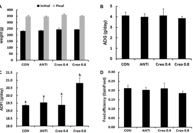
크레오소트 첨가 급여에 따른 성장 성적을 Fig. 1에 나타내 었다. 흰 쥐의 증체량을 분석한 결과 그룹 간에 유의적인 차이 를 나타내지 않았으며(Fig. 1A), 일당 증체량 역시 그룹 간의 유의적인 차이는 나타나지 않았다(Fig. 1B). 일당사료섭취량은 Creo 0.8 그룹이 CON과 ANTI 그룹보다 유의적으로 높은 것 으로 나타났으며(Fig. 1C) (p<0.05), 사료 효율은 그룹 간의 차 이가 없는 것으로 나타났다(Fig. 1D). 크레오소트는 구아야콜 (guaiacol)과 크레졸(creosol) 등을 포함하는 페놀 화합물로써 특유의 향을 가지고 있다[17]. 지난 20여년 동안 방향유(essen- tial oils), 방향성 허브(aromatic herbs)와 같은 식물성 사료
A B
C D
Fig. 1. Effect of increasing percentage of dietary creosote on growth performance in rats. (A) Body weight. (B) Average daily gain.
(C) Average daily feed intake. (D) Feed efficiency. a-bMeans are significantly different in each group (p<0.05). Data represent means ± SD of 4 replicates. CON: control, ANTI: antibiotics 0.5%, Creo 0.4: creosote 0.4%, Creo 0.8: creosote 0.8%.
Table 2. Intestinal microbiota richness and diversity was increased with creosote
Group Subjects Analyzed reads1) Normalized reads Observed OTUs2) Chao 1 index3) Shannon diversity index4)
CON 1
2 3
7436 6779 7982
6779 6779 6779
1088 890 1092
2419 2050 2250
4.98 4.49 4.76
ANTI 1
2 3
7810 7969 7478
6779 6779 6779
1201 673 1390
2490 1293 3415
5.05 4.50 5.28
Creo 0.4 1
2 3
8393 10335
8483
6779 6779 6779
1326 1236 1589
2967 2439 3618
5.04 4.80 5.23
Creo 0.8 1
2 3
7551 8603 7593
6779 6779 6779
1867 1672 1970
4882 3714 5083
5.90 5.70 6.05
1)Analyzes reads were obtained from pyrosequencing. 2)Operational Taxonomic Unit. 3)Species richness. 4)Species diversity.
CON: control, ANTI: antibiotics 0.5%, Creo 0.4: creosote 0.4%, Creo 0.8: creosote 0.8%.
첨가제는 동물의 사료 첨가제로서 많이 사용되어 왔으며[9], 사료의 풍미와 기호성 증진 및 성장 촉진 효과를 나타내었다 [35]. 흰 쥐에서 크레오소트 섭취로 인한 체중 증가는 나타나지 않았지만, Sodhi 등[38]의 연구에서 크레오소트를 섭취한 돼지 의 성장률이 증가하였으며, Salmonella typhimurium으로 설사 를 유발 한 후 처리한 결과 크레오소트가 항생제 보다 빨리
설사 증상이 회복되는 것으로 보고 하였다.
장내 미생물의 다양성 분석
흰 쥐 장내 미생물의 16S rDNA sequence를 이용하여 다양 성을 분석하였다(Table 2). Reads 간의 유사도를 근거로 각 그룹에 대한 OTUs 값을 분석한 결과 CON과 ANTI 그리고
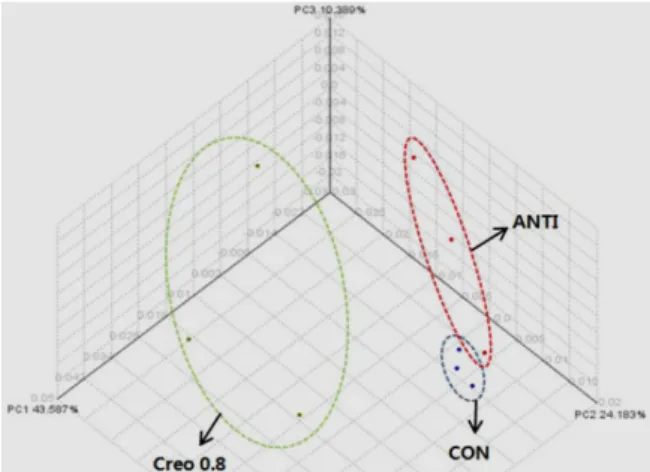
Fig. 2. Principal coordinates analysis (PCoA) of community structures using a fast unifrac distance matrix. Percent- age of variation explained by principal component 1 (PC1) (43.6%). Percentage of variation explained by principal component 2 (PC2) (24.2%). Percentage of var- iation explained by principal component 3 (PC3) (10.4
%). CON: control, ANTI: antibiotics 0.5%, Creo 0.4: cre- osote 0.4%, Creo 0.8: creosote 0.8%.
Creo 0.4 그룹 간에는 차이가 나타나지 않았지만 Creo 0.8 그 룹에서 관찰된 OTUs의 수가 높게 나타났다. Shannon 다양성 지수를 이용하여 장내 미생물 종 다양성을 분석한 결과 Creo 0.8 그룹에서 높게 나타났으며, Chao1 분석을 이용하여 분석 한 종 풍부도 역시 Creo 0.8 그룹에서 높게 나타났다. 일반적으 로 장내 미생물 다양성 변화는 숙주의 건강과 연관되어 있으 며, 서구화된 식습관으로 인해 비만 및 만성 질환을 가지고 있는 사람에 비해 수렵 생활을 하는 사람들의 장내 미생물 다양성이 증가된다[18]. 식이섬유 섭취량 저하로 인한 장내 미 생물의 다양성 감소는 미생물의 주요 대사산물인 short chain fatty acid (SCFA)의 생성을 감소시키고 그로 인해 장 점막 면역 개선에 부정적인 영향을 미친다[14]. 반면 장내 미생물 다양성 증가에 의해 생성된 SCFA는 chemokine의 생성을 억 제하고 단핵구/대식세포, 호중구의 동원(動員, recruitment)을 억제하는 항염증 효과를 가진다[2]. 따라서 장내 미생물 다양 성 증가는 면역 체계의 변화와 연관되어 있으며, 장내 미생물 다양성의 감소는 숙주의 건강 장애와 관련되어 있다.
장내 미생물 군집에 대한 주좌표 분석
주좌표(PCoA: Principal Coordinates Analysis) 분석법으로 장내 미생물 군집 구조에 미치는 영향을 분석한 결과 미생물 군집이 3개 처리 그룹에 대해 각각 다르게 분포하는 것으로 나타났다(Fig. 2). ANTI 그룹은 그래프 우측 CON 그룹 미생물 군집에 가까이 위치하는 것으로 나타난 반면, Creo 0.8 그룹은 그래프 좌측에 위치하여 별도의 그룹으로 나타났다. 이러한 결과는 크레오소트를 0.8% 이상 섭취하였을 때 미생물의 군집 이 변화할 수 있다는 것을 의미하여 이를 뒷받침하기 위한
각 그룹간의 미생물 조성을 확인하였다.
장내 미생물 변화 분석
동물 이유시기에 장내 미생물의 불균형은 설사로 인한 폐사 율에 직접적인 영향을 미칠 수 있으며[27], 장내 미생물의 변화 는 환경, 식이, 항생제 복용 등과 같은 다양한 환경 요인에 의해 영향을 받는다[6]. 또한 대장 내 공생 미생물의 비정상적 불균형은 염증성 장 질환이나 과민성 대장 증후군과 같은 질 병 발생을 야기할 수 있지만[12], 숙주와의 상호작용 하는 장내 유익균 증가와 병원성 미생물 감소는 다양한 장 질환의 예방 과 치료에 긍정적인 영향을 미칠 수 있다[11]. 이전의 연구에서 크레오소트가 장내 수분 분비 조절 및 장 운동성 억제를 통한 설사병 완화 효과에 대하여 보고가 되었지만, 장내 미생물 변 화에 미치는 효과에 대해서는 연구가 보고 되어 있지 않았다.
따라서 본 연구에서는 크레오소트 첨가 급여가 장내 미생물 군집에 미치는 영향을 분석하였다(Fig. 3, Table 3). 장내 미생 물을 문(phylum) 수준에서 분석한 결과 모든 그룹에서 Firmi- cutes와 Bacteroidetes가 전체 장내 미생물의 90% 이상을 차지하 는 것으로 나타났으며(Fig. 3A), Creo 0.8 그룹에서 Firmicutes 가 유의적으로 감소하고, Bacteroidetes는 유의적으로 증가하여 F/B 비율을 감소시키는 것으로 나타났다(Fig. 3B, Table 3) (p<0.05). 이러한 결과는 크레오소트를 섭취한 숲 쥐(woodrats) 에서 Firmicutes가 상대적으로 낮은 비율로 나타나고 Bacteroi- detes가 높은 비율로 나타난다는 Kohl와 Dearing [25]의 연구 결과와 일치한다. 장내 미생물은 체중과도 연관되어 있으며 고지방 식이를 섭취할 경우 Firmicutes가 증가하고 Bacteroi- detes는 감소한다[42]. 크레오소트에 의해 우점하는 문(phylum) 미생물의 다양성과 풍부도가 증가하는 것으로 나타났지만 [25], 이러한 변화에 대한 작용 기전은 아직 명확하게 밝혀지지 않았으며 더 많은 연구가 필요하다.
Firmicutes에 속하는 장내 미생물을 과(family) 수준에서 분 석한 결과, butyrate를 생산하는 미생물 Lachnospiraceae가 Creo 0.8 그룹에서 유의적으로 증가하는 것으로 나타났으며(p<0.01), Lachnospiraceae에 속하는 미생물의 하위그룹을 분석한 결과 Eubacterium 속(genus)이 높은 수준으로 나타났다. Eubacterium 역시 butyrate 를 생산하는 미생물로 알려져 있다[43]. 크레오 소트는 구아야콜(guaiacol)을 포함하는 페놀 화합물이며[17], Kazemi-Bonchenari 등[20]에 의하면 구아야콜(guaiacol)이 butyrate의 생산을 증가시킨다고 보고 하였다. Ruminococcaceae 는 Creo 0.8 그룹에서 증가하였지만 유의적인 차이는 없었다.
장내 공생 미생물의 90~99%는 Firmicutes 및 Bacteroidetes로 구성되어 있으며[3], Firmicutes는 정상인에 비해 설사와 같은 장 질환에서 증가한다[44]. Butyrate는 결장 상피 세포에서 흡 수되어 막관통단백질(transmembrane protein) 중의 하나인 claudin 단백질 발현을 증가시켜 장 상피세포의 밀착연접 (tight junctions)을 강화하고[45], 염증성 장 질환을 완화시키
Table 3. Pyrosequencing analysis of the fecal microbiota composition in rats
Treatments
CON ANTI Creo 0.4 Creo 0.8
n=3 Mean % (SD)
n=3 Mean % (SD)
n=3 Mean % (SD)
n=3 Mean % (SD) Firmicutes
Clostridia Clostridiaceae Clostridium
Clostridium disporicum Lachnospiraceae
Peptostreptococcaceae Ruminococcaceae Bacilli
Lactobacillaceae Lactobacillus Turicibacter_f Turicibacter Bacteroidetes Proteobacteria Desulfovibrio
99.07 (0.27) 60.14 (3.95) 34.79 (1.16) 32.43 (1.17) 24.87 (0.82) 10.57 (1.94) 8.91 (1.92) 1.60 (0.19) 38.91 (3.69) 28.52 (7.43) 25.43 (7.74) 8.21 (3.60) 6.21 (2.75) 0.13 (0.03) 0.35 (0.06) 0.16 (0.03)
99.03 (0.68) 71.82 (8.07) 44.05 (3.06)**
41.33 (4.14)* 31.97 (5.15) 12.11 (7.72) 8.96 (0.07) 1.97 (1.27) 27.16 (8.75) 14.75 (7.01) 12.55 (6.15) 10.81 (2.54) 8.19 (2.18) 0.67 (0.63) 0.07 (0.07)**
0.01 (0.01)**
98.36 (0.65) 75.04 (7.17)* 37.29 (3.05) 34.34 (2.97) 26.17 (2.98) 19.16 (8.60) 11.09 (3.97) 2.17 (0.80) 23.73 (7.44)* 15.07 (7.12) 12.90 (5.75) 6.94 (2.26) 5.34 (1.99) 0.61 (0.42) 0.43 (0.29) 0.26 (0.24)
96.86 (0.89)* 64.99 (12.44) 14.66 (8.20)* 12.55 (7.37)**
8.68 (5.02)**
33.15 (6.72)**
4.63 (3.22) 4.52 (2.89) 31.82 (12.39) 27.67 (12.72) 24.04 (11.04) 1.59 (1.30)* 1.31 (1.09)* 2.01 (0.94)* 0.52 (0.20) 0.20 (0.12)
**p<0.01 versus the control group. *p<0.05 versus the control group. Data represent means ± SD of 3 replicates. CON: control, ANTI:
antibiotics 0.5%, Creo 0.4: creosote 0.4%, Creo 0.8: creosote 0.8%.
A
B
Fig. 3. Pyrosequencing analysis of the fecal microbiota composition from rats fed the creosote. (A) Relative abundance of microbial phyla identified in intestine of rats. Left: Percentage of 3 replicates. Right: Percentage of 3 replicates average. (B) Abundance ratio of Firmicutes and Bacteroidetes in faecal microbiota. a-bMeans are significantly different in each group (p<0.05). Data represent means ± SD of 3 replicates. CON: control, ANTI: antibiotics 0.5%, Creo 0.4: creosote 0.4%, Creo 0.8: creosote 0.8%.
는데 효과적이다[37]. 또한 butyrate는 장내에서 수분 및 이온 흡수를 통해 설사 증상 완화에 효과적이다[5].
Firmicutes에 속하는 장내 미생물을 속(genus) 수준에서 분 석한 결과 Turicibacter가 Creo 0.8 그룹에서 1.31%로 대조구의 6.21%에 비해 유의적으로 감소하는 경향으로 나타났으며(p<
0.01), 종(species) 수준에서는 Clostridium disporicum이 대조구 의 24.87%에 비해 Creo 0.8 그룹에서 8.68%로 유의적으로 감 소하였다(p<0.01). Turicibacter는 궤양성 대장염 환자의 장내에 서 증가하고 장 질환을 가진 새끼 돼지에서 발견된다[4]. 최근 장내 미생물은 다양한 장 질환의 원인으로 주목 받고 있으며 [15], 크론병과 같은 염증성 장질환은 결장과 소장에서 발생하 여 혈변 및 체중 감소를 유발한다[36].
Ataka K 등[1]에 의하면 크레오소트가 장내 항-분비(anti- secretory) 활성과 대장 내 항-운동성(anti-motility)에 기인하 여 설사 완화 효과를 나타내며, 살균 활성(microbicidal activ- ity)은 나타나지 않는다고 하였으나, 본 연구에서는 크레오소 트가 장내 병원성 미생물을 감소시켜 살균 활성을 가지는 것 으로 나타났다.
ANTI 그룹은 문(phylum) 수준에서 대조구에 비해 Proteo- bacteria가 유의적으로 감소하였으며(p<0.01), Proteobacteria과 (familly)에 속하는 미생물 중에서 Desulfovibrio가 유의적으로 감소하였다(p<0.01). Proteobacteria 문에는 많은 염증성 미생물 이 포함되어 있으며, Desulfovibrio는 결장의 병원성 미생물로 알려져 있다[31, 39]. 또한 크레오소트는 주로 Firmicutes (phylum)에 속하는 gram positive 병원성 미생물을 감소시키 고, apramycin 항생제는 Proteobacteria (phylum)에 속하는 gram negative 병원성 미생물을 감소시킨다는 것으로 나타났 다. 항생제는 다양한 작용기전을 통하여 E. coli나 Salmonella 등과 같은 장내 병원성 미생물을 억제 시키며, apramycin은 gram-negative bacteria 감염의 치료에 효과를 나타낸다[24, 28].
크레오소트와 항생제는 모두 동물의 설사 증상을 완화 시킬 수 있지만 그 작용 기전은 다르게 나타났다. 결론적으로 너도 밤나무 추출물인 크레오소트가 장내 병원성 미생물 감소를 통해 항생제 대체제로서 대안이 될 수 있다는 것을 나타내며, 설사 완화에 대한 작용기전은 더 많은 연구가 필요할 것으로 사료된다.
감사의 글
이 논문은 2017년도 경남과학기술대학교 대학회계 연구비 지원에 의하여 연구되었음.
References
1. Ataka, K., Ito, M. and Shibata, T. 2005. New views on anti- diarrheal effect of wood creosote: is wood creosote really
a gastrointestinal antiseptic? Yakugaku Zasshi 125, 937-950.
2. Bermon, S., Petriz, B., Kajėnienė, A., Prestes, J., Castell, L.
and Franco, O. L. 2015. The microbiota: an exercise im- munology perspective. Exerc. Immunol. Rev. 21, 70-79.
3. Biagi, E., Candela, M., Fairweather-Tait, S., Franceschi, C.
and Brigidi, P. 2012. Aging of the human metaorganism:
the microbial counterpart. Age (Dordr) 34, 247-267.
4. Breton, J., Massart, S., Vandamme, P., De Brandt, E., Pot, B. and Foligné, B. 2013. Ecotoxicology inside the gut: impact of heavy metals on the mouse microbiome. BMC. Pharmacol.
Toxicol. 14, 62.
5. Canani, R. B., Terrin, G., Cirillo, P., Castaldo, G., Salvatore, F., Cardillo, G., Coruzzo, A. and Troncone, R. 2004. Butyrate as an effective treatment of congenital chloride diarrhea.
Gastroenterology 127, 630-634.
6. Carding, S., Verbeke, K., Vipond, D. T., Corfe, B. M. and Owen, L. J. 2015. Dysbiosis of the gut microbiota in disease.
Microb. Ecol. Health Dis. 2, 26191.
7. Chao, A. 1984. Nonparametric estimation of the number of classes in a population. Scand. J. Statist. 11, 265-270.
8. Duncan, D. B. 1995. Multiple range and multiple F tests.
J. Biometrics 11, 1-42.
9. Franz, C., Baser, K. H. C. and Windisch, W. 2010. Essential oils and aromatic plants in animal feeding – a European perspective. A review. Flavour Fragr. J. 25, 327-340.
10. Gallois, M., Rothkötter, H. J., Bailey, M., Stokes, C. R. and Oswald, I. P. 2009. Natural alternatives to in-feed antibiotics in pig production: can immunomodulators play a role?
Animal 3, 1644-1661.
11. Ghaisas, S., Maher, J. and Kanthasamy, A. 2016. Gut micro- biome in health and disease: Linking the microbiome- gut-brain axis and environmental factors in the pathogenesis ofsystemic and neurodegenerative diseases. J. Pharmacol.
Ther. 158, 52-62.
12. Gophna, U. 2011. Microbiology. The guts of dietary habits.
Science 334, 45-46.
13. Hah, K. H., Lee, C. W., Jin, S. K., Kim, I. S., Song, Y. M., Hur, S. J., Kim, H. Y., Lyou, H. J. and Ha, J. H. 2005. Effect of feeding probiotics on physico-chemical properties and sensory evaluation of pork. Kor. J. Food Sci. An. 25, 295-303.
14. Han, M., Wang, C., Liu, P., Li, D., Li, Y. and Ma, X. 2017.
Dietary Fiber Gap and Host Gut Microbiota. Protein Pept.
Lett. 24, 388-396.
15. He, Q., Gao, Y., Jie, Z., Yu, X., Laursen, J. M., Xiao, L., Li, Y., Li, L., Zhang, F., Feng, Q., Li, X., Yu, J., Liu, C., Lan, P., Yan, T., Liu, X., Xu, X., Yang, H., Wang, J., Madsen, L., Brix, S., Wang, J., Kristiansen, K. and Jia, H. 2017. Two dis- tinct metacommunities characterize the gut microbiota in Crohn's disease patients. Gigascience 6, 1-11.
16. Hill, T. C., Walsh, K. A., Harris, J. A. and Moffett, B. F.
2003. Using ecological diversity measures with bacterial communities. J. FEMS. Microbiol. Ecol. 43, 1-11.
17. Hiramoto, K., Yamate, Y., Kobayashi, H., Ishii, M., Miura, T., Sato, E. F. and Inoue, M. 2012. Effect of the smell of Seirogan, a wood Creosote, on dermal and intestinal mu- cosal immunity and allergic inflammation. J. Clin. Biochem.
Nutr. 51, 91-95.
18. Hur, K. Y. 2017. Gut microbiota and metabolic disorders.
J. Kor. Diabetes 18, 63-70.
19. Jang, I. S. 2015. Effects of by-products of herbal medicine on performance, intestinal microbial population, blood bio- chemical profiles and immunological parameters in broiler chicks. Kor. J. Poult. Sci. 42, 307-314.
20. Kazemi-Bonchenar, M., Falahati, R., Poorhamdollah, M., Heidari, S. R. and Pezeshki, A. 2018. Essential oils improved weight gain, growth and feed efficiency of young dairy calves fed 18 or 20% crude protein starter diets. J. Anim.
Physiol. Anim. Nutr. (Berl) 17, doi: 10.1111/jpn.12867.
21. Kim, D. W., Kim, J. H., Kim, S. K., Kang, G. H., Kang, H.
K., Lee, S. J. and Kim, S. H. 2009. A study on the efficacy of dietary supplementation of organic acid mixture in broil- er chicks. J. Anim. Sci. Technol. (Kor.) 51, 207-216.
22. Kim, D. W., Kim, S. H., Yu, D. J., Kang, G. H., Kim, J. H., Kang, H. G., Jang, B. G., Na, J. C., Suh, O. S., Jang, I. S.
and Lee, K. H. 2007. Effects of single or mixed supplements of plant extract, fermented medicinal plants and lactobacil- lus on growth performance in broilers. Kor. J. Poult. Sci. 34, 187-196.
23. Kim, O. S., Cho, Y. J., Lee, K., Yoon, S. H., Kim, M., Na, H., Park, S. C., Jeon, Y. S., Lee, J. H., Yi, H., Won, S. and Chun, J. 2012. Introducing EzTaxon-e: a prokaryotic 16S rRNA gene sequence database with phylotypes that repre- sent uncultured species. Int. J. Syst. Evol. Microbiol. 62, 716- 721.
24. Ko, Y. H., Yang, H. Y., Kang, S. Y., Kim, E. S. and Jang, I. S. 2007. Effects of a bend of prunus mume extract as an alternative to antibiotics on growth performance, activity of figestive enzymes and microflora population in broiler chickens. J. Anim. Sci. Technol. (Kor.) 49, 611-620.
25. Kohl, K. D. and Dearing, M. D. 2012. Experience matters:
prior exposure to plant toxins enhances diversity of gut mi- crobes in herbivores. Ecol. Lett. 15, 1008-1015.
26. Lee, C. H., Jo, I. H., Shon, J. C. and Lee, S. H. 2009. Effect of dietary supplementation of organic acid and antibiotics mixture on growth performances and blood metabolites in growing pigs. Organic Agriculture 17, 237-251.
27. Lee, S. B. and Choi, S. H. 2006. Isolation and Identification of Probiotic Lactobacillus Isolates for Calf Meal Supple- ments. Kor. J. Food Sci. An. 26, 106-112.
28. Leitner, G., Waiman, R. and Heller, E. D. 2001. The effect of apramycin on colonization of pathogenic Escherichia coli in the intestinal tract of chicks. Vet. Q. 23, 62-66.
29. Li, W. and Godzik, A. 2006. Cd-hit: a fast program for clus- tering and comparing large sets of protein or nucleotide sequences. J. Bioinformatics 22, 1658-1659.
30. Lim, S. K., Nam, H. M., Lee, H. S., Kim, A. R., Jang, G.
C., Jung, S. C. and Kim, T. S. 2013. Prevalence and character- ization of apramycin-resistant Salmonella enterica serotype Typhimurium isolated from healthy and diseased pigs in Korea during 1998 through 2009. J. Food Prot. 76, 1443-1446.
31. McOrist, A. L., Warhurst, M., McOrist, S. and Bird, A. R.
2001. Colonic infection by Bilophila wadsworthia in pigs.
J. Clin. Microbiol. 39, 1577-1579.
32. Moriguchi, N., Sato, A., Shibata, T. and Yoneda, Y. 2011.
A historical review of the therapeutic use of wood creosote.
Part II: Original plant source of crude drug wood creosote.
Yakushigaku Zasshi 46, 13-20.
33. Ogata, N., Baba, T. and Shibata, T. 1993. Demonstration of antidiarrheal and antimotility effects of wood Creosote. J.
Pharmacology 46, 173-180.
34. Quynh, A. N., Sharma, N., Cho, K. K., Yeo, T. J., Kim, K.
B., Jeong, C. Y., Min, T. S., Kim, J. Y., Kim, J. N. and Jeong, D. K. 2014. Efficacious rat model displays non-toxic effect with Korean beechwood Creosote: a possible antibiotic substitute. J. Biotechnol. Biotechnol. Equip. 28, 447-454.
35. Reyer, H., Zentek, J., Männer, K., Youssef, I. M. I., Aumiller, T., Weghuber, J., Wimmers, K. and Mueller, A. S. 2017.
Possible molecular mechanisms by which an essential oil blend from star anise, rosemary, thyme, and oregano and saponins increase the performance and ileal protein digesti- bility of growing broilers. J. Agric. Food Chem. 65, 6821-6830.
36. Salaritabar, A., Darvishi, B., Hadjiakhoondi, F., Manayi, A., Sureda, A., Nabavi, S. F., Fitzpatrick, L. R., Nabavi, S. M.
and Bishayee, A. 2017. Therapeutic potential of flavonoids in inflammatory bowel disease: A comprehensive review.
World J. Gastroenterol. 23, 5097-5114.
37. Simeoli, R., Mattace Raso, G., Pirozzi, C., Lama, A., Santoro, A., Russo, R., Montero-Melendez, T., Berni Canani, R., Calignano, A., Perretti, M. and Meli, R. 2017. An orally ad- ministered butyrate-releasing derivative reduces neutrophil recruitment and inflammation in dextran sulphate sodium- induced murine colitis. Br. J. Pharmacol. 174, 1484-1496.
38. Sodhi, S. S., Kim, J. H., Sharma, N., Cho, K. K., Kim, J. Y., Kim, K. B., Jeong, C. Y., Yoon, M. Y., Oh, S. J. and Jeong, D. K. 2014. Korean Beechwood Creosote as a substitute to an antibiotic for post weaning diarrhea in piglets. Pak. Vet.
J. 34, 341-346.
39. Sun, J., Huang, T., Chen, C., Cao, T. T., Cheng, K., Liao, X. P. and Liu, Y. H. 2017. Comparison of fecal microbial composition and antibiotic resistance genes from swine, farm workers and the surrounding villagers. Sci. Rep. 7, 4965.
40. Tang, Z. R., Yin, Y. L., Nyachot,i C. M., Huang, R. L., Li, T. J., Yang, C., Yang, X. J., Gong, J., Peng, J., Qi, D. S., Xing, J. J., Sun, Z. H. and Fan, M. Z. 2005. Effect of dietary supple- mentation of chitosan and galacto-mannan-oligosaccharide on serum parameters and the insulin-like growth factor-I mRNA expression in early-weaned piglets. Domest. Anim.
Endocrinol. 28, 430-441.
41. Kuge, T., Meerveld, B. G. V. and Sokabe, M. 2006. Stress-in- duced breakdown of intestinal barrier function in the rat:
Reversal by wood creosote. Life Sci. 79, 913-918.
42. Turnbaugh, P. J., Backhed, F., Fulton, L. and Gordon, J. I.
2008. Diet-induced obesity is linked to marked but rever- sible alterations in the mouse distal gut microbiome. Cell Host Microbe 3, 213-223.
43. Vernocchi, P., Del Chierico, F., Quagliariello, A., Ercolini, D., Lucidi, V. and Putignani, L. 2017. A metagenomic and
초록:너도밤나무 크레오소트가 흰 쥐의 장내 미생물 변화에 미치는 영향
김정아1․유다윤1․김인성1․이철영1․정동기2․이상석3․최인순4․조광근1*
(1경남과학기술대학교 동물소재공학과, 2제주대학교 생명공학부, 3순천대학교 동물자원과학과, 4신라대학교 생명
과학과)
설사는 경제동물의 이유시기 폐사율을 일으키는 가장 높은 요인이다. 크레오소트는 전통 의약품으로 오랜 세월 동안 지사제로 사용되어 왔다. 본 연구는 흰 쥐 모델에서 크레오소트 급여가 동물의 성장 효율 및 장내 미생물에 미치는 영향을 구명할 목적으로 수행되었다. 4주령의 수컷 흰쥐 24마리를 임의로 대조구, 항생제 그룹, 크레오소 트 0.4% 그룹, 크레오소트 0.8% 그룹으로 배치하였다. 대조구는 기초사료, 항생제 그룹은 apramycin 0.5%, 크레오 소트 그룹은 크레오소트 0.4%와 0.8% 수준으로 하여 예비시험 기간 1주일, 본시험 기간 4주일 동안 급여하였다.
일당증체량은 실험구간 차이가 없었으나, 사료 섭취량은 Creo 0.8 그룹에서 유의적으로 증가하였다(p<0.05). 장내 미생물에 대한 문(phylum) 수준 분석 결과 Creo 0.8 그룹에서 Firmicutes가 감소하고 Bacteroidetes가 증가하여 F/B 비율을 감소시키는 것으로 나타났다(p<0.05). 과(family) 수준에서는 Lachnospiraceae가 크레오소트 0.8% 수준에서 증가되었으며(p<0.01), 속(genus) 수준에서는 Turicibacter가 감소되었다(p<0.01). 종(species) 수준에서는 Clostridium disporicum이 감소되었다(p<0.01). 이상의 결과는 쥐에 크레오소트 급여는 사료 섭취량을 증가시키고 장내 미생물 의 변화에 영향을 미칠 수 있다는 것을 시사한다.
in silico functional prediction of gut microbiota profiles may concur in discovering new cystic fibrosis patient-targeted probiotics. Nutrients 9, 1342.
44. Wang, A., Ling, Z., Yang, Z., Kiela, P. R., Wang, T., Wang, C., Cao, L., Geng, F., Shen, M., Ran, X., Su, Y., Cheng, T.
and Wang, J. 2015. Gut microbial dysbiosis may predict di-
arrhea and fatigue in patients undergoing pelvic cancer ra- diotherapy: a pilot study. PLoS One 10, e0126312.
45. Yan, H. and Ajuwon, K. M. 2017. Butyrate modifies in- testinal barrier function in IPEC-J2 cells through a selective upregulation of tight junction proteins and activation of the Akt signaling pathway. PLoS One 12, e0179586.