6
서 론
유방암은 서구사회 여성에서 가장 흔한 암으로 보고되고 있고, 우리나라의 경우 1998년 보건복지부 암등록보고서에 따르면 위암에 이어 두 번째로 많은 여성암으로 전체 여성 암의 14.1%를 차지하고 있으며 점점 증가하는 추세에 있 다.(1)
유방암 및 여타 암의 발암기전에 대해 현재까지 완전히 밝혀진 기전은 없으나 여러 가지 발암 관련 인자들의 자극
유방암 발암과 진행에 있어 c-erbB-2 수용체 발현과 Mitogen-activated Protein Kinases 활성도의 역할
연세대학교 의과대학 1외과학교실, 2Brain Korea 21 Project, 3한림대학교 의과대학 외과학교실 및 4포천중문의과대학 외과학교실
박병우1,2․허민규2․김기석2․고승상1․김승일3․이경식4
책임저자:박병우, 서울시 서대문구 신촌동 134 ꂕ 120-752, 연세대학교 의과대학 외과학교실 Tel: 02-361-5564, Fax: 02-313-8289
E-mail: [email protected]
접수일:2002년 9월 17일, 게재승인일:2002년 12월 30일 본 연구는 1999년도 연세대학교 의과대학 교수연구비의 지원으로 이루어졌음.
Mitogen-activated Protein Kinases Activities and c-erbB-2 Expression in Breast Cancer Carcino- genesis and Progression
Byeong-Woo Park, M.D.1,2, Min-Kyu Heo, B.S.2, Ki-Suk Kim, B.S.2, Seung-Sang Ko, M.D.1, Seung-Il Kim, M.D.3 and Kyong Sik Lee, M.D.4
Purpose: Increased level mitogen-activated protein kinase (MAPK) and activation of MAPK have been reported in human breast cancers, especially in breast cancers with HER2/neu overexpression. To understand the relationship between the MAPK protein expressions and other clinico- pathological parameters, we examined the status of MAPKs in 20 breast cancers compared to those of paired normals.
Methods: A total of 20 breast cancers and paired normal breast tissues were included in this study. Tissues were obtained at the operation room and stored at -80oC. Tissue proteins were extracted and the concentration was deter- mined by Bio-Rad protein assay method. Western blot analysis were performed to determine the level of MAPKs expressions using 100 ug of tissue protein in 8%, 10%, or 12% sodium dodecyl sulphate polyacrylamide gel electro- phoresis (SDS-PAGE). MAPK assays were carried out by a non-radioactive method developed by Cell Signaling Tech.
as recommened by the manufacturer. Clinico-pathological information was provided from the Breast Cancer Registry of Department of Surgery, Yonsei University College of Medicine.
Results: The levels of MAPKs were higher in 95% of breast
cancers compared to those of paired normals. The levels of ERK1/2 were significantly higher in cancer tissues com- pared to paired normals but the activated forms were not.
The levels of JNK, p38, and MKP1 proteins were significantly increased in the cancer tissue compared to the paired nor- mals. The levels of ERK1/2 and activated ERK1/2 proteins were not different between tumor stages. There were no significant differences of the levels of ERK1/2 and activated ERK1/2 proteins between HER2-negative and HER2- positive cancers. There were significantly higher levels of activated ERK1/2 proteins in ER-positive cancers than those in ER-negative cancers (P<0.05).
Conclusion: The levels of MAPKs, but not the activated forms, seem to be increased in breast cancer tissues compared to those of paired normals. The levels of activated MAPKs seem to be associated with estrogen receptor expression in cancer tissues. (J Korean Surg Soc 2003;64:6-13) Key Words: Mitogen-activated protein kinase, HER2/neu,
Estrogen receptor, Breast cancer, Stage 중심 단어: 맵카이나제, HER2/neu, 에스트로젠 수용
체, 유방암, 종양 병기
ꠏꠏꠏꠏꠏꠏꠏꠏꠏꠏꠏꠏꠏꠏꠏꠏꠏꠏꠏꠏꠏꠏꠏꠏꠏꠏꠏꠏꠏꠏꠏꠏꠏꠏꠏꠏꠏꠏꠏꠏꠏꠏꠏꠏꠏꠏꠏꠏꠏꠏ Department of 1Surgery and 2Brain Korea 21 Project, Yonsei Uinversity College of Medicine, Department of Surgery, 3Hal- lym University and 4Pochon Chungmun University College of Medicine
박병우 외:유방암 발암과 진행에 있어 c-erbB-2 수용체 발현과 Mitogen-activated Protein Kinases 활성도의 역할 7 ꠏꠏꠏꠏꠏꠏꠏꠏꠏꠏꠏꠏꠏꠏꠏꠏꠏꠏꠏꠏꠏꠏꠏꠏꠏꠏꠏꠏꠏꠏꠏꠏꠏꠏꠏꠏꠏꠏꠏꠏꠏꠏꠏꠏꠏꠏꠏꠏꠏꠏꠏꠏꠏꠏꠏꠏꠏꠏꠏꠏꠏꠏꠏꠏꠏꠏꠏꠏꠏꠏꠏꠏꠏꠏꠏꠏꠏꠏꠏꠏꠏꠏꠏꠏꠏꠏꠏꠏꠏꠏꠏꠏꠏꠏꠏꠏꠏꠏꠏꠏꠏꠏꠏꠏꠏꠏꠏꠏꠏꠏꠏꠏꠏꠏꠏ 에 의해 발암유전자 혹은 발암억제유전자의 돌연변이 또는
과발현에 의해 세포의 이상증식 또는 세포고사(apoptosis)의 감소로 종양을 형성하는 것으로 알려져 있다.
유방암에서 c-erbB-2 암유전자의 과발현은 전체 유방암 중 약 30%에서 발견되고 이 암유전자는 유방암의 발암과 진행에 관여하는 것으로 알려져 있으며, 암유전자수용체의 과발현은 고도악성종양의 표현형으로 조기재발 등 불량한 예후와 관계있다고 알려져 있으며,(2,3) Mitogen-actiovated protein kinases (MAPKs)는 세포증식 및 세포고사에 중추적 인 역할을 하며 다양한 세포 밖 신호와 암유전자 산물에 의해 조절되고 가역적인 단백인산화를 통하여 그 활성도가 조절되는 것으로 알려져 있다.
다수의 신세포암(renal cell carcinoma)에서 MAPK의 항상 성활성화(constitutive activation)가 관찰되어 MAPK 신호전 달계의 항상성 활성화는 신세포암 발생기전에 중요한 역할 을 하며,(4) MAPK mRNA 발현의 증가는 전이능(metastatic potential)이 높은 유방암 세포주에서 관찰되었고 사람의 유 방암조직 및 전이된 림프절에서 tyrosyl residue의 인산화 및 MAP kinae 활성도가 양성종양에 비해 5∼10배 증가되어 있 어 이러한 과발현이 여러 형태의 사람유방암의 발생과 진 행 및 전이에 결정적인 요인이 될 수 있다고 보고되었다.(5) 성장인자나 그 수용체는 물론이고 그 밖의 단백들이 궁 극적으로 MAP kinase를 활성화시킴으로써 많은 상피세포 암에서 결정적 암유전자(dominant oncogenes)로 역할을 하 게 되는데, 이러한 MAP kinase를 비활성화시키는 MAP ki- nase phosphatase (MKP)-1이 배양세포에서 과발현되면 ras에 의한 MAPK-mediated mitogenic effect는 저하되고(6) 세포의 분화를 억제한다.(7) 따라서 MKP-1은 종양억제인자의 역할 을 담당할 가능성이 있을 것으로 추측되나, MKP-1의 과발 현은 전립선 암, 결장암, 방광암의 발생과정 초기에 관찰되 고 종양의 분화가 악화된 경우나 전이암에서는 그 발현이 저하되며, 이 때 ERK-1의 활성은 MKP-1의 발현에 관계없 이 증가되어 있었고 MKP-1을 포함하는 5q35-ter locus의 소 실(loss)이 없는 것으로 보아 MKP-1의 발현은 사람의 여러 상피세포암의 초기 징표(marker)일 뿐 종양억제인자로 역할 을 하는 것은 아닌 것으로 보고되었다.(8) 그러나 유방암의 경우 저분화암이나 말기까지도 상당한 MKP-1발현을 보이 는 것으로 볼 때, 종양진행의 말기까지도 MAP kinase 의존 적으로 남아 있는 경향이다.(8)
MAPK 활성화를 유도하는 똑같은 신호가 이 MKP-1의 발 현을 유도하고, 성장인자에 의한 MKP-1 mRNA의 급격한 유도는 ERK의 비활성화를 위한 것이 아니라 JNK의 비활성 화를 위해 필요한 것이다. 전립선암에서 JNK-1 효소능은 MKP-1의 발현과 역관계인 반면 ERK-1활성도는 MKP-1발 현과 상관관계가 없는 것으로 볼 때, 사람의 종양세포에서 MKP-1은 JNK의 탈인산화에 치중하여 비활성화시킨다고 여겨진다.(9) 이는 세포증식 및 세포고사체계의 동시 활성
화로 세포내 혼란이 초래되는 데 대해(아마도 MKP-1에 의 한) 선택적인 세포고사경로(apoptotic pathway)의 억제는 mi- togenic signal이 적절한 반응을 유도하는 데 필수적일 수 있 다.(9)
따라서 사람의 유방암에서도 암유전자산물의 과발현의 결과로 혹은 여타 다른 경로의 활성화를 통하여, 혹은 이와 관계없이 항상성 있는 MAPK 활성화가 중요한 발암기전일 가능성이 있다. 그러므로 사람 유방암조직에서 MAPK의 활 성도를 측정하고 발암유전자로 알려진 c-erbB-2와의 상관 성 및 c-erb-2 유전자 발현에 따른 각각의 MAPK 활성도를 비교분석하여 유방암에서 MAPK의 역할과 MAPK 활성화 에 관계되는 환경적 요인을 밝히고자 하였다.
방 법 1) 대상 환자 및 조직의 보관
대상 환자는 1기 유방암 환자 6명, 2기 9명 및 3기 5명의 환자였고, 정상 및 암 조직을 이용하여 실험하였다. 수술대 에서 적출된 조직은 즉시 냉동하여 필요 시까지 -80oC에 보관하였다. 대상 환자의 병리조직학적 정보는 본 교실 유 방암 등록 데이터베이스를 이용하였다.
2) 조직단백질 추출
실험에 사용할 조직을 liquid nitrogen에 얼린 채 소독된 약사발에 분쇄하여 차가운 분해용액(ice-cold lysis buffer) (70 mM B-glycerophosphate pH 7.2, 1 mM each mea- and ortho- vanadate, 2 mM manesium chloride, 1 mM EGTA, 1 mM dithiothreitol (DTT), 0.5% Triton X-100, 0.2 mM phenylmethylsulphonyl fluoride (PMSF) and 5 ug ml-1 each of pepstatin A, shymostatin, leupeptin and peptin)으로 용해시킨 후 어름속에서 20분간 방치하였다. 각 표본(sample)은 약 30 초 동안 소니케이션한 후(sonicate) 4oC 23,000 g에서 약 15 분간 원심분리 한 후 조직불순물을 제거하고 다시 한 번 원심 분리하여 상층액을 획득하였다. 이렇게 획득된 포본은 분주하 여 -80oC에 동결 보관하였고 단백질 농도는 Bradford 시약 (Bio-Rad Laboratories, Richmond, CA, USA)을 이용한 Bio-Rad 단백분석법을 이용하여 결정하였다.
3) Western blot analysis
추출된 단백질용액은 100 ug씩 단백질의 분자량에 따라 8%, 10%, 12% sodium dodecyl sulphate polyacrylamide gel (SDS-PAGE)에 전기 영동을 실시한 후 단백질을 Protran Ni- trocellulose Membrane (Schleicher and Schuell Corporation, Dassel, Germany)에 transfer하였다. Blots은 blocking buffer (20 mM Tris-Cl, pH 7.5, 15 mM sodium chloride, 0.05%
Tween-20 (TBST) containing 5% non-fat Carnation milk)을 이 용하여 처리하였다. 면역blot (Immunoblot)은 TBST로 세척
ꠏꠏꠏꠏꠏꠏꠏꠏꠏꠏꠏꠏꠏꠏꠏꠏꠏꠏꠏꠏꠏꠏꠏꠏꠏꠏꠏꠏꠏꠏꠏꠏꠏꠏꠏꠏꠏꠏꠏꠏꠏꠏꠏꠏꠏꠏꠏꠏꠏꠏꠏꠏꠏꠏꠏꠏꠏꠏꠏꠏꠏꠏꠏꠏꠏꠏꠏꠏꠏꠏꠏꠏꠏꠏꠏꠏꠏꠏꠏꠏꠏꠏꠏꠏꠏꠏꠏꠏꠏꠏꠏꠏꠏꠏꠏꠏꠏꠏꠏꠏꠏꠏꠏꠏꠏꠏꠏꠏꠏꠏꠏꠏꠏꠏꠏ 한 후, 적적량의 1차 항체를 함유한 TBST milk로 실온에서
2시간 또는 4oC에서 밤새 방치하였다. Blots은 TBST로 세척 한 후 적정량의 2차항체를 함유한 TBST milk로 약 1∼2시 간 동안 incubation 하였고 그 후 blots은 Amersham ECL kit (Amersham International, Buckinghamshire, UK)을 이용하여 현상하였다. Western band의 단백량을 비교하기 위해 감광 된 film을 GS-690 imaging Densitometer를 사용하여 측정하 였다.
4) Antibodies
본 연구에 사용된 1차 항체는 Phospho-p44/42 MAP Kinase (Thr202/Tyr204) (Cell Signaling Tech., MA, USA), p44/ 42 MAP Kinase (Cell Signaling Tech., MA, USA), Phospho- p38 MAP Kinase (Thr180/Tyr182) (Cell Signaling Tech., MA, USA), p38 MAP Kinase (Cell Signaling Tech., MA, USA), Phospho-SAPK/JNK (Cell Signaling Tech., MA, USA), SAPK/
JNK (Cell Signaling Tech., MA, USA), 및 MKP-1 (Santa Cruze Biotech., CA USA)을 사용하였으며 p44/42 MAP Kinase As- say Kit (Cell Signaling Tech., MA, USA)을 이용하여 In vitro MAPK assay를 시행하였다. 2차 항체는 Anti-mouse IgG-HRP (Santa Cruze Biotech., CA USA) 및 Anti-rabbit IgG-HRP
(Santa Cruze Biotech., CA USA)를 사용하였다.
5) In Vitro MAPK assays
MAPK assays는 Cell Signaling Tech. (MA, USA)가 개발한 방법을 제조사의 교범에 따라 수행하였다. MAPKs는 300 ug의 조직추출액과 phospho-specific ERK1/2 MAPK (Thr202/
Tyr204) 단클론항체를 면역침전(immunoprecipitation)시킨 후 kinase assays는 ATP 및 substrate인 Elk-1의 존재하에 수행하 였다. phosph-Elk-1 단백발현은 phospho-specific Elk-1 (Ser 383) 항체를 이용하여 Western blot analysis로 분석하였다.
이 phospho Elk-1 항체는 Ser383에 인산화된 Elk-1 단백질을 특이적으로 인식한다.
6) 통계분석
모든 통계분석은 컴퓨터 통계 프로그램인 SPSS 10.0을 이용하여 수행하였다. 연속변수 간의 비교는 SPSS 프로그 램의 independent sample t-test를 이용하였으며 이산변수 간 의 비교는 crosstab을 이용하였다. P<0.05의 범위를 유의하 다고 판정하였다.
Fig. 1. Levels of MAPKs in the representative breast cancers and paired normals. The ERK1/2 proteins were detected by Western blot analysis by using ERK1/2 polyclonal antibody (upper panel). The phospho-ERK1/2 proteins were detected by Western blot analysis by using phospho-specific MAPK (pERK1/2) antibody(lower panel). The levels of ERK1/2 were significantly higher in cancer tissues compared to paired normals but the activated forms were not. N and C represent normal and carcinoma, respectively.
Phospho-ERK
P44P42
Phospho-P44 Phospho-P42 ERK
6 7 11 18
N C N C N C N C
Fig. 2. Levels of JNK, p38, and MKP1 in the representative breast cancers and paired normals. Each protein was detected by Western blot analysis by using JNK1/2 polyclonal antibody (uppermost panel), p38 polyclonal antibody (middle panel), and MKP1 polyclonal antibody (lowest panel). The levels of JNK, p38, and MKP1 proteins were significantly increased in the cancer tissue compared to the paired normals. N and C represent normal and carcinoma, respectively.
4 5 8 9
N C N C N C N C
JNK
P38
MKP-1
54 46 38 42 38
박병우 외:유방암 발암과 진행에 있어 c-erbB-2 수용체 발현과 Mitogen-activated Protein Kinases 활성도의 역할 9 ꠏꠏꠏꠏꠏꠏꠏꠏꠏꠏꠏꠏꠏꠏꠏꠏꠏꠏꠏꠏꠏꠏꠏꠏꠏꠏꠏꠏꠏꠏꠏꠏꠏꠏꠏꠏꠏꠏꠏꠏꠏꠏꠏꠏꠏꠏꠏꠏꠏꠏꠏꠏꠏꠏꠏꠏꠏꠏꠏꠏꠏꠏꠏꠏꠏꠏꠏꠏꠏꠏꠏꠏꠏꠏꠏꠏꠏꠏꠏꠏꠏꠏꠏꠏꠏꠏꠏꠏꠏꠏꠏꠏꠏꠏꠏꠏꠏꠏꠏꠏꠏꠏꠏꠏꠏꠏꠏꠏꠏꠏꠏꠏꠏꠏꠏ
결 과
1) MAPKs proteins and activated ERK1/2 in cancer tissues and paired normals
ERK1/2의 발현은 유방암조직에서 정상조직에서보다 1예 (5%)를 제외하고 유의하게 증가하여 평균 4.3배 증가(0.7∼
12.2배)된 결과를 보여 유방암조직에서 정상조직에서보다 ERK1/2 단백질의 발현이 유의하게 증가되었다. 그러나 활 성화된 ERK1/2의 발현은 6예(30%)에서는 정상조직보다 낮 았고 8예(40%)는 차이가 없었으며 6예(30%)에서는 증가 된 소견을 보여 유방암조직에서 정상조직보다 평균 1.17배 (0.21∼5.6배) 증가된 결과를 보였다. in vitro MAPK assay에 의한 MAPK 활성화 결과는 Western blot에 의한 활성화 MAPK의 발현 결과와 동일하였다(Data not shown). 그 외 JNK 단백질은 유방암조직에서 평균 1.89배(0.83∼3.34) 증 가하였고, p38 단백질은 평균 5.06배(0.97∼25.34) 증가하였 으며 MAPK 활성화 억제에 관여하는 MKP1 단백질은 유방 암조직에서 평균 1.71배(0.5∼3.26) 증가하여 MAPK 신호전 달계 내 단백질의 발현은 유방암 조직에서 정상조직에 비 해 증가하는 소견이었다(Table 1, Fig. 1, 2).
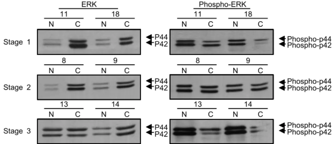
2) Levels of ERK1/2 and activated ERK1/2 according to the axillary lymph node status and tumor stages
액와림프절 전이 여부에 따른 MAPK 및 활성화 정도는 림프절 전이 양성군에서 약간 감소하는 경향은 있었으나 통계적으로 유의한 차이는 없었으며, 병기의 변화에 따른 MAPKs의 차이는 3기 유방암에서 활성화 ERK1/2 증가의 경향은 있었으나 통계적으로 유의한 수준은 아니었다.
(Table 1, Fig. 3)
3) Levels of ERK1/2 and activated ERK1/2 according to HER2 and ER expressions
유방암 조직에서 HER2 발현 유무에 따른 MAPK 단백질 및 활성화 정도의 차이는 발견되지 않았으며(Table 1, Fig.
4), ER 발현에 따라서는 ER 양성군에서 ERK1/2 단백질이 증가한 경향이 있었고 특히 활성화 ERK1/2의 유의한(P<
0.05) 증가를 보였다(Table 1, Fig. 5).
4) Levels of ERK1/2 and activated ERK1/2 according to histologic grade and MKP1 levels
종양의 분화도에 따른 ERK1/2 및 활성화 정도의 차이는
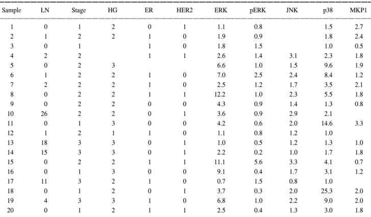
Table 1. Clinico-pathological characteristics and the ratios of MAPKs in cancer tissues over paired normals
ꠚꠚꠚꠚꠚꠚꠚꠚꠚꠚꠚꠚꠚꠚꠚꠚꠚꠚꠚꠚꠚꠚꠚꠚꠚꠚꠚꠚꠚꠚꠚꠚꠚꠚꠚꠚꠚꠚꠚꠚꠚꠚꠚꠚꠚꠚꠚꠚꠚꠚꠚꠚꠚꠚꠚꠚꠚꠚꠚꠚꠚꠚꠚꠚꠚꠚꠚꠚꠚꠚꠚꠚꠚꠚꠚꠚꠚꠚꠚꠚꠚꠚꠚꠚꠚꠚꠚꠚꠚꠚꠚꠚꠚꠚꠚꠚꠚꠚꠚꠚꠚꠚꠚꠚꠚꠚꠚꠚꠚꠚꠚꠚꠚꠚꠚ
Sample LN Stage HG ER HER2 ERK pERK JNK p38 MKP1
ꠏꠏꠏꠏꠏꠏꠏꠏꠏꠏꠏꠏꠏꠏꠏꠏꠏꠏꠏꠏꠏꠏꠏꠏꠏꠏꠏꠏꠏꠏꠏꠏꠏꠏꠏꠏꠏꠏꠏꠏꠏꠏꠏꠏꠏꠏꠏꠏꠏꠏꠏꠏꠏꠏꠏꠏꠏꠏꠏꠏꠏꠏꠏꠏꠏꠏꠏꠏꠏꠏꠏꠏꠏꠏꠏꠏꠏꠏꠏꠏꠏꠏꠏꠏꠏꠏꠏꠏꠏꠏꠏꠏꠏꠏꠏꠏꠏꠏꠏꠏꠏꠏꠏꠏꠏꠏꠏꠏꠏꠏꠏꠏꠏꠏꠏ
1 0 1 2 0 1 1.1 0.8 1.5 2.7
2 1 2 2 1 0 1.9 0.9 1.8 2.4
3 0 1 1 0 1.8 1.5 1.0 0.5
4 2 2 1 1 2.6 1.4 3.1 2.3 1.8
5 0 2 3 6.6 1.0 1.5 9.6 1.9
6 1 2 2 1 0 7.0 2.5 2.4 8.4 1.2
7 2 2 2 1 0 2.5 1.2 1.7 3.5 2.1
8 0 2 2 1 1 12.2 1.0 2.3 5.5 1.8
9 0 2 2 0 0 4.3 0.9 1.4 1.3 0.8
10 26 2 2 0 1 3.6 0.9 2.9 2.1
11 0 1 3 0 0 4.2 0.6 2.0 14.6 3.3
12 1 2 1 1 0 1.1 0.8 1.2 1.0
13 18 3 3 0 1 1.0 0.5 1.2 1.3 1.0
14 15 3 3 0 1 2.2 0.2 1.0 1.7 1.8
15 0 2 2 1 1 11.1 5.6 3.3 4.1 0.7
16 0 1 3 0 0 9.1 0.4 1.7 3.1 1.2
17 11 3 2 1 0 0.7 1.5 0.8 1.0
18 0 1 2 0 1 3.7 0.3 2.0 25.3 2.0
19 4 3 3 1 0 6.8 1.0 2.2 9.0 2.0
20 0 1 2 1 1 2.5 0.4 1.3 3.0 1.8
ꠏꠏꠏꠏꠏꠏꠏꠏꠏꠏꠏꠏꠏꠏꠏꠏꠏꠏꠏꠏꠏꠏꠏꠏꠏꠏꠏꠏꠏꠏꠏꠏꠏꠏꠏꠏꠏꠏꠏꠏꠏꠏꠏꠏꠏꠏꠏꠏꠏꠏꠏꠏꠏꠏꠏꠏꠏꠏꠏꠏꠏꠏꠏꠏꠏꠏꠏꠏꠏꠏꠏꠏꠏꠏꠏꠏꠏꠏꠏꠏꠏꠏꠏꠏꠏꠏꠏꠏꠏꠏꠏꠏꠏꠏꠏꠏꠏꠏꠏꠏꠏꠏꠏꠏꠏꠏꠏꠏꠏꠏꠏꠏꠏꠏꠏ LN, HG, and ER represents the number of metastatic lymph nodes in the axilla, hitologic grade, and estrogen receptor, respectively. The values of MAPKs were calculated by denomination of the optical density of Western blots of Cancer tissue over that of paired normal tissue.
ꠏꠏꠏꠏꠏꠏꠏꠏꠏꠏꠏꠏꠏꠏꠏꠏꠏꠏꠏꠏꠏꠏꠏꠏꠏꠏꠏꠏꠏꠏꠏꠏꠏꠏꠏꠏꠏꠏꠏꠏꠏꠏꠏꠏꠏꠏꠏꠏꠏꠏꠏꠏꠏꠏꠏꠏꠏꠏꠏꠏꠏꠏꠏꠏꠏꠏꠏꠏꠏꠏꠏꠏꠏꠏꠏꠏꠏꠏꠏꠏꠏꠏꠏꠏꠏꠏꠏꠏꠏꠏꠏꠏꠏꠏꠏꠏꠏꠏꠏꠏꠏꠏꠏꠏꠏꠏꠏꠏꠏꠏꠏꠏꠏꠏꠏ
없었으며(Table 1) MKP1 발현이 증가한 유방암군에서 HER2 발현이 상대적으로 높았고(60% vs 33%) ERK단백질의 발 현은 유사하였으나 통계적 유의성은 없었으며 활성화
ERK1/2의 감소가 경계적 유의성(P=0,086)을 보였다(Table 1, Fig. 6).
Fig. 3. Levels of MAPKs in the representative breast cancers and paired normals according to tumor stages. The ERK1/2 proteins were detected by Western blot analysis by using ERK1/2 polyclonal antibody (left panel). The phospho-ERK1/2 proteins were detected by Western blot analysis by using phospho-specific MAPK (pERK1/2) antibody (right panel). Uppermost panel represents stage 1, middle one represents stage 2, and lowest panel represents stage 3. The levels of ERK1/2 and activated ERK1/2 proteins were not different between stages. N and C represent normal and carcinoma, respectively.
Stage 1 P44
P42
P44P42
P44P42
Phospho-p44 Phospho-p42
Phospho-p44 Phospho-p42
Phospho-p44 Phospho-p42 Stage 2
Stage 3
11 18 11 18
N C N C N C N C
8 9 8 9
N C N C N C N C
13 14 13 14
N C N C N C N C
ERK Phospho-ERK
Fig. 4. Levels of MAPKs in the representative breast cancers and paired normals according to HER2 expressions. The ERK1/2 proteins were detected by Western blot analysis by using ERK1/2 polyclonal antibody (upper panel). The phospho-ERK1/2 proteins were detected by Western blot analysis by using phospho-specific MAPK (pERK1/2) antibody (lower panel). There were no significant differences of the levels of ERK1/2 and activated ERK1/2 proteins between HER2-negative (left panel) and HER2-positive cancers.
N and C represent normal and carcinoma, respectively.
2 3 13 14
N C N C N C N C
HER2(-) HER2(+)
ERK P44
P42 Phospho-p44 Phospho-p42 P44P42
Phospho-p44 Phospho-p42 Phospho-ERK
Fig. 5. Levels of MAPKs in the representative breast cancers and paired normals according to ER expressions. The ERK1/2 proteins were detected by Western blot analysis by using ERK1/2 polyclonal antibody (upper panel). The phospho-ERK1/2 proteins were detected by Western blot analysis by using phospho-specific MAPK (pERK1/2) antibody (lower panel). There were significantly higher levels of activated ERK1/2 proteins ER-positive (left panel) cancers than those in ER-negative cancers (P<0.05). N and C represent normal and carcinoma, respectively.
6 7 11 18
N C N C N C N C
ER(+) ER(-)
ERK P44
P42
Phospho-p44 Phospho-p42 Phospho-ERK
박병우 외:유방암 발암과 진행에 있어 c-erbB-2 수용체 발현과 Mitogen-activated Protein Kinases 활성도의 역할 11 ꠏꠏꠏꠏꠏꠏꠏꠏꠏꠏꠏꠏꠏꠏꠏꠏꠏꠏꠏꠏꠏꠏꠏꠏꠏꠏꠏꠏꠏꠏꠏꠏꠏꠏꠏꠏꠏꠏꠏꠏꠏꠏꠏꠏꠏꠏꠏꠏꠏꠏꠏꠏꠏꠏꠏꠏꠏꠏꠏꠏꠏꠏꠏꠏꠏꠏꠏꠏꠏꠏꠏꠏꠏꠏꠏꠏꠏꠏꠏꠏꠏꠏꠏꠏꠏꠏꠏꠏꠏꠏꠏꠏꠏꠏꠏꠏꠏꠏꠏꠏꠏꠏꠏꠏꠏꠏꠏꠏꠏꠏꠏꠏꠏꠏꠏ
고 찰
MAPK의 활성화는 세포증식과 밀접한 상관성이 있으며 섬유아세포(fibroblast)에 MAPK kinase (MEK)의 발현을 통 해 MAPK를 활성화시키면 oncogenicity를 유도할 수 있 다.(10) 정상유방조직에서보다 유방암조직에서 MAPK 발현 의 증가와 활성화 MAPK (activated form)의 증가의 보고가 많다. MAPK 활성도는 유방암의 악성도에 필수적이지만 MAPK 단백 발현정도와 MAPK 활성도와 상관성은 일부 종 양에서만 관찰되며 이는 MAPK 과발현이 곧 활성도의 증가 를 의미하는 것은 아니며, MAPK 단백질 발현정도보다는 활성화 MAPK의 존재가 세포증식능을 나타낸다.(11) 본 연 구에서도 유방암조직에서 ERK뿐 아니라 JNK 및 p38 등 같 은 신호전달계 내의 knase의 발현이 정상조직보다 유방암 조직에서 현저함이 관찰되었다. 그러나 activated form의 과 발현은 전체 발현보다는 현저하지 않았으며 이는 Mueller(11) 등이 보고한 MAPK 단백발현이 MAPK 활성도 증가와 항상 일치하지는 않으며 일부 종양에서만 관찰된다는 보고와 일 치하는 소견이었다.
MAPK는 세포증식에 중추적 역할을 하며 다양한 세포 외 신호 및 여러 암유전자 산물에 의해 조절되며,(12-15) 성장 인자, 호르몬 및 신경전달물에 의해 급속히 인산화되어 활 성화되며 방사선조사, 과산화수소(hydrogen peroxide) 및 자 외선에 의해서도 활성화될 수 있다.(16) 펩티드 성장인자는 ERK1/2의 주 조절자(major regulator)이며,(17) HER1 또는 HER2 수용체 과발현 유방암 세포에서는 ras 및 MAPK의 발현이 증가하는 데,(18) 종양의 높은 악성도의 표현형(an aggressive tumor phenotype)일 수 있다.(14) 최근의 연구결과 유방암의 경우 활성화 MAPK의 세포분포가 높은 것으로 보 고되고 있으며,(19) 에스트로젠 및 프로제스테론 역시 MAPK 활성화에 관여하는 것으로 보고되고 있다.(18) 그러나 세포 증식과 세포고사는 본질적으로 연결되어 있으며, potent mi- togenic stimuli는 생존인자가 존재하는 환경에서는 세포증
식을 유도하지만 그렇지 않은 환경에서는 세포고사를 초래 한다.(20-23) 즉 MAPK 신호전달계의 활성화를 통하여 세포 의 환경조건에 따라 세포증식 또는 세포고사가 일어날 수 있음을 시사한다. 본 연구의 결과에서 활성화 ERK1/2의 증 가는 HER2의 발현과는 유의한 상관성을 보이지 않았고, ER 발현 유방암조직에서 현저하였는데(P<0.05), 이는 에스 트로젠 의존성 환경과 MAPK 활성화의 상관 및 세포증식과 의 상관성을 시사한다고 하겠다. 따라서 향후 ER 발현과 HER2 발현 및 MAPK 신호전달계의 상호연관성에 대한 연 구도 필요할 것으로 생각된다.
에스트라디올 의존성 종양세포의 MAPK 활성화 기전은 첫째, MAPK에 의한 ER의 인산화 및 전사의 강화; 둘째, 에 스트라디올의 성장인자 자극을 통한 MAPK의 증가; 셋째, 에스트라디올의 세포막 ER을 이용하는 경로의 활성화 등 이 제기되고 있으나,(19) 여러 연구 결과(24-26) 에스트라디 올은 MAPK 활성화와 관계없이 유방암세포 증식을 자극하 는 것으로 보고되고 있다.
ERK 활성도는 액와림프절전이 유방암에서 증가하는 경 향(11)이며 추적기간 중 재발을 보인 환자군에서 높은 MAPK 활성도를 보여 원발종양의 MAPK 활성도가 무병생존율의 예후인자로 작용할 가능성을 제시하였다.(11) 본 연구에서 는 액와림프절 전이유무에 따라 ERK 및 JNK 발현의 차이 는 없었으며 p38의 발현은 액와림프절 양성 유방암조직에 서 감소하였고, 종양의 에스트로젠수용체 발현 유무에 따 라서는 ER 양성군에서 ERK1/2의 발현 및 활성도가 증가하 였으며 p38 발현은 감소하는 경향을 보인 반면 JNK 발현의 차이는 없었다. 이는 에스트로젠 의존성 유방암의 경우 세 포증식과 관련있는 ERK1/2의 활성화에 좋은 환경을 제공 하는 것을 시사하고, 따라서 세포고사와 관계있는 JNK단백 의 발현의 변화가 없었던 것으로 생각된다. 또한 병기의 진 행에 따라 병기가 진행될수록 ERK1/2의 발현 및 활성도의 유의한 차이는 없었다.
이상의 결과를 종합하면 유방암조직에서 ERK1/2 단백질 총량은 정상조직에 비해 현저히 증가하나(평균 4.31배: 20 Fig. 6. Levels of MAPKs in the representative breast cancers and paired normals according to MKP1 expression. There were no significant
differences in ERK1/2 and activated forms between higher MKP1 (samples 4, 5, and 8) and Lower MKP1 groups (sample 9).
N and C represent normal and carcinoma, respectively.
4 5 8 9
N C N C N C N C
ERK
4238
P44P42
Phospho-ERK Phospho-p44
Phospho-p42 MKP-1
ꠏꠏꠏꠏꠏꠏꠏꠏꠏꠏꠏꠏꠏꠏꠏꠏꠏꠏꠏꠏꠏꠏꠏꠏꠏꠏꠏꠏꠏꠏꠏꠏꠏꠏꠏꠏꠏꠏꠏꠏꠏꠏꠏꠏꠏꠏꠏꠏꠏꠏꠏꠏꠏꠏꠏꠏꠏꠏꠏꠏꠏꠏꠏꠏꠏꠏꠏꠏꠏꠏꠏꠏꠏꠏꠏꠏꠏꠏꠏꠏꠏꠏꠏꠏꠏꠏꠏꠏꠏꠏꠏꠏꠏꠏꠏꠏꠏꠏꠏꠏꠏꠏꠏꠏꠏꠏꠏꠏꠏꠏꠏꠏꠏꠏꠏ 예 중 19예에서 1.01∼12.21배 증가) 활성화 ERK1/2의 발현
은 ER 발현과 유의한 상관성(P<0.05)을 보여 ERK1/2의 활 성화는 에스트로젠 의존성 환경의 영향을 받고있음을 시사 한다고 하겠고, 기존의 보고와는 달리 HER2의 발현에 따른 영향은 상대적으로 적은 것으로 보인다. 이는 유방암의 발 생과 진행과정에 에스트로젠의 영향과 에스트로젠과 MAPK 신호전달계의 상호작용의 가능성을 시사한다고 하겠다. 또 stress-activated protein kinase로 알려진 p38 및 JNK 단백질의 역할은 완전히 규명되지 않고 있는데, 이에 대한 연구가 병 행되어야 할 것으로 생각된다.
유방암 원발병소 및 전이병소에 MAPK의 과발현이 나타 났다. 림프절에 전이된 유방암세포의 전이를 함유한 림프 절에 mapk의 과발현이 나타나고 유방암의 경우 양성종양 의 경우에 비해 약 5∼10배의 MAPK 활성이 증가하였다.(5) 이렇게 mapk 발현이 약 5∼20배 증가하는 것이 유방암 발 생 또는 전이에 결정적인 역할을 할 수 있다.(5) MAPK mRNA의 발현은 전이도(metastatic potential)이 높은 유방암 세포주에 더욱 증가되어 있으며,(27) 특히 MAPK의 활성화 는 종양의 병기 및 조직분화도와 유의한 상관성을 보였 다.(4) 본 연구의 결과로 볼 때 ERK1/2의 발현과 활성화는 유방암 조직의 조직분화도와 유의한 상관성을 보이지 않았 다. 이에 대한 결론은 추후 대상 환자 수의 증가를 통해 검 증하여야 할 것이고 MAPK의 활성화와 생존율과 상관성 역 시 추적결과를 통해 검증하여야 하겠다.
MKP1은 MAPK의 탈인산화를 통하여 MAPK의 비활성화 에 관여하는 효소로 mitogenic signal에 의해 유발되며, 배양 세포에 과발현시키면 MAPK에 의한 mitogenic effect를 감소 시키고,(6) 분화를 억제한다(block differentiation).(7) MKP1 은 mitogenic signals, 즉 MAPK 활성화를 유도하는 신호들에 의해 발현이 유도된다.(28) MKP1은 전립선암, 결장암 및 방 광암 발암의 초기단계에서 과발현되며 분화도가 커지고 전 이가 될수록 지속적으로 발현을 상실한다. 한편 유방암에 서는 MKP1 발현이 현저하고 특히 저분화암이나 말기에 이 르러서도 MKP1 발현이 뚜렷하다. MKP1의 과발현과 관계 없이 ERK1의 효소적 활성도는 증가되어 있었다. 이러한 사 실은 MKP1은 다양한 상피세포암의 초기표지자임을 나타 낼 뿐 MKP1이 상피세포암 발암을 억제하는 종양억제자로 활동하는 것은 아님을 시사한다 하겠다.(8) MKP1 mRNA의 증가와 함께 대부분의 종양에서 ERK1, 2의 증가를 보였다.
전립선암, 결장암 및 방광암에서 MKP1의 발현은 종양의 분화도 및 병기와 역상관관계를 보여 저분화암(high-grade tumor) 및 전이암에서 MKP1 및 ERKs 발현이 상실되는데, 이는 종양의 진행에 따른 MAPK 의존성의 탈피현상(MAPK independence)이 나타남을 시사한다.(29,30) 반면 유방암의 경우 종양진행의 후기까지도 MAPK 의존성으로 남는 경향 이 있다.(8) 본 연구의 결과 MKP1의 발현과 HER2 발현, ER 발현, 조직분화도, 종양의 병기, ERK1/2 발현 및 활성화
ERK1/2와 유의한 상관성은 보이지 않았다. 이는 유방암조 직에서 정상조직에 비해 MKP1의 발현정도가 높은 결과와 더불어 MKP1의 발현은 MAPK 발현증가에 따른 반작용으 로 상피세포암의 초기표지자 역할(8)을 시사하는 것으로 생 각되나 MKP1 발현이 증가한 군에서 활성화 ERK1/2의 발 현이 감소하는 경향(P=0.086)을 보여 향후 이에 대한 보다 정확한 연구가 필요할 것으로 생각된다.
REFERENCES
1) The Korean Breast Cancer Society. Clinical chatracteristics of Korean brest cancer patients in 1998. J Korean Med Sci 2000;
15:569-79.
2) Slamon DJ, Clark GM, Wong SG, Levin WJ, Ullrich A, McGuire WL. Human breast cancer: correlation of relapse and survival with amplification of the HER-2/neu oncogene.
Science 1987;235:177-82.
3) Slamon DJ, Godolphin W, Jones LA, Holt JA, Wong SG, Keith DE, et al. Studies of the HER-2/neu proto-oncogene in human breast and ovarian cancer. Science 1989;244:707-12.
4) Oka H, Chatani Y, Hoshino R, Ogawa O, Kakehi Y, Terachi T, et al. Constitutive activation of mitogen-activated protein (MAP) kinases in human renal cell carcinoma. Cancer Res 1995;55(18):4182-7.
5) Sivaraman VS, Wang H, Nuovo GJ, Malbon CC. Hyperex- pression of mitogen-activated protein kinase in human breast cancer. J Clin Invest 1997;99:1478-83.
6) Sun H, Tonks NK, Bar-Sagi D. Inhibition of Ras-induced DNA synthesis by expression of the phosphatase MKP-1. Science 1994;266:285-8.
7) Yao H, Labudda K, Rim C, Capodieci P, Loda M, Stork PJ.
Cyclic adenosine monophosphate can convert epidermal growth factor into a differentiating factor in neuronal cells. J Biol Chem 1995;270:20748-53.
8) Loda M, Capodieci P, Mishra R, Yao H, Corless C, Grigioni W, et al. Expression of mitogen-activated protein kinase phos- phatase-1 in the early phases of human epithelial carcino- genesis. Am J Pathol 1996;149:1553-64.
9) Magi-Galluzzi C, Mishra R, Fiorentino M, Montironi R, Yao H, Capodieci P, et al. Mitogen-activated protein kinase phos- phatase 1 is overexpressed in prostate cancers and is inversely related to apoptosis. Lab Invest 1997;76:37-51.
10) Brunet A, Pages G, Pouyssegur J. Constitutively active mutants of MAP kinase kinase (MEK1) induce growth factor-relaxation and oncogenicity when expressed in fibroblasts. Oncogene 1994;9:3379-87.
11) Mueller H, Flury N, Eppenberger-Castori S, Kueng W, David F, Eppenberger U. Potential prognostic value of mitogen- activated protein kinase activity for disease-free survival of primary breast cancer patients. Int J Cancer 2000;89:384-8.
12) Blenis J. Signal transduction via the MAP kinases: proceed at
박병우 외:유방암 발암과 진행에 있어 c-erbB-2 수용체 발현과 Mitogen-activated Protein Kinases 활성도의 역할 13 ꠏꠏꠏꠏꠏꠏꠏꠏꠏꠏꠏꠏꠏꠏꠏꠏꠏꠏꠏꠏꠏꠏꠏꠏꠏꠏꠏꠏꠏꠏꠏꠏꠏꠏꠏꠏꠏꠏꠏꠏꠏꠏꠏꠏꠏꠏꠏꠏꠏꠏꠏꠏꠏꠏꠏꠏꠏꠏꠏꠏꠏꠏꠏꠏꠏꠏꠏꠏꠏꠏꠏꠏꠏꠏꠏꠏꠏꠏꠏꠏꠏꠏꠏꠏꠏꠏꠏꠏꠏꠏꠏꠏꠏꠏꠏꠏꠏꠏꠏꠏꠏꠏꠏꠏꠏꠏꠏꠏꠏꠏꠏꠏꠏꠏꠏ
your own RSK. Proc Natl Acad Sci USA 1993;90:5889-92.
13) Cobb MH, Goldsmith EJ. How MAP kinases are regulated.
J Biol Chem 1995;270:14843-6.
14) Janes PW, Daly RJ, deFazio A, Sutherland RL. Activation of the Ras signalling pathway in human breast cancer cells over- expressing erbB-2. Oncogene 1994;9:3601-8.
15) Callans LS, Naama H, Khandelwal M, Plotkin R, Jardines L.
Raf-1 protein expression in human breast cancer cells. Ann Surg Oncol 1995;2:38-42.
16) Stevenson MA, Pollock SS, Coleman CN, Calderwood SK.
X-irradiation, phorbol esters, and H2O2 stimulate mitogen- activated protein kinase activity in NIH-3T3 cells through the formation of reactive oxygen intermediates. Cancer Res 1994;
54:12-5.
17) Pearson G, Robinson F, Beers Gibson T, Xu BE, Karandikar M, Berman K, et al. Mitogen-activated protein (MAP) kinase pathways: regulation and physiological functions. Endocr Rev 2001;22:153-83.
18) von Lintig FC, Dreilinger AD, Varki NM, Wallace AM, Casteel DE, Boss GR. Ras activation in human breast cancer.
Breast Cancer Res Treat 2000;62:51-62.
19) Santen RJ, Song RX, McPherson R, Kumar R, Adam L, Jeng MH, et al. The role of mitogen-activated protein (MAP) kinase in breast cancer. J Steroid Biochem Mol Biol 2002;80:239-56.
20) Adams JM, Cory S. The Bcl-2 protein family: arbiters of cell survival. Science 1998;281:1322-6.
21) Datta SR, Dudek H, Tao X, Masters S, Fu H, Gotoh Y, Greenberg ME. Akt phosphorylation of BAD couples survival signals to the cell-intrinsic death machinery. Cell 1997;91:
231-41.
22) Kauffmann-Zeh A, Rodriguez-Viciana P, Ulrich E, Gilbert C, Coffer P, Downward J, et al. Suppression of c-Myc-induced
apoptosis by Ras signalling through PI(3)K and PKB. Nature 1997;385:544-8.
23) Nass SJ, Dickson RB. Defining a role for c-Myc in breast tumorigenesis. Breast Cancer Res Treat 1997;44:1-22.
24) Cicatiello L, Addeo R, Altucci L, Belsito Petrizzi V, Boccia V, Cancemi M, et al. The antiestrogen ICI 182,780 inhibits proliferation of human breast cancer cells by interfering with multiple, sequential estrogen-regulated processes required for cell cycle completion. Mol Cell Endocrinol 2000;165:199-209.
25) Addeo R, Altucci L, Battista T, Bonapace IM, Cancemi M, Cicatiello L, et al. Stimulation of human breast cancer MCF-7 cells with estrogen prevents cell cycle arrest by HMG-CoA reductase inhibitors. Biochem Biophys Res Commun 1996;
220:864-70.
26) Altucci L, Addeo R, Cicatiello L, Dauvois S, Parker MG, Truss M, et al. 17beta-Estradiol induces cyclin D1 gene tran- scription, p36D1-p34cdk4 complex activation and p105Rb phosphorylation during mitogenic stimulation of G(1)-arrested human breast cancer cells. Oncogene 1996;12:2315-24.
27) Pencil SD, Toh Y, Nicolson GL. Candidate metastasis-associated genes of the rat 13762NF mammary adenocarcinoma. Breast Cancer Res Treat 1993;25:165-74.
28) Sun H, Charles CH, Lau LF, Tonks NK. MKP-1 (3CH134), an immediate early gene product, is a dual specificity phos- phatase that dephosphorylates MAP kinase in vivo. Cell 1993;
75:487-93.
29) Fearon ER, Vogelstein B. A genetic model for colorectal tumorigenesis. Cell 1990;61:759-67.
30) Kastrinakis WV, Ramchurren N, Rieger KM, Hess DT, Loda M, Steele G, et al. Increased incidence of p53 mutations is associated with hepatic metastasis in colorectal neoplastic progression. Oncogene 1995;11:647-52.