*Corresponding author. Tel: +82-33-760-2860; Fax: +82-33-760- 5224; E-mail: [email protected]
http://dx.doi.org/10.5483/BMBRep.2012.45.10.120
Received 5 June 2012, Revised 27 June 2012, Accepted 11 July 2012 Keywords: JNK, Leukotactin-1, Macrophage, Mycobacterium tuber-culosis, p38 MAPK
ISSN: 1976-6696 (print edition) / 1976-670X (electronic edition)
Copyright ⓒ 2012 by the The Korean Society for Biochemistry and Molecular Biology
c-Jun N-terminal kinase (JNK) and p38 mitogen-activated
protein kinase (p38 MAPK) are involved in Mycobacterium
tuberculosis-induced expression of Leukotactin-1
Jang-Eun Cho
1, Sangjung Park
2, Sang-Nae Cho
3, Hyeyoung Lee
2& Yoon Suk Kim
2,*
1Department of Biomedical Laboratory Science, Daegu Health College, Daegu 702-722, 2Department of Biomedical Laboratory Science,
College of Health Scineces, Yonsei University, Wonju 220-710, 3Department of Microbiology, Yonsei University College of Medicine, Seoul
120-752, Korea
Leukotactin(Lkn)-1 is a CC chemokine and is upregulated in macrophages in response to Mycobacterium tuberculosis (MTB) infection. We investigated whether mitogen-activated protein kinases (MAPKs) are involved in MTB-induced expression of Lkn-1. The up-regulation of Lkn-1 by infection with MTB was inhibited in cells treated with inhibitors specific for JNK (SP600125) or p38 MAPK (SB202190). Since the up-regulation of Lkn-1 by MTB has been reported to be mediated by the PI3-K/PDK1/Akt signaling, we examined whether JNK and/or p38 MAPK are also involved in this signal pathway. MTB-induced Akt phosphorylation was blocked by treatment with JNK- or p38 MAPK-specific inhibitors implying that p38 and JNK are upstream of Akt. In addition, treatment with the PI3-K-specific inhibitor inhibited MTB-stimulated activation of JNK or p38 MAPK implying that PI3-K is upstream of JNK and p38 MAPK. These results collectively suggest that JNK and p38 MAPK are involved in the signal pathway responsible for MTB-induced up-regulation of Lkn-1. [BMB Reports 2012; 45(10): 583-588]
INTRODUCTION
Mycobacterium tuberculosis (MTB) is an intracellular pathogen
and is the causative agent of pulmonary tuberculosis which is one of the main causes of morbidity and mortality worldwide (1). Infection of human macrophages with MTB induces release of a variety of cytokines which participate in protective and im-mune host responses during human tuberculosis (2-6). Down- regulation of cytokine receptors in T cells has been reported to result in ineffective control of persisting pathogens such as MTB
(7). MTB induces the expression of a variety of chemokines such as monocyte chemotactic prote1 (MCP-1), macrophage in-flammatory protein-1α (MIP-1α), macrophage inin-flammatory pro-tein-1β (MIP-1β) and regulated upon activation, normal T-cell ex-pressed and secreted (RANTES) (8-12). These induced chemo-kines have been reported to be involved in the inhibition of in-tracellular MTB growth, formation of granulomas, and the re-cruitment of lymphocytes and monocytes to pleural cavity in TB patients (13-18).
Mycobacterial infection induced-expression of a variety of chemokines is known to be mediated via various signaling path-ways (19, 20). However, the signaling pathpath-ways by which MTB induces production of a variety of cytokines in macrophages are not yet fully understood. Recent studies have suggested that the mitogen-activated protein kinase (MAPK) pathway is an im-portant component of mycobacterial pathogenesis (21). The MAPK signaling pathway is a highly conserved pathway that is important in diverse aspects of the immune response (22-24). The MAPK family includes ERK, p38 MAPK, and stress-activated protein kinase/c-Jun N-terminal kinase (JNK) (25, 26). Activation of p38 MAPK has been reported to be induced in monocytes fol-lowing infection with MTB (27). In addition, the MAPK signaling pathway was shown to be essential for the mycobacterium-in-duced production of proinflammatory cytokines, such as tumor necrosis factor alpha (TNF-α), IL-1β, and MCP-1 (28, 29). Recently, it has been demonstrated that leukotactin-1 (Lkn-1), a member of the CC chemokine family, is induced during myco-bacterial infection implying the association of Lkn-1 with the de-velopment of tuberculosis. In addition, it has been shown that MTB-induced upregulation of Lkn-1 is mediated by the PI3-K/ PDK1/Akt signaling pathway (30, 31). However, involvement of additional signaling pathways in MTB-induced Lkn-1 regulation is unknown. In the present study, we aimed to elucidate the in-volvement of the MAPK pathway in MTB-induced expression of Lkn-1. We demonstrated that p38 MAPK and JNK are associated with regulation of Lkn-1 expression in response to MTB infection in macrophages.
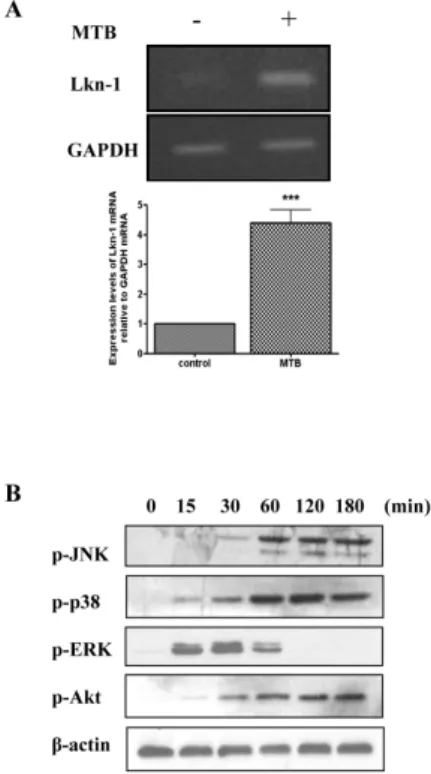
Fig. 1. MTB enhances expression of Lkn-1 and activates MAPKs. (A) THP-1 cells were treated with 100 nM PMA for 48 h, and infected with MTB (10 MOI) for 4 h. Total RNA was extracted and cDNA was prepared. PCR analysis was performed using Lkn-1-specific pri-mers and the PCR products were resolved by 1.8% agarose gel (upper panel). GAPDH was used as an internal control. Densitomet-ric analysis was performed (lower panel). Data are expressed as mean ± SD and are presented as expression levels of Lkn-1 mRNA relative to GAPDH mRNA (The expression level of Lkn-1 relative to GAPDH in the absence of mycobacterial infection was set to 1.0). The data represent results from three independent experiments. Statistical analysis was performed by Student’s t-test (***P < 0.001 relative to uninfected control). (B) Differentiated THP-1 cells were starved for 16 h and infected with MTB (10 MOI) for the indicated times (0, 15, 30, 60, 120, 180 min) before cell lysates were pre-pared, Cell lysates were resolved on 12% SDS-polyacrylamide gels and transferred to nitrocellulose membranes. Phosphorylation of JNK, p38 MAPK, ERK, and Akt was detected by Western blotting using anti-phospho-JNK, p38 MAPK, ERK, or Akt, respectively. Beta-actin was used as an internal control.
RESULTS AND DISCUSSION
MTB enhances expression of Lkn-1 and activates MAPKs
Our previous study reported the involvement of Lkn-1 in the im-mune response of macrophages against MTB and revealed that MTB infection causes increased expression and secretion of Lkn-1 in differentiated THP-1 cells (31). In the present study, we reconfirmed the infection of THP-1 cells with MTB induced up-regulation of Lkn-1 mRNA (Fig. 1A). Next, we examined whether infection with MTB activates protein kinases included in the MAPK family which is known to be associated with
MTB-in-duced production of cytokines (22-24). THP-1 cells were treated with PMA for 48 h and incubated with MTB for the indicated times. Phosphorylation of MAP kinases was detected by Western blotting. As shown in a previous report (31), MTB infection acti-vated Akt (Fig. 1B). In addition, infection of MTB caused phos-phorylation of MAP kinases including JNK, p38, and ERK (Fig. 1B). Data showed that MTB-induced phosphorylation of JNK and p38 MAPK was detectable 30 min after infection and persisted for at least 180 min after MTB infection. This pattern is similar to MTB-induced phosphorylation pattern of Akt. In contrast, phos-phorylation of ERK was detectable 15 min after infection but de-clined thereafter and was undetectable at 120 min after MTB in-fection (Fig. 1B). These results suggest that in addition to the pre-viously identified Akt, MAP kinases may be also involved in MTB-induced up-regulation of Lkn-1.
p38 MAPK and JNK are involved in MTB-stimulated up-regulation of Lkn-1
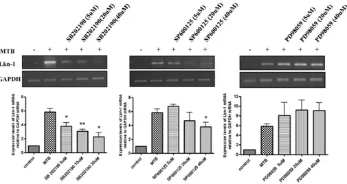
Based on the results above, we determined whether MAP kin-ases such as JNK, p38, and ERK are associated with MTB-in-duced expression of Lkn-1. PMA-treated THP-1 cells were pre-treated with the JNK inhibitor (SP600125), the p38 inhibitor (SB202190), or the MEK1 inhibitor (PD98059) for 45 min before exposure to MTB for 4 h. The mRNA level of Lkn-1 was detected by semi-quantitative PCR analysis. As shown in Fig. 2, treatment with SP600125 (an inhibitor of JNK) or SB202190 (an inhibitor of p38 MAPK) abolished MTB-induction of Lkn-1 in an inhibitor dose-dependent manner. However, pre-treatment with PD98059 (an inhibitor of MEK1) did not influence mRNA induction of Lkn-1 by MTB (Fig. 2). These data suggests that the p42 and p44 extracellular signal-regulated kinases 1 and 2 (ERK1/2) pathways are not associated with MTB-induced up-regulation of Lkn-1 since MEK1 is an upstream signaling molecule of ERK1/2. These results imply that although JNK, p38, and ERK were all activated in response to MTB infection, only JNK and p38 showed associa-tion with MTB-induced Lkn-1 expression. As shown in Fig. 1, al-though JNK, p38, and ERK were all activated by infection with MTB, the pattern of activation was different between JNK/p38 MAPK and ERK. Activation of JNK and p38 MAPK persisted for at least 3 h after MTB infection which is similar to the activation pattern of Akt. However, phosphorylation of ERK disappeared at 120 min after MTB infection (Fig. 1B). It is possible that pro-longed activation (for at least more than 3 h) of signaling mole-cules is required for MTB-induced expression of Lkn-1.
Lkn-1 induction by infection with MTB is mediated through PI3-K to p38/JNK to Akt signaling pathway
We previously demonstrated that the up-regulation of Lkn-1 caused by MTB infection was mediated by PI3-K, PDK1 and Akt (31). Here we determined whether JNK and p38 MAPK activated during MTB-induction of Lkn-1 are separate from or part of the PI3-K/PDK1/Akt signaling pathway. As shown in Fig. 3A, pre-treatment with a JNK inhibitor (SP600125) or p38 inhibitor (SB202190) before infection with MTB diminished MTB-induced
Fig. 2. p38 MAPK and JNK mediates MTB-induced expression of Lkn-1. PMA-treated THP-1 cells were pre-incubated with the inhibitors SB202190 (5 μM, 20 μM, 40 μM), SP600125 (5 μM, 20 μM, 40 μM), PD98059 (5 μM, 20 μM, 40 μM) for 45 min, followed by myco-bacterial infection (10 MOI) for 4 h. cDNA was prepared from total RNA extracted from treated cells. PCR analysis was performed using Lkn-1-specific primers. PCR products were analyzed by 1.8% agarose gel (upper panel) to detect Lkn-1 expression. GAPDH was used as an internal control. Densitometric analysis was performed (lower panel). Data are expressed as mean ± SD, and are presented as ex-pression levels of Lkn-1 mRNA relative to GAPDH mRNA. (The level of Lkn-1 relative to GAPDH in the absence of mycobacterial in-fection was set to 1.0). The data are results from three independent experiments. Statistical analysis was performed by Student’s t-test. (*P < 0.05, **P < 0.01 relative to uninfected control).
Fig. 3. MTB-stimulated expression of Lkn-1 is partially mediated through PI3-K to p38/JNK to Akt signaling pathway. (A) PMA-treated THP-1 cells were starved for 16 h, and treated with SP600125 (20 μM) or SB202190 (20 μM) for 45 min. Subsequently, cells were in-fected with MTB (10 MOI) for the indicated times before cell lysates were prepared, resolved on 12% SDS-polyacrylamide gels and trans-ferred to nitrocellulose membranes. Akt phosphorylation was determined by Western blotting using anti-phopho-Akt antibody. Differentiated THP-1 cells were starved for 16 h, and treated with Ly294002 (20 μM) or OSU03012 (5 μM) for 45 min. Subsequently, cells were in-fected with MTB (10 MOI) for the indicated times before cell lysates were prepared. Phosphorylation of p38 MAPK (B) or JNK (C) was determined by Western blotting using anti-phopho-p38 MAPK and anti-phopho-JNK antibody, respectively. Beta-actin was used as an in-ternal control.
phosphorylation of Akt. The levels of phosphorylated p38 MAPK and JNK were not affected by pre-treatment with the Akt in-hibitor before stimulation with MTB (data not shown). These re-sults show that JNK and p38 are upstream molecules of Akt in
the signaling pathway.
In addition, differentiated THP-1 cells were pre-treated with Ly294002 (PI3-K inhibitor) or OSU03012 (PDK1 inhibitor) be-fore exposure to MTB and phosphorylation of JNK and p38
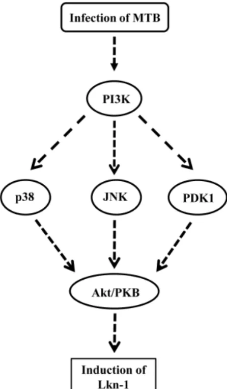
Fig. 4. The schematic model showing signal transduction pathway associated with MTB-induced expression of Lkn-1.
MAPK was assessed. Ly294002 treatment partially prevented phosphorylation of JNK and p38 MAPK (Fig. 3B and C, middle panel). In contrast, inhibition of PDK1 did not influence the lev-els of phosphorylated JNK and p38 MAPK (Fig. 3B and C, right panel). These data suggest that PI3-K is an upstream kinase of p38 MAPK and JNK in the signal pathway associated with up-regulation of Lkn-1 by MTB infection. In contrast, PDK1 does not seem to be an upstream molecule of p38 MAPK or JNK in this signal pathway. These results imply that increased ex-pression of Lkn-1 in response to MTB infection is mediated via three pathways: the PI3-K/PDK1/Akt, PI3-K/p38 MAPK/Akt, and the PI3-K/JNK/Akt signaling pathways (Fig. 4). Further studies are needed to determine if JNK and p38 MAPK are upstream mole-cules of PDK1.
In conclusion, in addition to the previously reported PI3-K/ PDK1/Akt pathway, we identified two additional signaling mole-cules, p38 MAPK and JNK, involved in MTB-induced Lkn-1 ex-pression in PMA-differentiated THP-1 cells. The elucidation of the signaling pathways involved in MTB-induced Lkn-1 ex-pression during tuberculosis development may provide useful in-formation which can be exploited for therapeutic intervention.
MATERIALS AND METHODS
Materials
Specific inhibitors of p38 MAPK (SB202190), MEK1 (PD98059),
JNK (SP600125), PI3-K (Ly294002) were purchased from Calbio-chem (San Diego, CA, USA). Specific inhibitors of PDK1 (OSU-03012) were purchased from Cayman (Ann Arbor, MI, USA). Dimetyl sulphoxide (DMSO) was obtained from Sigma-Aldrich (St. Louis, MO, USA).
Preparation of mycobacteria
MTB H37Rv (ATCC 27294) used in this study was grown for about four weeks at 37°C as a surface pellicle on Sauton me-dium enriched with 0.4% some-dium glutamate and 3.0% glycerol. The surface pellicles were collected and disrupted by gentle vor-texing with 6 mm glass beads. After clumps were settled, the up-per suspension was collected and aliquots were stored at −80oC. Before infection, aliquots were thawed and quantitated for viable colony-forming units (CFU) on Middlebrook 7H10 agar (Difco, Detroit, MI, USA).
Cell culture and infection of MTB
The THP-1 cells were maintained in RPMI 1640 medium with 2 mM glutamine, 10% heat inactivated fetal bovine serum, 100 U/ml penicillin and 100 μg/ml streptomycin (Gibco-BRL, Grand Island, NY, USA) at 37oC under 5% CO2. THP-1 cells were
seed-ed in six-well plates and treatseed-ed with 100 nM phorbol-12-myr-istate-13-acetate (PMA; Sigma) for 48 h to induce differentiation into macrophage-like cells, then washed three times with RPMI 1640 medium. Before infection, differentiated THP-1 cells were reconstituted in antibiotic free RPMI 1640 medium with 10% FBS. PMA-differentiated THP-1 cells were pretreated with in-hibitors for 45 min before stimulation with MTB H37Rv for 4 h at MOI 10.
RNA extraction and semi-quantitative reverse transcriptase PCR (RT-PCR)
After removing nonphagocytosed bacilli, total RNA was extracted from cultured cells using Trizol reagent (Invitrogen, Carlsbad, CA, USA) according to the manufacturer’s instructions. cDNA was synthesized by reverse transcription with 2 μg total RNA, 0.25 μg of random hexamer (Invitrogen) and 200 unit of Murine Molony Leukemia Virus Reverse Transcriptase (MMLV-RT; Invitrogen) for 50 min at 37oC and 15 min at 70oC. Subsequent PCR amplifica-tion using 0.2 units of Taq polymerase (Cosmo Genetech, Seoul, Korea) was performed in a thermocycler (Applied Biosystems, Foster city, CA, USA) for 40 cycles (94oC for 30s, 55oC for 30s,
72oC for 30s) using the Lkn-1 primer (sense 5´-CCTCTCCTG CCTCATGCTTA-3´, antisense 5´-ACTGGGTTTGGCACAGACTT- 3´). GAPDH was amplified as an internal control with primers (sense 5´-CGGGAAGCTTGTGATCAATGG-3´, antisense 5´-GG CAGTGATGGCATGGACTG-3´). PCR products were electro-phoresed on 1.8% (w/v) agarose gels containing 0.5 μg/ml ethi-dium bromide, and the sizes of the products were determined by comparison to the 100 bp DNA ladder marker (Bioneer, Daejeon, Korea). The intensity of each band amplified by RT-PCR was ana-lyzed using Gel Doc EQ Quantity One (version 4.5, Bio-Rad, Milan, Italy) and normalized to GAPDH mRNA in corresponding
samples.
Western blot analysis
PMA-differentiated THP-1 cells were grown in six-well plates and were starved for 16 h, pretreated with inhibitors for 45 min, then infected with MTB for 0, 15, 30, 60, 120 or 180 min. After incubation, cells were washed twice in ice-cold phosphate-buf-fered saline (PBS) and solubilzed in lysis buffer containing 10 mM Tris (pH 7.0), 140 mM NaCl containing proteinase in-hibitors (Roche, Basel, Switzerland) and phophatase inhibitor(s) (Thermo scientific, Waltham, MA, USA). Samples of equal amounts of protein (40 ug) were subjected to sodium dodecyl sulphate polyacrylamide gel electrophoresis (SDS-PAGE), and transferred to a nitrocellulose membrane. The membrane was blocked in Tris- buffered saline with 0.1% Tween-20 (TBST) buf-fer containing 5% non- fat milk. Also, immunoblotting was per-formed with anti-phospho-Akt (Ser473), p38 MAPK (Thr180/ Tyr182), JNK (Thr183/Tyr185), or ERK (Thr202/Tyr204) (Cell Signaling Technology, Danvers, MA, USA) (32). Immunoreac-tive proteins were detected by horseradish peroxidase-con-jugated secondary antibody (Jackson Immunoresearch Laborato-ries, West Grove, PA, USA). The blot was developed with a chemiluminescent system (Pierce, Rockford, IL, USA).
Statistics
All values are given as mean ± standard deviation (SD). When a significant difference was detected, further analysis was per-formed using a Student’s t-test. A P value of less than 0.05 was considered significant (GraphPad Prism 4 Software, SanDiego, CA, USA)
Acknowledgements
This work was supported by a grant of the Korean Health 21 R&D Project, Ministry of Health & Welfare, Republic of Korea (A010381).
REFERENCES
1. World Health Organization. WHO report 2007: Global tuberculosis control; surveillance, planning, financing. p. 277, Geneva: WHO, 2007.
2. Law, K., Weiden, M., Harkin, T., Tchou-Wong, K., Chi, C. and Rom, W. N. (1996) Increased release of interleukin-1 beta, interleukin-6, and tumor necrosis factor-alpha by bronchoalveolar cells lavaged from involved sites in pul-monary tuberculosis. Am. J. Respir. Crit. Care. Med. 153, 799-804.
3. Cooper, A. M. and Khader, S. A. (2008) The role of cyto-kines in the initiation, expansion, and control of cellular immuniy to tuberculosis. Immunol. Rev. 226, 191-204. 4. Fenton, M. J. (1998) Macrophages and tuberculosis. Curr.
Opin. Hematol. 5, 72-78.
5. Ferrara, G., Bleck, B., Richeldi, L., Reibman, J., Fabbri, L. M., Rom, W. N. and Condos, R. (2008) Mycobacterium
tuberculosis induces CCL18 expression in human
macro-phages. Scand. J. Immunol. 68, 668-674.
6. Peters, W. and Ernst, J. D. (2003) Mechanisms of cell re-cruitment in the immune response to Mycobacterium
tuberculosis. Microbes Infect. 5, 151-158.
7. Jin, H. T., Jeong, Y. H., Park, H. J. and Ha, S. J. (2011) Mechanism of T cell exhaustion in a chronic environ-ment. BMB Rep. 44, 217-231.
8. Majumder, N., Bhattacharjee, S., Bhattacharyya (Majum-dar), S., Dey, R., Guha, P., Pal, N. K. and Majumdar, S. (2008) Restoration of impaired free radical generation and proinflammatory cytokines by MCP-1 in mycobacterial pathogenesis. Scand. J. Immunol. 67, 329-339.
9. Lee, J. S., Song, C. H., Lim, J. H., Lee, K. S., Kim, H. J., Park, J. K., Paik, T. H., Jung, S. S. and Jo, E. K. (2003) Monocyte chemotactic protein-1 production in patients with active pulmonary tuberculosis and tuberculous pleurisy. Inflamm. Res. 52, 297-304.
10. Algood, H. M., Chan, J. and Flynn, J. L. (2003) Chemo-kines and tuberculosis. Cytokine Growth Factor Rev. 14, 467-477.
11. Saukkonen, J. J., Bazydlo, B., Thomas, M., Strieter, R. M., Keane, J. and Kornfeld, H. (2002) Beta-chemokines are in-duced by Mycobacterium tuberculosis and inhibit its growth. Infect. Immun. 70, 1684-1693.
12. Mendez-Samperio, P., Trejo, A. and Perez, A. (2008)
Mycobacterium bovis Bacillus Calmette-Guerin induces
CCL5 secretion via the Toll-like receptor 2-NF-kappaB and -Jun N-terminal kinase signaling pathways. Clin. Vaccine
Immunol. 15, 277-283.
13. Davis, J. M. and Ramakrishnan, L. (2009) The role of the granuloma in expansion and dissemination of early tuber-culous infection. Cell 136, 37-49.
14. Stegelmann, F., Bastian, M., Swoboda, K., Bhat, R., Kiessler, V., Krensky, A. M., Roellinghoff, M., Modlin, R. L. and Stenger, S. (2005) Coordinate expression of CC che-mokine ligand 5, granulysin, and perforin in CD8+ T cells provides a host defense mechanism against
Mycobacte-rium tuberculosis. J. Immunol. 175, 7474-7483.
15. Moser, B., Wolf, A. and Loetscher, P. (2004) Chemokine; multiple levels of leukocyte migration control. Trends
Immunol. 25, 75-84.
16. Riedel, D. D. and Kaufmann, S. H. (1997) Chemokine se-cretion by human polymorphonuclear granulocytes after stimulation with Mycobacterium tuberculosis and lip-oarabinomannan. Infect. Immun. 65, 4620-4623.
17. Karashima, K., Mujaida, N., Fujimura, M., Yasui, M., Nakazumi, Y., Matsuda, T. and Matsushima, K. (1997) Elevated chemokine levels in bronchoalveolar lavage fluid of tuberculosis patients. Am. J. Respir. Crit. Care Med.
155, 1474-1477.
18. Sadek, M. I., Sada, E., Toossi, Z., Schwander, S. K. and Rich, E. A. (1998) Chemokines induced by infection of mononuclear phagocytes with mycobacteria and present in lung alveoli during active pulmonary tuberculosis. Am.
J. Respir. Cell Mol. Biol. 19, 513-521.
19. Zhang, Y. and Dong, C. (2007) Regulatory mechanisms of mitogen-activated kinase signaling. Cell Mol. Life Sci. 64, 2771-2789.
20. Mendez-Samperio, P., Trejo, A. and Miranda, E. (2005) Mycobacterium bovis BCG induces CXC chemokine li-gand 8 secretion via the MEK-dependent signal pathway
in human epitheial cells. Cell Immunol. 234, 9-15. 21. Roach, S. K. and Schorey, J. S. (2002) Differential
regu-lation of the mitogen-activated protein kinases by patho-genic and nonpathopatho-genic mycobacteria. Infect. Immun.
70, 3040-3052.
22. Méndez-Samperio, P., Trejo, A. and Pérez, A. (2008) Mycobacterium bovis Bacillus Calmette-Guérin (BCG) stimulates IL-10 production via the PI3K/Akt and p38 MAPK pathways in human lung epithelial cells. Cell
Immunol. 251, 37-42.
23. A, S. K., Bansal, K., Holla, S., Verma-Kumar, S., Sharma, P. and Balaji, K. N. (2012) ESAT-6 induced COX-2 ex-pression involves coordinated interplay between PI3K and MAPK signaling. Mol. Immunol. 49, 655-663.
24. Lee, S. H., Kim, D. W., Back, S. S., Hwang, H. S., Park, E. Y., Kang, T. C., Kwon, O. S., Park, J. H., Cho, S. W., Han, K. H., Park, J., Eum, W. S. and Choi, S. Y. (2011) Transduced Tat-Annexin protein suppresses inflammation- associated gene expression in lipopolysaccharide (LPS)-stimulated Raw 264.7 cells. BMB Rep. 44, 484-489. 25. Obata, T., Brown, G. E. and Yaffe, M. B. (2000) MAP
kin-ase pathways activated by stress: the p38 MAPK pathway.
Crit. Care Med. 28, 67-77
26. Zhang, W. and Liu, H. T. (2002) MAPK signal pathways in the regulation of cell proliferation in mammalian cells.
Cell Res. 12, 9-18.
27. Surewicz, K., Aung, H., Kanost, R. A., Jones, L., Hejal, R. and Toossi, Z. (2004) The differential interaction of p38 MAP kinase and tumor necrosis factor-alpha in human
al-veolar macrophages and monocytes induced by Mycobac-terium tuberculosis. Cell Immunol. 228, 34-41.
28. Song, C. H., Lee, J. S., Lee, S. H., Lim, K., Kim, H. J., Park, J. K., Paik, T. H. and Jo, E. K. (2003) Role of mitogen-acti-vated protein kinase pathway in the production of tumor necrosis factor-alpha, interleukin-10, and monocyte chemo-tactic protein-1 by Mycobacterium tuberculosis H37Rv-in-fected human monocytes. J. Clin. Immunol. 23, 194-201. 29. Lee, H. M., Shim, D. M., Kim, K. K., Lee, J. S., Paik, T. H.
and Jo, E. K. (2009) Roles of reactive oxygen species in CXCL8 and CCL2 expression in response to the 30-kDa antigen of Mycobacterium tuberculosis. J. Clin. Immunol.
29, 45-56.
30. Youn, B. S., Zhang, S. M., Lee, E. K., Park, D. H., Broxmeyer, H. E., Murphy, P. M., Locati, M., Pease, J. E., Kim, K. K., Antol, K. and Kwon, B. S. (1997) Molecular cloning of leukotactin-1: a novel human beta-chemokine, a chemoattractant for neutrophils, monocytes, and lym-phocytes, and a potent agonist at CC chemokine receptors 1 and 3. J. Immunol. 159, 5201-5205.
31. Cho, J. E., Kim, Y. S., Park, S., Cho, S. N. and Lee, H. (2010) Mycobacterium tuberculosis-induced expression of Leukotactin-1 is mediated by the PI3-K/PDK1/Akt signal-ing pathway. Mol. Cells 29, 35-39.
32. Jeong, J. H., Ryu, D. S., Suk, D. H. and Lee, D. S. (2011) Anti-inflammatory effects of ethanol extract from Orosta-chys japonicus on modulation of signal pathways in LPS-stimulated RAW 264.7 cells. BMB Rep. 44, 399-404.