INTRODUCTION
Ovarian cancer is the most lethal and prime cause of death from gynecological malignancies in the Western world. In statistics, 27,000 new cases are diagnosed and 16,000 women die from the disease per annum in North America, suggesting that this disease has a high fatality rate because of lack of effective screening methods and paucity of symptoms and signs in early stage of the disease.1 Despite intense research efforts, the mechanism of transformation and development of ovarian cancer is not well elucidated. A majority of ovarian tumors appear to arise from the ovarian surface epithelium (OSE), which is a simple squamous-to-cuboidal mesothelium covering the ovary with few distinguishing features.1The etiology
and early events in the progression of these carcinomas are poorly understood because there are no appropriate animal models, and because methods to culture OSE have become available only recently. Much research is concentrated on the oncogenic signaling pathways to search therapeutic strategies for treatment of ovarian cancer. In particular, the oncogenic signaling pathways including mitogen-activated protein kinases (MAPKs) and phosphatidylinositol 3-kinases (PI3Ks) have been involved in the progression of ovarian cancer in the recent studies.
The MAPKs and PI3K cascades can be activatedvia receptor tyrosine kinases (RTKs) and G protein-coupled receptors (GPCRs), which include the receptors of hormones, growth factors, and cytokines. An importance
Oncogenic Pathways of Mitogen-activated Protein Kinases and Phosphatidylinositol 3-kinase in Epithelial Ovarian Cancer
Eui-Bae Jeung,† Se-Hyung Park,* Peter C. K. Leung,* Hoe-Saeng Yang,‡ Jae-Chul Sim,‡ Kyung-Tai Kim,∥ Kyung-Chul Choi*,§
*Department of Obstetrics and Gynaecology, BC Research Institute Children’s and Women's Health,
University of British Columbia, Vancouver, British Columbia, Canada V6H 3V5; †Laboratory of Veterinary Biochemistry and Molecular Biology, College of Veterinary Medicine and Research Institute of Veterinary Medicine,
Chungbuk National University, Cheongju, Chungbuk, Republic of Korea;
‡Department of Obstetrics and Gynaecology, College of Medicine, Dongguk University, Korea,
§Department of Obstetrics and Gynaecology, University of British Columbia,
∥Department of Obstetrics and Gynaecology, College of Medicine, Hanyang University, Korea
Mitogen-activated protein kinases (MAPKs) are a group of serine/threonine kinases which are activated in response to a diverse array of extracellular stimuli, and mediate signal transduction from the cell surface to the nucleus. The chemotherapeutic agents, growth factors and reproductive hormones have been demonstrated to activate MAPKs, suggesting that the MAPK signaling pathway plays an important role in the regulation of proliferation, apoptosis, survival and differentiation in response to these external stimuli in ovarian cancer. In addition to MAPKs as an oncogenic pathway, phosphatidylinositol 3-kinase (PI3K) plays a role in a various range of important cellular processes associated with malignant characterestics including cell growth, survival and migration, which is a member of lipid kinases subfamily that phosphorylates and dephosphorylates the 3-position of the inositol ring of phosphoinositides in a membrane. In this review, recent results of the MAPK and PI3K signaling cascades by external stimuli, and potential roles of these oncogenic pathways as an oncogenic pathway are summarized in epithelial ovarian cancer.
Key Words : MAPK, PI3K, Signaling pathway, Ovarian cancer
책임저자 : 최경철
and significance of MAPKs and PI3K cascades have been enlightened since last 5 years in ovarian cancer to elucidate the mechanism of involved factors. Thus, in this review, we summarize MAPKs and PI3K activation and their signaling cascades caused by chemotherapeutic agents, growth factors and reproductive hormones in normal and (pre) neoplastic OSE cells based on experimental observations. In addition, a significance of MAPKs and PI3K pathways in these cells is discussed in the development of therapeutic strategies to cure ovarian cancer patients.
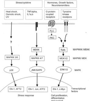
MAPKs CASCADE IN OVARIAN CANCER MAPKs are a group of serine/threonine kinases which are activated in response to a diverse array of extracellular stimuli, and mediate signal transduction from the cell surface to the nucleus.2 Three MAPK family members have been well-characterized including extracellular signal-regulated kinases (ERK1 and ERK2), c-jun terminal kinase/stress-activated protein kinases (JNK/SAPK), and p38 as indicated in Fig. 1.3,4 In addition to 3 groups of MAPK, other MAPK family members, ERK3, 4 and 5, four p38-like kinases, and p57 MAPK have been cloned, but a biological role of these MAPKs is not well known.4 As early discussed, the MAPK cascade is activated via two distinct classes of cell surface receptors, RTKs and GPCRs. The cellular signals transmitted through this cascade result in an activation of diverse molecules which regulate cell growth, survival and differentiation. ERK1 (p44 MAPK) and ERK2 (p42 MAPK) activated by mitogenic stimuli are a group of the most extensively studied members, whereas JNK/SAPK and p38 are activated in response to stress such as heat shock, osmotic shock, cytokines, protein synthesis inhibitors, antioxidants, ultra-violet, and DNA-damaging agents.5,6 MAPK family members are directly regulated by the kinases known as MAPK kinases (MAPKKs), which activate the MAPKs by phosphorylation of tyrosine and threonine residues.4,6 Currently, seven different MAPKKs have been cloned and characterized.4 The first MAPKKs cloned were MAPK/ERK kinase 1 and 2 (MEK 1/2), which specifically activate ERKs. MKK3 and 6 specifically
activate p38, whereas MKK5 stimulates the phosphorylation of ERK5. The MKK4 and 7 are known to activate JNK. The MAPKKs are activated by a rapidly expanding group of kinases called MAPKK kinases (MAPKKKs), which activate the MAPKKs by phosphorylation of serine and threonine residues.4,6 The activated MAPKs phosphorylate a large number of both cytoplasmic and nuclear proteins, exerting their specific functions. For example, activated ERK1/2 phosphorylate ternary complex factor (TCF) proteins such as Elk-1 and SAP-1, which form transcriptional complexes with serum response factor (SRF) in the promoter region of early response genes (e.g. c-fos, egr-1, junB) and thereby regulate their expression as seen Fig. 1.7 Many of these nuclear proteins, as a result of their ability to modulate expression of other proteins, are potential candidates for critical factors involved in the cellular response to stimuli.
Fig. 1. The MAPK signalling transduction pathways.
1. MAPK activation in neoplastic cells
Expression levels of kinases have been compared in normal, immortalized and neoplastic OSE cells.8 The expression of casein kinase II (CK2), p38 MAPK, cyclin-dependent kinase, and the phosphatidylinositol
3-kinase (PI3K) effectors Akt2 and p70 S6 kinase (S6K) were several-fold higher in neoplastic OSE than in normal OSE, whereas no significant difference was observed in the expression of ERK1/2.8 Interestingly, c-Jun NH2-kinase, not PI3K resulted in telomerase activity and an inhibition of JNK by a specific inhibitor reversed telomerase activity, and the expression of JNK induced an activation of a reporter gene fused to the hTERT promoter sequence at the transcriptional level.
These results suggest that JNK plays a critical role in the regulation of telomerase activity and may provide possible therapeutic insights to cure ovarian cancer patients.9 In addition, the correlation between BRCA1 and stress-associated MAPK has been proposed in ovarian cancer cells. The overexpression of BRCA1 induced the JNK, an apoptotic signaling pathway, and BRCA1 enhanced the signaling pathways that sequentially involved H-Ras, MEKK4, JNK, Fas ligand/Fas interactions, and caspase-9 activation.10
Recently, gamma-synuclein is significantly up- regulated in the majority of late-stage breast and ovarian cancers and its overexpression may be related with enhanced tumorigenicity. Furthermore, it has been shown that overexpression of this gene resulted in a constitutive activation of ERK1/2 and down-regulation of JNK1 in response to environmental stress signals, including UV, arsenate, and heat shock.11 The cross-talk between signaling pathways and multidrug resistant protein MDR-1 has been suggested in the control of sensitivity or resistance to chemotherapy. A constitutive activation of the ERK1/2 pathway was observed, whereas the level of active JNK and p38 was unchanged in the taxol resistant cells. Inhibition of the ERK1/2 pathway by specific inhibitors, UO126 or PD098059, induced a re- sensitization in the taxol resistant cells.12
There is some evidence that MAPK pathway may play a role in the metastasis of ovarian cancer. Treatment of ovarian cancer cells with MEK1 inhibitors, U0126 and PD98059, induced a significant suppression of the MMP-9 secretion activated by fibronectin (FN), implying an importance of these signaling molecules as a chemotherapeutic target for cancer to prevent metastasis.13 The urokinase-type plasminogen activator receptor
(u-PAR) has been implicated in tumor progression, and the expression of this gene is strongly up-regulated by phorbol 12-myristate 13-acetate (PMA), a PKC activator.
Treatment with PMA resulted in a rapid (5 min) activation of JNK pathway in the u-PAR-deficient OVCAR-3 ovarian cancer cells. These results suggest that the PMA- or c-Ha-Ras-dependent stimulation of u-PAR gene expression requires a JNK1-dependent signaling module.14
2. Activation of MAPKs by chemotherapeutic agents
Cisplatin has been widely used as a chemotherapeutic agent to treat ovarian cancers, whereas its use is limited because of cisplatin-resistance. The molecular mechanism of cisplatin-induced biological effect in ovarian cancer is not well understood in the regulation of MAPK pathways.
Recent data indicate that cisplatin caused a late and prolonged induction of both ERK1/2 and JNK1 activity in a dose-dependent manner, whereas no significant difference was observed in p38 activity in SKOV-3 cells.
These findings suggest that ERK and other signal transduction pathways may play an important role in the response to cisplatin for the development of new strategies to enhance the therapeutic use of platinum drugs.15 In the cisplatin-resistant CaOV-3 and cisplatin- sensitive A2780 ovarian cancer cells, cisplatin induced an activation of both ERK and JNK in a different time and dose patterns.16 Cisplatin-induced JNK activation was neither extracellular and intracellular Ca2+ nor protein kinase C-dependent, whereas cisplatin-induced ERK activation was extracellular and intracellular Ca2+- dependent and protein kinase C-dependent.16In regarding to the regulation of apoptosis by cisplatin, treatment with cisplatin activated a robust apoptotic pathway involved in the activation of JNK and p38 MAPK in cisplatin- sensitive ovarian cancer cells, whereas it fails to elicit the response in cisplatin-resistant 2008/C13 cells.17In cisplatin-resistant cells, the proteolytic activation of MEKK1 by caspase-3 is deficient, implying that inadequate caspase-3 processing and MEKK1 activation may lead to a cisplatin-resistant phenotype.17
The cisplatin-induced activation of two MAP kinases,
JNK1 and ERK1/2 was compared in the cisplatin- sensitive ovarian carcinoma cell line A2780 and its derivative cisplatin-resistant cell lines CP70 and C200.18 The different patterns of cisplatin-induced JNK1 and ERK1/2 activation were observed in these cell lines with different levels of cisplatin sensitivity. It appears that inhibition of cisplatin-induced ERK1/2 activation enhanced sensitivity to cisplatin in both cisplatin-sensitive and cisplatin-resistant cell lines, suggesting that these MAPK pathways may be important in the cisplatin- resistance in ovarian cancer and the mechanism of these pathways can be used as a possible therapeutic strategy.18 The most recent results indicate that cisplatin differentially induced JNK and p38 pathways, with the cisplatin-sensitive cells showing prolonged (8-12 h) activation and the cisplatin-resistant cells showing only transient (1-3 h) activation of JNK and p38.19In addition, the inhibition of cisplatin-induced JNK and p38 activation blocked cisplatin-induced apoptosis and persistent activation of JNK resulted in an increase in the phosphorylation of c-Jun transcription factor, which stimulated a transcription of an immediate downstream target, a death inducer Fas ligand (FasL) in cisplatin- sensitive cells. Thus, JNK-c-Jun-FasL-Fas cascade plays an important role in the regulation of cisplatin-induced apoptosis in ovarian cancer cells, and the length of JNK activation may be essential in the determination of survival or apoptosis in ovarian cancer cells.19
Taxol, a microtubule stabilizer, is a useful therapeutic agent for ovarian cancer treatment. Treatment with taxol resulted in an activation of ERK1/2 and p38 MAPK in human ovarian carcinoma cells with distinct kinetics.20 The low concentrations of taxol (1-100 nM) activated ERK1/2 within 0.5-6 h, whereas a longer exposure (24 h) at low concentrations abrogated ERK1/2 phosphorylation/
activation. Higher concentrations (1-10 mM) of taxol resulted in a sharp inhibition of ERK1/2 activity, whereas these high concentrations activated p38 at 2-24 h, suggesting that the activation of MAPK may be dependent on the dose and exposure time of chemothe- rapeutic agent.20 It is of interest that treatment with paclitaxel resulted in a phosphorylation of p70S6K (T421/S424) and this paclitaxel-induced phosphorylation
requires both de novo RNA and protein synthesis via multiple signaling pathways including ERK1/2, JNK, PKC, Ca2+, PI3K, and mammalian target of rapamycin (mTOR). These results imply that paclitaxel is able to induce p70S6K phosphorylation and exert its antitumor effect via multiple signaling pathways, especially inhibition of p70S6K.21
3. Activation of MAPK by growth factors and cytokines
Endothelin-1 (ET-1), a possible autocrine factor, has been implicated in an activation of MAPK in ovarian cancer. Treatment of OVCA 433 ovarian cancer with ET-1 resulted in a phosphorylation of ERK-2 and mitogenic responses, and epidermal growth factor receptor (EGF-R)/ras-dependent pathway may contribute to the activation of ERK-2 and mitogenic signaling induced by ET-1 in these cells. These results suggest that ET-1 induced activation of MAPK is mediated in part by signaling pathways that are initiated by transactivation of the EGF-R.22As an autocrine regulator, lysophosphatidic acid (LPA) and sphingosine-1-phosphate (S1P) have been demonstrated to activate MAPK kinase (MEK) and p38 MAPK via AKT pathway in HEY ovarian cancer cells, the kinase activity and S473 phosphorylation of Akt induced by LPA and S1P requires both MEK and p38 MAPK, and MEK is likely to be upstream of p38, in these cells, suggesting that the requirement for both MEK and p38 is cell type- and stimulus-specific.23In addition, treatment with extracellular calcium induced an activation of MAPK in the response to cell proliferation in rhesus ovarian surface epithelial cells in culture.24 Human interleukin-8 (IL-8) rapidly activated ERK-1/-2 pathway via stimulation of the CXCR-1/2 receptors.25 By using inhibitors genestein and herbimycin A, tyrosine kinases are involved in the IL-8 activation of ERK-1/-2 in SKOV-3 cells, suggesting an important “cross-talk”
between chemokine and growth factor pathways, which may involved in the cell migration and proliferation in ovarian cancer.25
In addition to an implication of ERK1/2, JNK pathway has been suggested to play a role in the cell proliferation and apoptosis in ovarian cancer. For instance, treatment
with tumor necrosis factor (TNF) alpha activated ERK1/2 at 10-20 min, and a maximum threefold induction of ERK1/2 activity was observed after 1 min of treatment.26 Inhibition of TNF alpha-induced ERK1/2 activity by PD98059 was associated with induction of apoptosis in the TNF alpha-resistant cell line UCI 101. Inhibition of TNF alpha-induced ERK1/2 activity was followed by a subsequent transient increase in TNF alpha-induced JNK1 activity. These results indicate that ERK1/2 activity may modulate cellular response to TNF alpha, suggesting that the balance between ERK1/2 and JNK1 activation may be pivotal in the cellular growth and apoptosis response to TNF alpha.26
4. Activation of MAPKs by hormonal factors There is increasing evidence that gonadotropin- releasing hormone (GnRH) and its agonists (GnRHa) may play a critical role in the inhibition of cell proliferation in gynecological cancers including ovarian and endometrial cancers. However, their biological mechanism remains uncovered. It is well known that GnRH receptor (GnRH-R) belongs to GPCRs, suggesting an involvement of MAPK by GnRH/GnRH-R system. Recently, the role of MAPK family in the antiproliferative effect of GnRHa in CaOV-3 ovarian cancer cell line has been demonstrated.27 Treatment of CaOV-3 cells with GnRHa resulted in an activation of ERK at 5 min, reached the highest activation at 3 h and sustained until 24 h, whereas GnRHa had no effect on the activation of the JNK. In addition, the ERK kinase was also activated and an increase in phosphorylation of son of sevenless (Sos), and Shc was observed following GnRHa treatment. Treatment with an inhibitor of mitogen-activated protein/ERK kinase, PD98059 reversed the antiproliferative effect of GnRHa and the GnRH- induced dephosphorylation of the retinoblastoma protein.
These results indicate that an activation of ERK may play an important role in the antiproliferative effect of GnRHa.27Furthermore, we have shown that an agonist of GnRH, (D-Ala6)-GnRH, induced a biphasic pattern of ERK-1/-2 activation. A low concentration of GnRHa (10-10 M) resulted in a significant decrease of MAPK activity, whereas high concentrations (10-7 and 10-6 M)
induced an activation of MAPK pathway.28
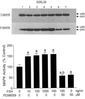
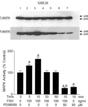
An involvement of gonadotropins, follicle-stimulating hormone (FSH) and luteinizing hormone (LH), has been proposed in the progression and metastasis of ovarian cancer. Expression of FSH receptor (FSH-R), one of GPCRs family, has been demonstrated in normal OSE,29 ovarian inclusions and epithelial tumors,30 implicating a possible role of FSH in these cells. In addition, treatment with FSH resulted in a growth-stimulation in ovarian cancer cells30,31 in a dose- and time-dependent mannerin
Fig. 2. Effect of FSH in the presence or absence of PD98059 on MAPK activation. The phosphorylated form (P-MAPK) normalized by total form (T-MAPK) was analyzed in IOSE-29 cells. Data are shown as the means of three individual experiments, and are presented as the mean±SD. a, P<0.05 vs. untreated control; b, P<0.05 vs.
FSH (100 ng/ml) treatment; c, P<0.05 vs. PD98059 (50 µM) treatment. 1, untreated control; 2, FSH (10 ng/ml) treatment; 3, FSH (100 ng/ml) treatment; 4, FSH (1000 ng/ml) treatment; 5, FSH (100 ng/ml) treatment; 6, FSH (100 ng/ml) plus PD98059 (50 µM) treatment; 7, PD98059 (50 µM) treatment. [Reproduced, permission with, from:
Choi KC, Kang SK, Tai CJ, Auersperg N, Leung PC;
“Follicle-stimulating hormone activates mitogen-activated protein kinase in preneoplastic and neoplastic ovarian surface epithelial cells” in: J Clin Endocrinol Metab 2002;
87:2245-2253. Copyright from Endocrine Society].
vitro. Despite these findings, the precise molecular mechanism of FSH in the control of growth stimulation and intracellular signaling in ovarian cancer remained unknown. Thus, we investigated the mechanism of growth-stimulatory effect, an involvement of MAPKs by FSH in pre-neoplastic OSE cells.32 Treatment with FSH resulted in the MAPK activation of immortalized OSE (IOSE-29) cells, whereas the stimulatory effect by FSH in the cellular proliferation and MAPK activation was
Fig. 3. Effect of FSH in the presence or absence of PD98059 on MAPK activation. The P-MAPK normalized by T-MAPK was analyzed in IOSE-29 cells. Data are shown as the means of three individual experiments, and are presented as the mean±SD. a, P<0.05 vs. untreated control; b, P<0.05 vs. FSH (100 ng/ml) treatment for 10 min. 1, untreated control; 2, FSH (100 ng/ml) treatment for 5 min; 3, FSH (100 ng/ml) treatment for 10 min; 4, FSH (100 ng/ml) treatment for 20 min; 5, FSH (100 ng/ml) treatment for 60 min; 6, FSH (100 ng/ml) plus PD98059 (50 µM) treatment for 10 min; 7, PD98059 (50 µM) treatment. [Reproduced, permission with, from: Choi KC, Kang SK, Tai CJ, Auersperg N, Leung PC; “Follicle- stimulating hormone activates mitogen-activated protein kinase in preneoplastic and neoplastic ovarian surface epithelial cells” in: J Clin Endocrinol Metab 2002;87:2245- 2253. Copyright from Endocrine Society].
completely abolished in the presence of PD98059, a MEK inhibitor, suggesting that growth stimulatory effect of FSH is mediated through MAPK activation in these OSE cells (Fig. 2). In a time-dependent study, FSH significantly increased MAPK activity at 5-10 min in IOSE-29 cells, and an activated MAPK declined to control level after 20 min in these cells (Fig. 3). In addition, treatment with FSH resulted in substantial phosphorylation of Elk-1, the Ets family transcriptional factor, confirming that FSH action is mediated via activation of MAPK. These results imply that that MAPK cascade may be involved in cellular function such as growth stimulation in response to FSH in pre-neoplastic OSE cells.32
In addition to FSH in the stimulation of MAPK activity, adenosine triphosphate (ATP) in our previous study has been implicated in the regulation of cell proliferation and activation of MAPK pathway in ovarian cancer cells.33 Extracellular ATP binds to heterotrimeric G protein-coupled P2 purinoceptors and plays a role in cellular proliferation and intracellular calcium concentrations (Ca2+) in ovarian cancer cells.34,35 In a previous study, treatment with ATP resulted in an activation of ERK-1/-2 in IOSE-29 cells, whereas the stimulatory effect of ATP in the cellular proliferation and MAPK activation was completely abolished in the presence of PD98059 and staurosporin (a PKC inhibitor), suggesting that the growth stimulatory effect of ATP is mediated via PKC-dependent MAPK activation in pre- neoplastic OSE cells (Fig. 4). In addition, treatment with ATP resulted in substantial phosphorylation of Elk-1, suggesting that the MAPK cascade may be involved in the growth stimulation in response to ATP in pre- neoplastic OSE cells as seen in Fig. 4.33
PI3K CASCADE IN OVARIAN CANCER PI3K is a member of lipid kinases subfamily that phosphorylates and dephosphorylates the 3-position of the inositol ring of phosphoinositides in a membrane.36 This cytosolic enzyme consists of an 85-kDa regulatory subunit and a 110-kDa catalytic subunit.37 It has been demonstrated that the PI3K play an important role as an oncogenic pathway in a diverse range of cellular
processes associated with malignant characteristic including cell growth, migration, survival, and neoplastic differentiation.
Fig. 4. Effect of ATP on ERK-1/-2 and Elk-1 in the absence or presence of PD98059 and staurosporin. To examine the role of ATP on MAPKs in IOSE-29, the cells were pretreated with 50 µM PD98059 or 0.1 µM staurosporin for 30 min, followed by treatment with 100 µM ATP for 10 min. [Reproduced, permission with, from:
Choi K-C, Tai C-J, Tzeng C-R, Auersperg N, Leung PC;
“Adenosine triphosphate (ATP) activates mitogen-activated protein kinases (MAPKs) in neoplastic ovarian surface epithelium (OSE) cells” in: Biol Reprod 2003;68:309-315.
Copyright from Society for the Study of Reproduction]
1. PI3K in cancer cell growth and apoptosis Treatment with paclitaxel resulted in a phosphorylation of p70S6K (T421/S424), a downstream pathway of PI3K, and decreased its activity in mitotic cells via multiple signaling pathways. These results suggest that paclitaxel- induced p70S6K (T421/S424) phosphorylation and kinase inactivation are differentially regulated as a chemotherapeutic role in the anti-proliferative effect as least in part.21 Inhibition of the PI3K pathway induced the suppression of telomerase activity in ovarian cancer cell lines, indicating that telomerase activity may be dependent on PI3K-mediated phosphorylation.38 An introduction of an active catalytic subunit (110-kDa) of PI3K into an ovarian cancer cell line confers resistance to the effects of paclitaxel, which its resistance to paclitaxel can be reversed by LY294002, an inhibitor of
PI3K.39 It is of interest that an inhibition of PI3K enhanced paclitaxel-induced apoptosis in human ovarian cancer by in vivo and in vitro morphological studies.
These findings suggest that a combination of a PI3K inhibitor and conventional chemotherapy may provide an potential approach to enhance the effect of chemotherapeutic agents and to minimize side effects such as a dermatological toxicity of specific inhibitors regarding to tumor growth and ascites production in ovarian cancer.39
In has been demonstrated that LY294002 induced enhanced nuclear pyknosis and diminished cytoplasmic volume in tumor cells, confirmed as an apoptosis.
Treatment with LY294002 significantly resulted in the inhibition of cell growth and ascites formation in ovarian carcinoma in vivo and markedly inhibited ovarian cancer cell proliferation in vitro, suggesting an important role of PI3K inhibitors as a novel therapeutic approach in ovarian cancer patients.40 In our recent study, treatment with LY294002 (0.1-10 μM) induced an inhibition of cell growth in neoplastic ovarian cancer cell lines, OVCAR-3 and SKOV-3, while no difference was observed by LY294002 in pre-neoplastic ovarian cancer cell line, IOSE-80PC, which was derived from normal OSE by transfecting SV40 T antigen (data not shown). These results demonstrate that the inhibition of PI3K pathway by a specific inhibitor is more potentially effective in neoplastic ovarian cancer cells, but not in normal or pre-neoplastic OSE cells, suggesting that PI3K pathway may be a selective targeting approach for ovarian cancer treatment. It is of interest that treatment with Wortmannin (0.01-1 μM) also inhibited cell growth more effectively in neoplastic ovarian cancer cell lines, OVCAR-3 and SKOV-3 (data not shown).
2. PI3K in cancer cell migration
Treatment of ovarian cancer cells with P13K inhibitors, Wortmannin and LY294002, strongly suppressed the fibronectin (FN)-dependent secretion of MMP-9 together with the inhibition of Akt activation, whereas GF109203X, a specific PKC inhibitor, failed to induce any inhibitory effect on the FN-dependent MMP-9 secretion.13 Moreover, both a MEK1 inhibitor and P13K
inhibitor, but not the PKC inhibitor, strongly decreased the invasiveness of NOM1 cells, suggesting that activation of dual signaling pathways, MEKI-MAPK and P13K-Akt, is required for the FN-dependent activation of MMP-9 secretion.13 It has been demonstrated that an inhibition of PI3K and MAPK activity impairs EGF- stimulated cell migration, in vitro invasion, pro-MMP-9 and MMP-9 production in ovarian cancer cells, supporting an expanded role of elevated PI3K activity in cellular responses associated with ovarian tumor metastasis. These results hypothesize that divergent kinase activities may regulate distinct cellular events associated with growth factor-induced invasion of ovarian cancer cells.41
3. PI3K up- and down-stream pathways Several signaling molecules of the PI3K pathway, including Akt2 and p70 S6K, were constitutively activated in FH-OSE (familial history of ovarian cancer) from six of six women but in NFH-OSE (non-familial history of ovarian cancer) from only four of eight women.42 PI3K and its downstream targets serine/
threonine kinases AKT1 and AKT2 are required for the full induction of p21 in A2780 cells treated with cisplatin or paclitaxel. The use of specific inhibitor, LY294002 or dominant negative AKT resulted in an inactivation of the PI3K/AKT signal pathway and inhibition of p21, whereas no difference was observed in the expression of the proapoptotic protein BAX by cisplatin and paclitaxel treatment.43 In addition, PI3K is involved in the expression of p21 induced by cisplatin or paclitaxel in OVCAR-10 cells, which have wild-type p53, but not in OVCAR-5 cells, which lack functional p53. These results suggest that the PI3K/AKT signal transduction pathway mediates p21 expression, and contributes to cell cycle regulation promoted by p53 in response to chemotherapeutic treatment.43 The presence of somatic mutations in the gene for the p85alpha regulatory subunit of PI3K (PIK3R1) has been shown in primary human colon and ovarian tumors, and cancer cell lines.41 Expression of a mutant protein with a 23 amino acid deletion leads to constitutive activation of PI3K, implying that p85alpha is a new oncogene involved in human
tumorigenesis.41
CONCLUDING REMARKS
Three well-characterized MAPK family, ERK1/2, JNK/SAPK, and p38, play an important role in the regulation of proliferation, survival and apoptosis in response to the external stimuli including hormones, growth factors, cytokines and chemotherapeutic chemicals in ovarian cancer. The proposed signaling pathways by reproductive hormones including FSH, ATP and GnRH have been suggested in the control of MAPK and transcriptional factors to exert their roles in the regulation of cell proliferation and apoptosis in ovarian cancer cells based on our previous results (Fig. 5). In addition to their roles abovementioned, MAPK pathway may contribute to the metastasis and chemoresistance of ovarian cancer.
However, exact mechanisms of these MAPKs need to be clarified in the metastasis and differentiation in normal and neoplastic OSE cells. In addition to MAPK pathway, it has been demonstrated that an inactivation of PI3K pathway by a specific inhibitor plays an important role in diverse cellular processes such as growth inhibition, induction of apoptosis and metastasis. An overexpression
Fig. 5. Activation of the MAPK signaling pathway by FSH, ATP and GnRH in ovarian cancer.
of catalytic or regulatory subunit results in an activation of a downstream pathway, AKT, and regulates cell proliferation and apoptosis, supporting that this PI3K may be involved in oncogenic signaling pathway. Thus, understanding of these oncogenic pathways regulated by external stimuli in these biological aspects will provide therapeutic insights to explore a novel approach for ovarian cancer treatment.
ACKNOWLEDGMENTS
PCKL is the recipient of a Distinguished Scholar Award from the Michael Smith Foundation for Health Research. This work was supported by the Canadian Institutes of Health Research.
REFERENCES
1. Auersperg N, Wong AS, Choi KC, Kang SK and Leung PC. Ovarian surface epithelium: biology, endocrinology, and pathology. Endocr Rev 2001; 22: 255-88.
2. Davis RJ. MAPKs: new JNK expands the group. Trends Biochem Sci 1994; 19: 470-3.
3. Cobb MH and Goldsmith EJ. How MAP kinases are regulated. J Biol Chem 1995; 270: 14843-6.
4. Fanger GR. Regulation of the MAPK family members:
role of subcellular localization and architectural organiza- tion. Histol Histopathol 1999; 14: 887-94.
5. Garrington TP and Johnson GL. Organization and regulation of mitogen-activated protein kinase signaling pathways. Curr Opin Cell Biol 1999; 11: 211-8.
6. Robinson MJ and Cobb MH. Mitogen-activated protein kinase pathways. Curr Opin Cell Biol 1997; 9: 180-6.
7. Wasylyk B, Hagman J and Gutierrez-Hartmann A. Ets transcription factors: nuclear effectors of the Ras-MAP- kinase signaling pathway. Trends Biochem Sci 1998; 23:
213-6.
8. Wong AS, Kim SO, Leung PC, Auersperg N and Pelech SL. Profiling of protein kinases in the neoplastic transformation of human ovarian surface epithelium.
Gynecol Oncol 2001; 82: 305-11.
9. Alfonso-De Matte MY, Yang H, Evans MS, Cheng JQ and Kruk PA. Telomerase is regulated by c-Jun NH2- terminal kinase in ovarian surface epithelial cells. Cancer Res 2002; 62: 4575-8.
10. Thangaraju M, Kaufmann SH and Couch FJ. BRCA1 facilitates stress-induced apoptosis in breast and ovarian cancer cell lines. J Biol Chem 2000; 275: 33487-96.
11. Pan ZZ, Bruening W, Giasson BI, Lee VM and Godwin AK. Gamma-synuclein promotes cancer cell survival and inhibits stress- and chemotherapy drug-induced apoptosis by modulating MAPK pathways. J Biol Chem 2002; 277:
35050-60.
12. Ding S, Chamberlain M, McLaren A, Goh L, Duncan I and Wolf CR. Cross-talk between signalling pathways and the multidrug resistant protein MDR-1. Br J Cancer 2001;
85: 1175-84.
13. Thant AA, Nawa A, Kikkawa F, Ichigotani Y, Zhang Y, Sein TT, et al. Fibronectin activates matrix metalloproteinase-9 secretion via the MEK1-MAPK and the PI3K-Akt pathways in ovarian cancer cells. Clin Exp Metastasis 2000; 18: 423-8.
14. Gum R, Juarez J, Allgayer H, Mazar A, Wang Y and Boyd D. Stimulation of urokinase-type plasminogen activator receptor expression by PMA requires JNK1- dependent and -independent signaling modules. Oncogene 1998; 17: 213-25.
15. Persons DL, Yazlovitskaya EM, Cui W and Pelling JC.
Cisplatin-induced activation of mitogen-activated protein kinases in ovarian carcinoma cells: inhibition of extracellular signal-regulated kinase activity increases sensitivity to cisplatin. Clin Cancer Res 1999; 5: 1007-14.
16. Hayakawa J, Ohmichi M, Kurachi H, Ikegami H, Kimura A, Matsuoka T, et al. Inhibition of extracellular signal- regulated protein kinase or c-Jun N-terminal protein kinase cascade, differentially activated by cisplatin, sensitizes human ovarian cancer cell line. J Biol Chem 1999; 274: 31648-54.
17. Gebauer G, Mirakhur B, Nguyen Q, Shore SK, Simpkins H and Dhanasekaran N. Cisplatin-resistance involves the defective processing of MEKK1 in human ovarian adenocarcinoma 2008/C13 cells. Int J Oncol 2000; 16:
321-5.
18. Cui W, Yazlovitskaya EM, Mayo MS, Pelling JC and Persons DL. Cisplatin-induced response of c-jun N- terminal kinase 1 and extracellular signal-regulated protein kinases 1 and 2 in a series of cisplatin-resistant ovarian carcinoma cell lines. Mol Carcinog 2000; 29: 219-28.
19. Mansouri A, Ridgway LD, Korapati AL, Zhang Q, Tian L, Wang Y, et al. Sustained Activation of JNK/p38 MAPK Pathways in Response to Cisplatin Leads to Fas Ligand Induction and Cell Death in Ovarian Carcinoma Cells. J Biol Chem 2003; 278: 19245-56.
20. Seidman R, Gitelman I, Sagi O, Horwitz SB and Wolfson M. The role of ERK 1/2 and p38 MAP-kinase pathways in taxol-induced apoptosis in human ovarian carcinoma cells. Exp Cell Res 2001; 268: 84-92.
21. Le XF, Hittelman WN, Liu J, McWatters A, Li C, Mills GB, et al. Paclitaxel induces inactivation of p70 S6 kinase
and phosphorylation of Thr421 and Ser424 via multiple signaling pathways in mitosis. Oncogene 2003; 22: 484- 97.
22. Vacca F, Bagnato A, Catt KJ and Tecce R.
Transactivation of the epidermal growth factor receptor in endothelin-1-induced mitogenic signaling in human ovarian carcinoma cells. Cancer Res 2000; 60: 5310-7.
23. Baudhuin LM, Cristina KL, Lu J and Xu Y. Akt activation induced by lysophosphatidic acid and sphingosine-1-phosphate requires both mitogen-activated protein kinase kinase and p38 mitogen-activated protein kinase and is cell-line specific. Mol Pharmacol 2002; 62:
660-71.
24. Wright JW, Toth-Fejel S, Stouffer RL and Rodland KD.
Proliferation of rhesus ovarian surface epithelial cells in culture: lack of mitogenic response to steroid or gonadotropic hormones. Endocrinology 2002; 143: 2198- 207.
25. Venkatakrishnan G, Salgia R and Groopman JE.
Chemokine receptors CXCR-1/2 activate mitogen- activated protein kinase via the epidermal growth factor receptor in ovarian cancer cells. J Biol Chem 2000; 275:
6868-75.
26. Yazlovitskaya EM, Pelling JC and Persons DL.
Association of apoptosis with the inhibition of extracellular signal-regulated protein kinase activity in the tumor necrosis factor alpha-resistant ovarian carcinoma cell line UCI 101. Mol Carcinog 1999; 25: 14-20.
27. Kimura A, Ohmichi M, Kurachi H, Ikegami H, Hayakawa J, Tasaka K, et al. Role of mitogen-activated protein kinase/extracellular signal-regulated kinase cascade in gonadotropin-releasing hormone-induced growth inhibition of a human ovarian cancer cell line. Cancer Res 1999; 59:
5133-42.
28. Kang SK, Tai CJ, Cheng KW and Leung PC.
Gonadotropin-releasing hormone activates mitogen- activated protein kinase in human ovarian and placental cells. Mol Cell Endocrinol 2000; 170: 143-51.
29. Zheng W, Magid MS, Kramer EE and Chen YT. Follicle- stimulating hormone receptor is expressed in human ovarian surface epithelium and fallopian tube. Am J Pathol 1996; 148: 47-53.
30. Zheng W, Lu JJ, Luo F, Zheng Y, Feng Y, Felix JC, et al. Ovarian epithelial tumor growth promotion by follicle- stimulating hormone and inhibition of the effect by luteinizing hormone. Gynecol Oncol 2000; 76: 80-8.
31. Wimalasena J, Dostal R and Meehan D. Gonadotropins, estradiol, and growth factors regulate epithelial ovarian cancer cell growth. Gynecol Oncol 1992; 46: 345-50.
32. Choi KC, Kang SK, Tai CJ, Auersperg N and Leung PC.
Follicle-stimulating hormone activates mitogen-activated protein kinase in preneoplastic and neoplastic ovarian surface epithelial cells. J Clin Endocrinol Metab 2002; 87:
2245-53.
33. Choi K-C, Tai C-J, Tzeng C-R, Auersperg N and Leung PCK. Adenosine Triphosphate Activates Mitogen- Activated Protein Kinase in Pre-Neoplastic and Neoplastic Ovarian Surface Epithelial Cells. Biol Reprod 2003; 68:
309-15.
34. Batra S and Fadeel I. Release of intracellular calcium and stimulation of cell growth by ATP and histamine in human ovarian cancer cells (SKOV-3). Cancer Lett 1994;
77: 57-63.
35. Schultze-Mosgau A, Katzur AC, Arora KK, Stojilkovic SS, Diedrich K and Ortmann O. Characterization of calcium-mobilizing, purinergic P2Y(2) receptors in human ovarian cancer cells. Mol Hum Reprod 2000; 6: 435-42.
36. Cantley LC, Auger KR, Carpenter C, Duckworth B, Graziani A, Kapeller R, et al. Oncogenes and signal transduction. Cell 1991; 64: 281-302.
37. Carpenter CL and Cantley LC. Phosphoinositide kinases.
Biochemistry 1990; 29: 11147-56.
38. Alfonso-De Matte MY, Cheng JQ and Kruk PA.
Ultraviolet irradiation- and dimethyl sulfoxide-induced telomerase activity in ovarian epithelial cell lines. Exp Cell Res 2001; 267: 13-27.
39. Hu L, Hofmann J, Lu Y, Mills GB and Jaffe RB.
Inhibition of phosphatidylinositol 3’-kinase increases efficacy of paclitaxel in in vitro and in vivo ovarian cancer models. Cancer Res 2002; 62: 1087-92.
40. Hu L, Zaloudek C, Mills GB, Gray J and Jaffe RB. In vivo and in vitro ovarian carcinoma growth inhibition by a phosphatidylinositol 3-kinase inhibitor (LY294002). Clin Cancer Res 2000; 6: 880-6.
41. Philp AJ, Campbell IG, Leet C, Vincan E, Rockman SP, Whitehead RH, et al. The phosphatidylinositol 3’-kinase p85alpha gene is an oncogene in human ovarian and colon tumors. Cancer Res 2001; 61: 7426-9.
42. Wong AS, Pelech SL, Woo MM, Yim G, Rosen B, Ehlen T, et al. Coexpression of hepatocyte growth factor-Met: an early step in ovarian carcinogenesis? Oncogene 2001; 20:
1318-28.
43. Mitsuuchi Y, Johnson SW, Selvakumaran M, Williams SJ, Hamilton TC and Testa JR. The phosphatidylinositol 3-kinase/AKT signal transduction pathway plays a critical role in the expression of p21WAF1/CIP1/SDI1 induced by cisplatin and paclitaxel. Cancer Res 2000; 60: 5390-4.