INTRODUCTION
In the renin-angiotensin-aldosterone systems, angiotensin II is well known to mediate progressive renal disease not only by elevating glomerular pressure, but also by non-hemody- namic effects on the kidney, such as proliferation of renal cells including mesangial cells, recruitment of mononuclear cells into the kidney by NF-κB-mediated transcription of chemo- kines, and stimulating renal TGF-β, fibronectin and type I collagen expression (1, 2). Angiotensin converting enzyme inhibitors and angiotensin receptor blockers have shown efficacy in slowing the progression of diabetic and non-dia- betic renal disease as well as in decreasing proteinuria (1).
Recently, emerging and convincing evidences have shown the independent role of aldosterone in the progression of renal disease. In the remnant kidney model using rat, aldosterone induced proteinuria, hypertension and glomerulosclerosis (3). In addition, inhibition of aldosterone by spironolactone not only slowed the development of glomerulosclerosis, but also induced the regression of existing glomerulosclerosis in the same model (4). Aldosterone infusion developed malig-
nant nephrosclerosis in saline-drinking stroke-prone hyper- tensive rats, independent of the effects of blood pressure (5) and induced severe inflammation and fibrosis by macrophage infiltration and cytokine up-regulation in saline-drinking rats (6).
The role of TGF-βin the aldosterone-induced renal injury has been suggested in animal models of various renal diseases.
In a rat model of chronic cyclosporine A (CsA) nephrotoxic- ity, spironolactone treatment reduced arteriolopathy and tubu- lointerstitial fibrosis by prevention of CsA-induced up-reg- ulation of TGF-β, collagen I and fibronectin (7), and also prevented the progression of renal injury in preexisting chron- ic CsA nephrotoxicity (8). Aldosterone also played a role in diabetic renal injury, which was demonstrated by increasing glomerular and tubulointerstitial macrophage infiltration, plasminogen activator inhibitor-1 and TGF-βexpression in streptozotocin-induced diabetic rats (9), and by increasing renal tissue and urinary levels of monocyte chemoattractant protein-1 as well as renal macrophage infiltration in type 2 diabetic rats (10). Juknevicius and colleagues demonstrated that aldosterone infusion for 3 days in normal rats increased
S195
Jeong-Sun Han, Bum-Soon Choi*, Chul-Woo Yang*, and Yong-Soo Kim* Renal Research Laboratory, Division of Nephrology*, Department of Internal Medicine, The Catholic University of Korea, College of Medicine, Seoul, Korea
Address for correspondence Yong-Soo Kim, M.D.
Division of Nephrology, Department of Internal Medicine, The Catholic University of Korea, College of Medicine, 505 Banpo-dong, Seocho-gu, Seoul 137-040, Korea
Tel : +82.2-590-2707, Fax : +82.2-599-3589 E-mail : [email protected] DOI: 10.3346/jkms.2009.24.S1.S195
Aldosterone-induced TGF- β β1 Expression is Regulated by Mitogen- Activated Protein Kinases and Activator Protein-1 in Mesangial Cells
Aldosterone has been shown to stimulate renal TGF-β1expression. However, the mechanisms for aldosterone-induced TGF-β1expression have not been clearly determined in mesangial cells. We examined the role of extracellular-signal regu- lated kinase 1 and 2 (ERK1/2), c-Jun N-terminal kinase (JNK) and activator pro- tein-1 (AP-1) in the aldosterone-induced TGF-β1expression in rat mesangial cells.
TGF-β1protein in the conditioned medium released from rat mesangial cells was measured by sandwich ELISA, TGF-β1mRNA expression was analyzed by North- ern blotting, AP-1 DNA binding activity was measured by EMSA and the ERK1/2, JNK activity was analyzed by western blotting. Aldosterone significantly stimulated TGF-β1protein production and TGF-β1mRNA expression in mesangial cells in a dose-dependent manner. Aldosterone significantly increased AP-1 DNA binding activity in mesangial cells. Pre-treatment of cells with AP-1 inhibitor, curcumin, block- ed aldosterone-induced AP-1 DNA binding activity as well as aldosterone-induced TGF-β1production. Aldosterone increased phosphorylation of ERK1/2 and JNK in mesangial cells. Pre-treatment of cells with ERK1/2 inhibitor, PD98059, or JNK inhi- bitor, SP600125 significantly inhibited aldosterone-induced ERK1/2 and JNK activi- ty and subsequently TGF-β1production, respectively. We conclude that aldosterone- induced TGF-β1expression in mesangial cells is regulated by the ERK1/ 2, JNK and AP-1 intracellular signaling pathways.
Key Words : Aldosterone; Transforming Growth Factor beta1; Extracellular Signal-Regulated MAP Kinases;
JNK Mitogen-Activated Protein Kinases; Transcription Factor AP-1
Received : 18 September 2008 Accepted : 17 December 2008
urinary TGF-βexcretion without changes in blood pressure or evidence of kidney damage (11).
The renal protective effects of inhibiting aldosterone were also demonstrated in chronic kidney disease patients. In a prospective randomized open-label study, spironolactone treat- ment for 1 yr in patients with chronic kidney disease already treated with angiotensin-converting enzyme inhibitors and/
or angiotensin type 1 receptor antagonists significantly dec- reased proteinuria without any change in glomerular filtra- tion rate (12). Similar results were observed in another clin- ical study (13).
Putting these results from various experimental studies to- gether, it is positive that aldosterone stimulates renal TGF-β expression in the course of progressive renal injury. Recent- ly, Han et al. (10) described the aldosterone stimulated NF- κB DNA binding activity and subsequently monocyte che- moattractant protein-1 expression in mesangial cells. How- ever, it has not been clearly determined if aldosterone-induced TGF-βexpression was mediated through the extracellular-sig- nal regulated-kinase 1 and 2 (ERK1/2), c-Jun N-terminal kinase (JNK) and activator protein-1 (AP-1) pathways in me- sangial cells. In this study, we examined the role of ERK1/2, JNK and activator protein-1 in the intracellular signaling pathways of aldosterone-induced TGF-β1expression in rat mesangial cells.
MATERIALS AND METHODS
Reagents
Recombinant human TGF-β1and anti-human TGF-β1 antibodies were purchased from R&D Systems (Minneapo- lis, MN, U.S.A.). Anti-c-Jun antibody, anti-c-Fos antibody and AP-1 consensus oligonucleotides (5′-CGC TTG ATG ACT CAG CCG GAA-3′) were purchased from Santa Cruz Biotechnology (Santa Cruz, CA, U.S.A.). JNK and ERK1/2 antibodies were purchased from Cell Signaling Technology (Beverly, MA, U.S.A.). The [32P]dCTP and [γ-32P]ATP were obtained from NEN (Boston, MA, U.S.A.). Aldosterone, cur- cumin and spironolactone were purchased from Sigma-Aldrich (Deisenhofen, Germany). PD98059 was purchased from Cal- biochem (Darmstadt, Germany). SP600125 was purchased from Biomol Research Laboratories (Plymouth Meeting, PA, U.S.A.). All supplements for cell culture were purchased from Life Technologies (Gaithersburg, MD, U.S.A.).
Cell culture
Rat mesangial cells were isolated from glomeruli of nor- mal renal cortex as described previously (14). Mesangial cells were grown in RPMI1640 medium supplemented with 10%
heat-inactivated FBS, 10 mM HEPES, and an antibiotic/anti- fungal solution.
TGF-ββ1ELISA
After a 24 hr starvation, confluent cells were stimulated with aldosterone (0.2 ng/mL, 1 ng/mL, 5 ng/mL) for 24 hr. The conditioned medium was collected, centrifuged at 10,000 xg (4℃) for 3 min and stored at -70℃until assayed. For the time course, confluent cells incubated in the serum free medi- um were incubated with or without aldosterone (5 ng/mL) for the indicated time points. For TGF-β1ELISA, the conditioned medium was incubated with 1N HCl for 60 min to activate latent TGF-β1, and then neutralized with 1N NaOH. Anti- TGF-β1Ab (1 μg/mL) and biotinylated Anti-TGF-β1Ab (300 ng/mL) were used. The TMB microwell peroxidase sub- strate system (Kirkegaard & Perry Laboratories, Gaithers- burg, MD, U.S.A.) was used as an enzyme substrate and the reaction was followed by measurement of OD450.
Isolation of RNA and TGF-ββ1Northern blot analysis Serum-starved confluent cells were stimulated with aldos- terone (5 ng/mL) for the indicated time points. RNA was isolated by one-step guanidium-thiocyanatephenol-chloro- form extraction. RNA quantification was determined by absorbance at 260 nm. Total RNA was fractionated by elec- trophoresis on a 1% agarose-formaldehyde gel, blotted by capillary transfer on a nylon membrane (Gene Screen; NEN, Boston, MA, U.S.A.), and cross-linked by UV irradiation (UV Strata-linker 1800; Stratagene, La Jolla, CA, U.S.A.).
The membrane was probed with rat TGF-β1cDNA labeled by random priming using a commercial kit (Amersham, Buckinghamshire, U.K.) and [32P]dCTP. The blot was pre- hybridized at 42℃for 2 hr in 50% formamide, 0.1% SDS, 2×Denhardt’s solution, 5×standard SSPE, and 0.1 mg/mL salmon sperm DNA. Probe (106cpm/mL) was added to the prehybridization solution, and the blot was hybridized over- night at 42℃. The blot was then washed twice for 10 min each at 55℃in 2×standard saline citrate and 0.1% SDS, and autoradiography was performed with Fuji radiograhpy film (Tokyo, Japan) and an intensifying screen at -70℃. The rat TGF-β1probe was then removed by boiling, and the same blot was rehybridized to a cDNA probe encoding for the GAPDH. The intensity of the blots was quantified by densitometric analysis.
Electrophoretic mobility shift assay (EMSA)
Confluent cells were starved for 24 hr and then were stimu- lated with aldosterone (5 ng/mL) for 15 min. Nuclear extracts were prepared from the treated cells that were pelleted (-500 xg, 10 min, 4℃) and resuspended in hypotonic buffer (10 mM HEPES, pH 7.9, 10 mM KCl, 0.1 mM EDTA, 0.1 mM EGTA, 1 mM DTT, 0.5 mM PMSF, complete protease inhib- it mixture tablets) and incubated for 10 min on ice. Cells were lyzed by the addition of Nonidet P-40 (final concentration
of 0.1%). The nuclear pellet was resuspended in extraction buffer (20 mM HEPES, pH 7.9, 0.4 mM KCl, 1 mM EDTA, 1 mM EGTA, 1 mM DTT, 1 mM PMSF, complete protease inhibit mixture tablets) and incubated for 10 min on ice, and the tubes were centrifuged at 14,000 xg (4℃) for 10 min.
The supernatants were stored at -70℃until used. The pro- tein content in nuclear extracts was measured by the Brad- ford method (Bio-Rad, Hercules, CA, U.S.A.). The oligonu- cleotide containing the AP-1 consensus binding sequence was radiolabeled with [γ-32P]ATP by T4 polynucleotide kinase (Roche, Mannheim, Germany) for 30 min at room temper- ature. Unincorporated label was removed with a Chroma spin STE-10 column (Clontech Laboratories, Palo Alto, CA, U.S.A.). The binding reaction was performed for 30 min at room temperature in a total volume of 20 μL containing bind- ing buffer (50 mM Tris, pH 7.5, 250 mM NaCl, 2.5 mM
EDTA, 20% glycerol, 2.5 mM DTT, and 5 mM MgCl2), 5 μg of nuclear protein extracts, 1 μL of 32P-labeled oligonu- cleotide, and 50 μg/mL poly (dI-dC) (Roche). The DNA- protein complexes were fractionated by electrophoresis over 6% polyacrylamide gels in 1x Tris-boric acid-EDTA buffer.
The gels were dried and exposed to radiograhy film for autora- diography.
Western blot analysis
Confluent cells were starved for 24 hr and then were stim- ulated with aldosterone (5 ng/mL) for 10 min. The treated cells were lyzed for 10 min on ice in lysis buffer (50 mM Tris, pH 7.5, 40 mM NaCl, 1% Triton X-100, 2 mM EDTA, 1 μg/mL leupeptin, 2 mM DTT, and 1 mM PMSF). Lysates were cleared by centrifugation at 14,000 xg (4℃) for 10 min.
Fig. 1. Aldosterone stimulates TGF-β1production in mesangial cells. (A) Mesangial cells were incubated with or without aldosterone (5 ng/
mL) for the indicated time points ( , untreated: , treated with aldosterone). *p<0.05 vs. untreated control. (B) Cells were incubated with various dose of aldosterone or angiotensin II (10-7M) or a combination of aldosterone and angiotensin II for 24 hr. *p<0.05 vs. untreated control. �p<0.05 vs. angiotensin II-treated cells. (C) Cells were pre-incubated with or without spironolactone (10-9M) for 60 min and then incubated with aldosterone (5 ng/mL) for 16 hr. *p<0.05 vs. untreated control. �p<0.05 vs. aldosterone-treated cells. In all of (A), (B) and (C) TGF-β1protein was measured by ELISA. Results from 5 independent experiments are shown as mean±SEM. (D) TGF-β1protein expression was measured by western blot analysis after cells were pre-incubated with or without spironolactone (10-9M) for 60 min, and then incubated with aldosterone. *p<0.01 vs. untreated control. �p<0.05 vs. aldosterone-treated cells.
TGF-ββ1(pg/104cells) 3
2
1
0
0 8 16 24 48
Hour
TGF-ββ1 Actin
*
*
*
*
* *
*,�
*,�
A TGF-ββ1(pg/104cells)
4
3
2
1
0
- 0.2 1 5 - 0.2 1 5
- - - - + + + +
Aldosterone (ng/mL) ANG II (10-7M)
B
Arbitrary unit (fold increase)
4
3
2
1
0
- - 0.2 1 5 5
- + - - - +
Aldosterone (ng/mL) Spironolactone
D
*
* *
� *
�
TGF-ββ1(pg/104cells) 3
2
1
0
- - + +
- + - +
Aldosterone Spironolactone
C
Total protein was quantified by the Bradford method. Equal amounts of lysates were fractionated by 10% SDS-PAGE and electrotransferred to Bio-Blot nitrocellulose membranes (Bio-Rad). The membranes were blocked with TBS (pH 7.6)/
5% nonfat dry milk/0.05% Tween 20, and were blotted with the indicated antibodies at 4℃overnight. The membranes were incubated with 1/1,000 diluted HRP-conjugated sec- ondary antibody (Amersham) at room temperature for 1 hr and visualized by an ECL kit (Amersham). The membranes were stripped in solution (62.5 mM Tris-HCl, pH 7.5, 20 mM DTT, and 1% SDS) for 5 min at 50℃with agitation and reprobed with anti-total JNK or anti-total ERK1/2 anti- body.
Statistical analysis
Data were expressed as the mean±SEM. Statistical com- parisons between multiple groups were performed by ANO- VA and Bonferroni’s method was applied to control for mul- tiple testing. Statistical comparisons between multiple time points were performed by repeated measure ANOVA, with a minimum value of p<0.05 considered to represent statisti- cal significance.
RESULTS
Aldosterone Stimulates TGF-ββ1Production
TGF-β1 protein was measured in the supernatants of rat mesangial cells stimulated with aldosterone (5 ng/mL) by
ELISA. TGF-β1was constitutively produced by rat mesangial cells and was stimulated by aldosterone, which was statistical- ly significant at 24 hr and 48 hr (n=5, p<0.05; Fig. 1A). We then measured TGF-β1protein in the supernatants of cells incubated with various concentration of aldosterone with or without angiotensin II (10-7M) for 24 hr. Aldosterone stim- ulated TGF-β1production in a dose-dependent manner. An- giotensin II also stimulated TGF-β1production as previously described in the same cells (15). Furthermore, a combina- tion treatment of the cells with aldosterone and angiotensin II additively stimulated TGF-β1 production significantly more than single treatment (n=5, p<0.05; Fig. 1B). To deter- mine if the mineralocorticoid receptor mediates the aldos- terone-induced TGF-β1production, cells were pre-incubat- ed with or without spironolactone (10-9M) for 60 min and then stimulated with aldosterone (5 ng/mL) for 24 hr. Spirono- lactone abolished aldosterone-induced TGF-β1 production (n=5, p<0.05, Fig. 1C).
We also examined the TGF-β1protein expression in the lysates of cells, which were stimulated with aldosterone for 24 hr by western blot analysis. Aldosterone significantly stimulated TGF-β1protein expression in a dose-dependent manner, which was blocked by pre-incubation of cells with spironolactone (10-9M) (n=5, p<0.01, Fig. 1D).
Aldosterone stimulates TGF-ββ1mRNA expression We measured TGF-β1mRNA in the cells incubated with various concentration of aldosterone with or without angio- tensin II (10-7M) for 16 hr. Aldosterone significantly stimu- lated TGF-β1mRNA expression in a dose-dependent manner.
Fig. 2. Aldosterone stimulates TGF-β1mRNA expression in mesangial cells. (A) Cells were incubated with various dose of aldosterone (5 ng/
mL) or angiotensin II (10-7M) or a combination of aldosterone and angiotensin II for 16 hr. *p<0.05 vs. untreated control. �p<0.05 vs. angiotensin II-treated cells. (B) Cells were pre-incubated with or without spironolactone (10-9M) for 60 min and then incubated with aldosterone (5 ng/mL) for 16 hr. *p<0.05 vs. untreated control. �p<0.05 vs. aldosterone-treated cells. In both (A) and (B), Upper panel, the autoradiograph is a representative of five independent experiments with similar results. Lower panel, the optical density of autoradiographic signals was quanti- fied and calculated as the ratio of TGF-β1to GAPDH mRNA. Results are expressed as fold increase over untreated (represented as 1) in densitometric arbitrary units. Each value represents the mean±SEM.
TGF-ββ1
TGF-ββ1/GAPDH
5 4 3 2 1 0
- 0.2 1 5 - 0.2 1 5
- - - - + + + +
Aldosterone (ng/mL)
ANG II (10-7M) A
*
* *
GAPDH
*,�
*,�
*,�
TGF-ββ1
TGF-ββ1/GAPDH 2
1
0 - - + +
- + - +
Aldosterone
Spironolactone B
* GAPDH
�
A combination treatment of the cells with aldosterone and angiotensin II additively stimulated TGF-β1 mRNA expres- sion significantly more than single treatment (n=5, p<0.05, Fig. 2A). When cells were pre-incubated with spironolactone (10-9M) for 60 min and then stimulated with aldosterone (5 ng/mL) for 16 hr, aldosterone-induced TGF-β1mRNA expres- sion was abolished (n=5, p<0.05, Fig. 2B).
Role for AP-1 activation in the aldosterone-induced TGF- β
β1expression
The 5′-flanking sequences of rat TGF-β1 gene contains AP-1 binding site (16), suggesting a potential role for AP-1 in the regulation of TGF-β1expression. However, it has not been clearly determined whether aldosterone stimulates AP- 1 activity and whether aldosterone-induced AP-1 activity triggers TGF-β1expression in mesangial cells. Therefore, we examined AP-1 activity in rat mesangial cells stimulated with
aldosterone by EMSA using AP-1 consensus oligonucleotides.
AP-1 DNA binding activity was significantly increased by aldosterone (5 ng/mL), which was maximal at 15 min (n=5, p<0.05, Fig. 3A). Supershift analysis indicated that the AP- 1 complexes induced by aldosterone contained c-Jun and c- Fos proteins (Fig. 3B). The specificity of the AP-1 binding activity was tested using both an unlabelled consensus AP-1 oligonucleotiedes (cold probe) and a mutant AP-1 oligonu- cleotides, which has two base changes within the AP-1 bind- ing motif (CA to TG), at the same molar quantity (50-fold excess) for competition. The specific band induced by aldos- terone was deleted by preincubation of nuclear extracts with excessive amounts of unlabelled AP-1 oligonucleotides, but not by preincubation of nuclear extracts with mutant AP-1 oligonucleotides (data not shown). When the cells were pre- incubated with c-Jun/AP-1 inhibitor, curcumin (12.5 μM), for 15 min, the aldosterone-induced AP-1 activity was com- pletely abolished (n=3, p<0.05, Fig. 3B). To examine whether
0.05 vs. aldosterone-treated cells. In some experiments, the nuclear extracts from aldosterone-stimulated mesangial cells were pre-incu- bated with or without an antibody to c-jun or c-fos for 1 hr at room temperature and were then analyzed for AP-1 binding activity. Pre-incu- bation of nuclear extracts with c-jun or c-fos antibody induced supershift of aldosterone-induced AP-1 activity (upper panel). In both (A) and (B) Left panel, the autoradiograph is a representative of five independent experiments with similar results. Right panel, the optical den- sity of autoradiographic signals was quantified and calculated as the ratio of aldosterone-treated cells to untreated control. Results are ex- pressed as fold increase over untreated control (represented as 1) in densitometric arbitrary units. Each value represents the mean±SEM.
(C) Inhibition of aldosterone-induced TGF-β1production by curcumin. Cells were pre-incubated with or without curcumin (12.5 μM) for 15 min, and then incubated with aldosterone (5 ng/mL) for 24 hr and TGF-β1protein was measured by ELISA. *p<0.05 vs. untreated control.
�p<0.05 vs. aldosterone-treated cells.
Fig. 3. Role for AP-1 activation in the aldosterone-induced TGF- β1expression. (A) Time course for AP-1 activation by aldosterone.
Cells were incubated with aldosterone (5 ng/mL) for the indi- cated time points. AP-1 DNA binding activity in the nuclear ex- tracts was measured by EMSA using AP-1 consensus oligonu- cleotides. *p<0.05 vs. untreated control. (B) Inhibition of aldos- terone-induced AP-1 activity by curcumin. Cells were pre-incu- bated with or without AP-1 inhibitor, curcumin (12.5 μM), for 15 min, and then stimulated with aldosterone (5 ng/mL) for 15 min, and EMSA was performed. *p<0.05 vs. untreated control. �p<
A AP-1 DNA binding activity (fold increase)
6 5 4 3 2 1 0
0 5 10 15 30 60 Probe
alone 0 5 6060 (min)
AP-1
10 15 30
B AP-1 DNA binding activity (fold increase)
4
3
2
1
0
- - + +
- + - +
Aldosterone Curcumin Min
AP-1
C-jun C-Fos Probe alone Untreated Curcumin Aldosterone Aldosterone+ Curcumin
*
*
*
�
*
�
TGF-ββ1(pg/104cells) 3
2
1
0
- - + +
- + - +
Aldosterone Curcumin
C
aldosterone-induced activation of AP-1 is responsible for the increased TGF-β1production, cells were pre-incubated with curcumin (12.5 μM) for 15 min and then were stimulated with aldosterone (5 ng/mL) for 24 hr and TGF-β1was mea- sured in the cell supernatants by ELISA. Curcumin completely inhibited the aldosterone-induced TGF-β1 production (n=
5, p<0.05, Fig. 3C).
Role for JNK pathway in the aldosterone-induced TGF- β
β1expression
The activation of JNK has been shown to be linked to AP- 1 activity particularly to the phosphorylation of c-Jun (17).
We examined the phosphorylation of JNK in the cells stim- ulated with aldosterone by western blot analysis. Aldosterone (5 ng/mL) phosphorylated both 46 and 54 kDa JNKs in mesangial cells from 2 to 10 min and the peak phosphory-
lation was seen at 10 min (n=5, p<0.05, Fig. 4A). When the cells were pre-incubated with spironolactone (10-9M) and then stimulated with aldosterone, aldosterone-induced JNK activity was significantly inhibited (n=5, p<0.05, Fig.
4B). When the cells were pre-incubated with JNK inhibitor, SP600125 (20 μM), and then stimulated with aldosterone (5 ng/mL), aldosterone-induced TGF-β1protein production was significantly inhibited (n=5, p<0.05, Fig. 4C).
Role for ERK1/2 pathway in the aldosterone-induced TGF-ββ1expression
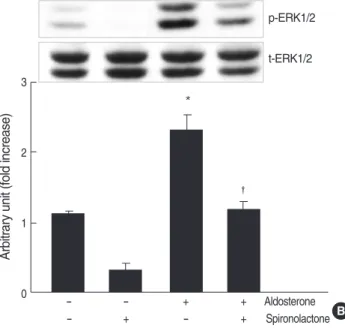
We examined the phosphorylation of ERK1/2 in the cells stimulated with aldosterone by western blot analysis. Aldos- terone (5 ng/mL) phosphorylated both 42 and 44 kDa ERK1/
2 in mesangial cells from 2 to 60 min and the peak phospho- rylation was seen at 10 min (n=3, p<0.05, Fig. 5A). When
Fig. 4. Role for JNK phosphorylation in the aldosterone-induced TGF-β1expression. (A) Cells were incubated with aldosterone (5 ng/mL) for various time points, and phosphorylated JNK was detect- ed by Western blot analysis. *p<0.05 vs. untreated control. (B) Inhi- bition of aldosterone-induced JNK phosphorylation by spironolac- tone. Cells were pre-incubated with or without spironolactone (10-9 M) for 60 min, and then incubated with aldosterone (5 ng/mL) for 10 min, and western blot was performed. *p<0.05 vs. untreated con- trol.�p<0.05 vs. aldosterone-treated cells. In both (A) and (B) Upper panel, the autoradiograph is a representative of five independent experiments with similar results. Lower panel, the optical density of autoradiographic signals was quantified and calculated as the ratio of aldosterone-treated cells to untreated control. Results are exp- ressed as fold increase over untreated control (represented as 1)
A B
Arbitrary unit (fold increase)
4
3
2
1
0 0 2 5 10 30 60
Min p-JNK
t-JNK
p-JNK
t-JNK
*
*
Arbitrary unit (fold increase)
4
3
2
1
0 - - + + Aldosterone
- + - + Spironolactone
*
C TGF-ββ1(pg/104cells)
3
2
1
0 - - + + Aldosterone
- + - + SP600125
*
�
�
in densitometric arbitrary units. Each value represents the mean±SEM. (C) Inhibition of aldosterone-induced JNK phosphorylation by SP600125. Cells were pre-incubated with or without SP600125 (20 μM) for 15 min, and then incubated with aldosterone (5 ng/mL) for 24 hr, and TGF-β1protein was measured by ELISA. *p<0.05 vs. untreated control. �p<0.05 vs. aldosterone-treated cells.
the cells were pre-incubated with spironolactone and then stimulated with aldosterone, aldosterone-induced ERK1/2 activity was significantly decreased (n=3, p<0.05, Fig. 5B).
In addition, when the cells were pre-incubated with ERK1/2 inhibitor, PD98059 (50 μM), and then stimulated with aldos- terone (5 ng/mL), aldosterone-induced TGF-β1protein pro- duction was inhibited (n=5, p<0.05, Fig. 5C).
DISCUSSION
In the present study, we demonstrated that aldosterone stimulated TGF-β1expression in rat mesangial cells, partly through the induction of ERK1/2 and JNK phosphorylation and AP-1 DNA binding activity. These results suggested a mechanism involved in the anti-fibrotic effect of spironolac- tone which was demonstrated in the animal models of vari-
ous renal injury as well as human chronic kidney diseases.
In our study, aldosterone induced transcription factor, AP- 1 DNA binding activity, which led to TGF-β1up-regulation in mesangial cells. Recently, the role of AP-1 activation in the regulation of TGF-β1expression in mesangial cells has been described in a few studies. A positive regulatory region in TGF-β15′-flanking sequence, which is responsible for the in- duction of TGF-β1, has an AP-1 binding site. Oxidized LDL has been shown to induce TGF-β1expression through the AP- 1 binding site (16). In a study done by Ahn et al. (18), trans- fection of mesangial cells with AP-1 decoy oligodeoxynucl- eotides strongly inhibited high glucose- and angiotensin II- induced TGF-β1expression. In addition, administration of AP-1 decoy oligodeoxynucleotides into streptozotocin-induced diabetic rat kidney using the hemagglutinating virus of Japan- liposome method also abolished TGF-β1and plasminogen activator inhibitor-1 expression.
Fig. 5. Role for ERK1/2 phosphorylation in aldosterone-induced TGF-β1expression. (A) Cells were incubated with aldosterone (5 ng/ mL) for various time points, and phosphorylated ERK1/2 was detected by Western blot analysis. *p<0.05 vs. untreated control.
(B) Inhibition of aldosterone-induced ERK1/2 phosphorylation by spironolactone. Cells were pre-incubated with or without spirono- lactone (10-9M) for 60 min, and then incubated with aldosterone (5 ng/mL) for 10 min, and western blot was performed. *p<0.05 vs.
untreated control. �p<0.05 vs. aldosterone-treated cells. In both a and b Upper panel, the autoradiograph is a representative of five independent experiments with similar results. Lower panel, the opti- cal density of autoradiographic signals was quantified and calcu- lated as the ratio of aldosterone-treated cells to untreated control.
Results are expressed as fold increase over untreated control (rep-
A B
Arbitrary unit (fold increase)
3
2
1
0 0 2 5 10 30 60
Min
p-ERK1/2
t-ERK1/2
*
*
*
*
*
Arbitrary unit (fold increase)
3
2
1
0 - - + + Aldosterone
- + - + Spironolactone
*
C TGF-ββ1 (pg/104cells)
3
2
1
0 - - + + Aldosterone
- + - + PD98059
*
�
�
resented as 1) in densitometric arbitrary units. Each value represents the mean±SEM. (C) Inhibition of aldosterone-induced ERK1/2 phos- phorylation by PD98059. Cells were pre-incubated with or without PD98059 (50 μM) for 15 min, and then incubated with aldosterone (5 ng/
mL for 24 hr, and TGF-β1protein was measured by ELISA. *p< 0.05 vs. untreated control. �p<0.05 vs. aldosterone-treated cells.
p-ERK1/2
t-ERK1/2
c-Jun contains a docking site for the JNK, which phos- phorylate serines 63 and 73, thereby enhancing its transcrip- tional potential and stability (19). Activation of JNK results in greatly enhanced c-Jun transcriptional activity and induc- tion of the AP-1 target gene (19). Most cells express two JNK isoforms, 45 kDa and 54 kDa in size and termed JNK1 and JNK2, which are highly similar in their modes of regulation (20). In our study, aldosterone rapidly induced phosphorylation of JNK and spironolactone abolished aldos- terone-induced phosphorylation of JNK. In addition, both JNK inhibitor, SP600125, and spironolactone suppressed aldosterone-induced TGF-β1 production, suggesting that blocking JNK activation contributed to the inhibition of c- jun/AP-1 activation, and finally TGF-β1expression.
In our study, aldosterone rapidly stimulated ERK1/2 phos- phorylation which led to subsequent increase in the TGF-β1 production. Aldosterone-induced ERK1/2 activation has been demonstrated in different in vitro and in vivo studies. Aldos- terone-induced ERK1/2 activation has been shown to stim- ulate collagen gene expression and synthesis in renal fibrob- lasts (21) to induce mesangial cell proliferation (22), and to increase mytogenic response synergistically with angiotensin II in vascular smooth muscle cells (23). However, a direct relationship between ERK1/2 activation and TGF-β1up- regulation in mesangial cells has not been clearly determined.
In an animal study using aldosterone/salt-induced hyper- tensive rats, chronic administration of spironolactone inhib- ited ERK1/2 activity and suppressed expression of TGF-β1, type I and III collagen, and monocyte chemoattractant pro- tein-1 mRNA, which resulted in the amelioration of perivas- cular fibrosis and coronary microvascular hyperplasia (24).
However, a direct relationship between ERK1/2 activation and TGF-β1up-regulation was not confirmed in that study.
Recently, the role for ERK1/2 in the stimulation of TGF-β1 expression was demonstrated in two different cells. In a study done by Huang et al. (25), renin induced rapid activation of cellular ERK1/2 in rat mesangial cells and an ERK kinase inhibitor significantly reduced the renin-induced ERK1/2 phosphorylation and the subsequent increase in TGF-β1 mRNA expression. Otsuka et al. (26) has reported that liposomes composed of phosphatidylserine activated ERK which led to the expression of TGF-β1in macrophages.
In summary, our data provide an evidence that aldosterone stimulates TGF-β1 expression in rat mesangial cells partly by enhancing ERK1/2 and JNK activities and subsequent AP-1 activity. This study also provides an understanding of a part of intracellular mechanisms for proliferative and fibrot- ic effect of aldosterone in the inflammatory renal diseases.
REFERENCES
1. Remuzzi G, Perico N, Macia M, Ruggenenti P. The role of renin- angiotensin-aldosterone system in the progression of chronic kidney
disease. Kidney Int 2005; 68: S57-65.
2. Ruster C, Wolf G. Renin-angiotensin-aldosterone system and pro- gression of renal disease. J Am Soc Nephrol 2006; 17: 2985-91.
3. Greene EL, Kren S, Hostetter TH. Role of aldosterone in the rem- nant kidney model in the rat. J Clin Invest 1996; 98: 1063-8.
4. Aldigier JC, Kanjanbuch T, Ma LJ, Brown NJ, Fogo AB. Regres- sion of existing glomerulosclerosis by inhibition of aldosterone. J Am Soc Nephrol 2005; 16: 3306-14.
5. Rocha R, Chander PN, Zuckerman A, Stier CT Jr. Role of aldos- terone in renal vascular injury in stroke-prone hypertensive rats.
Hypertension 1999; 33: 232-7.
6. Blasi ER, Rocha R, Rudolph AE, Blomme EA, Polly ML, McMa- hon EG. Aldosterone/salt induces renal inflammation and fibrosis in hypertensive rats. Kidney Int 2003; 63: 1791-800.
7. Feria I, Pichardo I, Juarez P, Ramirez V, Gonzalez MA, Uribe N, Garcia-Torres R, Lopez-Casillas F, Gamba G, Bobadilla NA. Ther- apeutic benefit of spironolactone in experimental chronic cyclospo- rine A nephrotoxicity. Kidney Int 2003; 63: 43-52.
8. Perez-Rojas J, Blanco JA, Cruz C, Trujillo J, Vaidya VS, Uribe N, Bonventre JV, Gamba G, Bobadilla NA. Mineralocorticoid recep- tor blockade confers renoprotection in preexisting chronic cyclos- porine nephrotoxicity. Am J Physiol Renal Physiol 2007; 292: F131-9.
9. Fujisawa G, Okada K, Muto S, Fujita N, Itabashi N, Kusano E, Ishi- bashi S. Spironolactone prevents early renal injury in streptozotocin- induced diabetic rats. Kidney Int 2004; 66: 1493-502.
10. Han SY, Kim CH, Kim HS, Jee YH, Song HK, Lee MH, Han KH, Kim HK, Kang YS, Han JY, Kim YS, Cha DR. Spironolactone pre- vents diabetic nephropathy through an anti-inflammatory mecha- nism in type 2 diabetic rats. J Am Soc Nephrol 2006; 17: 1362-72.
11. Juknevicius I, Segal Y, Kren S, Lee R, Hostetter TH. Effect of aldos- terone on renal transforming growth factor-beta. Am J Physiol Renal Physiol 2004; 286: F1059-62.
12. Bianchi S, Bigazzi R, Campese VM. Long-term effects of spirono- lactone on proteinuria and kidney function in patients with chronic kidney disease. Kidney Int 2006; 70: 2116-23.
13. Sato A, Hayashi K, Saruta T. Antiproteinuric effects of mineralocor- ticoid receptor blockade in patients with chronic renal disease. Am J Hypertens 2005; 18: 44-9.
14. Kreisberg JI, Karnovsky MJ. Glomerular cells in culture. Kidney Int 1983; 23: 439-47.
15. Kagami S, Border WA, Miller DE, Noble NA. Angiotensin II stim- ulates extracellular matrix protein synthesis through induction of transforming growth factor-βexpression in rat glomerular mesan- gial cells. J Clin Invest 1994; 93: 2431-7
16. Wu Z, Zhou Q, Lan Y, Wang Y, Xu X, Jin H. AP-1 complexes medi- ate oxidized LDL-induced overproduction of TGF-beta(1) in rat mesangial cells. Cell Biochem Funct 2004; 22: 237-47.
17. Karin M. The regulation of AP-1 activity by mitogen-activated pro- tein kinases. J Biol Chem 1995; 270: 16483-6.
18. Ahn JD, Morishita R, Kaneda Y, Lee KU, Park JY, Jeon YJ, Song HS, Lee IK. Transcription factor decoy for AP-1 reduces mesangial cell proliferation and extracellular matrix production in vitro and in vivo. Gene therapy 2004; 11: 916-23.
19. Smeal T, Binetruy B, Mercola D, Grover-Bardwick A, Heidecker G,
′ ′ ′
′
Rapp UR, Karin M. Oncoprotein-mediated signalling cascade stim- ulates c-Jun activity by phosphorylation of serines 63 and 73. Mol Cell Biol 1992; 12: 3507-13.
20. Guan Z, Tetsuka T, Baier LD, Morrison AR. Interleukin-1 beta acti- vates c-jun NH2-terminal kinase subgroup of mitogen-activated pro- tein kinases in mesangial cells. Am J Physiol 1996; 270: F634-41.
21. Nagai Y, Miyata K, Sun GP, Rahman M, Kimura S, Miyatake A, Kiyomoto H, Kohno M, Abe Y, Yoshizumi M, Nishiyama A. Aldos- terone stimulates collagen gene expression and synthesis via activa- tion of ERK1/2 in rat renal fibroblasts. Hypertension 2005; 46: 1039- 45.
22. Nishiyama A, Yao L, Fan Y, Kyaw M, Kataoka N, Hashimoto K, Nagai Y, Nakamura E, Yoshizumi M, Shokoji T, Kimura S, Kiy- omoto H, Tsujioka K, Kohno M, Tamaki T, Kajiya F, Abe Y. Involve- ment of aldosterone and mineralocorticoid receptors in rat mesan- gial cell proliferation and deformability. Hypertension 2005; 45:
710-6.
23. Min LJ, Mogi M, Li JM, Iwanami J, Iwai M, Horiuchi M. Aldosterone and angiotensin II synergistically induce mitogenic response in vas- cular smooth muscle cells. Circ Res 2005; 97: 434-42.
24. Nakano S, Kobayashi N, Yoshida K, Ohno T, Matsuoka H. Cardio- protective mechanisms of spironolactone associated with the angi- otensin-converting enzyme/epidermal growth factor receptor/extra- cellular signal-regulated kinases, NAD(P)H oxidase/lectin-like oxi- dized low-density lipoprotein receptor-1, and Rho-kinase pathways in aldosterone/salt-induced hypertensive rats. Hypertens Res 2005;
28: 925-36.
25. Huang Y, Noble NA, Zhang J, Xu C, Border WA. Renin-stimulated TGF-β1 expression is regulated by a mitogen-activated protein kinase in mesangial cells. Kidney Int 2007; 72: 45-52.
26. Otsuka M, Negishi Y, Aramaki Y. Involvement of phosphatidyli- nositol-3-kanase and ERK pathways in the production of TGF-β1 by macrophages treated with liposomes composed of phosphatidylser- ine. FEBS Letters 2007; 581: 325-30.