Original Article: Food Science/Microbiology
Immune-stimulatory Effects of Fomes fomentarius Extract in Murine Macrophages
Young Hoon Kim · Eun Gyu Park · Dulamjav Batsuren · Jamsranjav Ganbaatar
· Chu Won Nho · Cheol-Ho Pan · Jae Kwon Lee*
말굽버섯 추출물의 대식세포 면역반응 증강 효과
김영훈 · 박은규 · Dulamjav Batsuren · Jamsranjav Ganbaatar · 노주원 · 판철호 · 이재권*
Received: 5 August 2014 / Accepted: 11 September 2014 / Published Online: 31 December 2014
© The Korean Society for Applied Biological Chemistry 2014
Abstract In this study, we demonstrated whether the extract of Fomes fomentarius (FFE; FF extract) could be used to stimulate macrophages (RAW 264.7 cells). All four doses of FFE (5, 10, 20, and 40 µg/mL) had no significant cytotoxicity during the entire experimental period. FFE potently increased the production of nitric oxide (NO). Consistent with these observations, inducible NO synthase levels were increased by FFE in a dose-dependent manner. Moreover, FFE increased the production of tumor necrosis factor- α, interleukin (IL)-1β, and IL-6 in the same cells.
These stimulating effects of FFE were found to be caused by the stimulation of phosphorylation of I κBα and MAP kinases (p38, ERK, and JNK). These results suggest that FFE may be used as new agents for wide application in the immune study of mushroom.
Keywords Fomes fomentarius · immune-stimulation · macrophages
서 론
말굽버섯(Fomes fomentarius)은 구멍장이버섯(Polyporaceae)과 말굽버섯속(Fomes)에 속하는 버섯으로 한국, 일본, 중국, 북아 메리카, 필리핀, 인도네시아 등에 널리 분포하며, 활엽수의 고 목, 생목에 발생하는 다년생이다(Park과 Lee, 1999). 주요 성분 으로는 alkaloid, ergosta-7, 22-dien-3-one, 22-dien-3β-stearate, 22-dien-3β-ol, betulin, polysaccaride, lectin, fomentariol, fomentaric acid, agaritinic acid, agariolesin, carboxymethylcellulase, protease, β-(1-3)-D-giucan, D-parnkshion, Ge-132로 그 효능으로는 항암, 그람양성 세균 증식억제, 항산화, 해열, 이뇨, 항당뇨로 알려져 있다(Park과 Lee, 1999). 하지만, 말굽버섯의 면역 증강 효과에 관련된 연구는 충분히 이루어 지지 않은 상황이다(Kim 등, 2005).
대식세포는 interferon (IFN), lipopolysaccharide (LPS) 등의 면역증강물질에 의해 활성이 증가되며, 면역반응을 증가시키는 tumor necrosis factor-α (TNF-α), interleukin (IL)-1β, IL-6, 산화질소(nitric oxide, NO) 등을 분비하여 암세포에 대한 세포 독성을 나타낸다(Thomas와 Edginton, 1984; Hibbs 등, 1998).
뿐만 아니라 감염 초기 가장 먼저 반응하여 식작용을 하고, cytokines을 분비하여 조력 T 세포와 자연살해세포를 활성화시 키고, B 세포의 성숙 및 클론을 촉진시켜 초기 면역작용에 중 요한 역할을 한다.
본 연구에서는 말굽버섯의 면역증강 효과를 검증하기 위하여 말굽버섯의 이차대사물질들을 메탄올로 추출한 후 이 추출물 Y. H. Kim · E. G. Park · J. K. Lee
Department of Biology Education, College of Education, Chungbuk National University, Chungcheongbuk-Do, 361-763, Republic of Korea C. W. Nho · C.-H. Pan
Natural Products Research Center, Korea Institute of Science and Technology (KIST), 679 Saimdang-ro, Gangneung 210-340, Republic of Korea
D. Batsuren · J. Ganbaatar
Institutes of Chemistry and Chemical Technology IV Build. MAS, Peace Avenue, Ulaanbaatar-51, Mongolia
*Corresponding author (J. K. Lee: [email protected])
This is an Open Access article distributed under the terms of the Creative Commons Attribution Non-Commercial License (http://creativecommons.
org/licenses/by-nc/3.0/) which permits unrestricted non-commercial use, distribution, and reproduction in any medium, provided the original work is properly cited.
여 면역활성과 관련된 산화질소나 cytokines (IL-1β, IL-6, TNF-α)의 분비에 미치는 영향을 확인하였다. 또한 활성 기전을 알아보기 위하여 면역활성을 중계하는 세포 신호전달 체계 중 NF-κB와 MAP Kinases를 확인하였다.
재료 및 방법
추출물의 제조. 말굽버섯을 음용수로 2−3회 씻어내고 건조기로 건조한 후 파쇄기를 이용하여 분쇄 후 냉동 보관(−20oC)하였다.
보관된 건조물 800 g에 80% 메탄올(MeOH) 20 L를 가하여 상 온에서 4일간 추출한 다음 추출물을 거즈로 1차 여과하였다.
3,000×g에서 3분간 원심분리하고, 상등액 만을 취하여 0.2 µm filter로 여과한 후 감압 농축하였다.
시약. LPS는 Sigma Aldrich (USA)에서 구입하였고, WST-8은 Takara Bio Inc. (Takara Bio Inc., Japan)에서 구입하였다.
IêB, p38, ERK, JNK, pERK, β-actin의 단클론 항체와 이차 항체는 Santa Cruz Biotechnology, Inc. (USA)에서 구입하였으 며, p-p38, pJNK는 Cell Signaling Technology, Inc. (USA)에 서 구입하였다. TNF-α, IL-1β, IL-6의 ELISA Kit는 R&D (USA)에서 구입하였다.
세포 배양. 쥐 대식세포 세포주인 RAW 264.7 세포는 한국세 포주은행(Korea)에서 분양 받았으며, Dulbecco’s modified Eagle’s medium (Hyclone Laboratories, USA)에 10% fetal bovine serum (Hyclone Laboratories), 100 U/mL penicillin, 100µg/
mL streptomycin(Gibco BRL, USA)을 혼합한 배지를 이용하여 37oC, 5% CO2 인큐베이터에서 배양하였다.
세포 독성 측정. 96 well plate에 배양된 RAW 264.7 세포를 100µL씩, 5×10⁵ cells/well이 되도록 준비한 후, 5, 10, 20, 40µg/mL의 농도로 준비된 FFE와 LPS 1 µg/mL를 농도 별로 각각 처리한 다음, 37oC, 5% CO2인큐베이터에서 배양하였다.
음성대조군(control)에는 아무런 처리를 하지 않았다. 24시간 후 암실에서 WST-8 시약을 처리하고 한 시간 동안 CO2 인큐베이 터에서 반응시킨 다음 microplate reader를 이용하여 450 nm에 서 흡광도를 측정하였다.
산화질소 생성량 측정. 세포 독성 측정과 동일한 과정을 통하 여 준비한 세포의 배양상등액을 well당 50 µL씩 새로운 96 well flat bottom으로 옮긴 후, 상등액 50 µL와 Griess 시약(5%
phosphoric acid에 녹인 1% sulfanilamide+H2O에 녹인 1% α- naphthylamide) 50µL를 혼합하여 10분 동안 반응시킨 다음 microplate reader를 이용하여 550 nm에서 흡광도를 측정하였다.
웨스턴 블럿(Western blot). RAW 264.7 세포를 6 well plate (1.5×10⁶ cells/mL)에 FFE 또는 LPS를 처리하여 일정 시간 배 양 후 원심분리 방법으로 세포를 수거하였다. 회수한 세포를 Proprep (Intron, Korea)으로 분쇄하여 세포 추출물을 준비한다.
준비된 시료는 bicinchoninic acid법으로 총 단백질량을 구하였 다. 정량 된 단백질을 동일한 농도로 12% sodium dodecyl sulfate polyacrylamide gel electrophoresis에 전기영동 한 후 polyvinylidene difluoride 막에 옮기고 나서 5% skim milk로 2시간 blocking 하였다. β-Actin, inducible NO synthase (iNOS), p38, ERK, JNK, p-p38, pERK, pJNK 단백질의 발현은 각각 의 단 클론 항체를 이용하여 확인하였다.
Cytokine 측정. Cytokine 량을 측정하기 위하여 배양된 Raw
274.7 세포를 6 well plate에 0.5×10⁶ cells/mL씩 분주하고 5, 10, 20, 40µg/mL 농도의 FFE와 LPS(1 ìg/mL)를 각각 처리한 후, 18시간 동안 37oC, 5% CO2 인큐베이터에서 배양하였다.
배양 후 상등액을 수거하여 cytokine 량을 측정하였다. TNF-α, IL-1β와 IL-6는 ELISA Kit (R&D, USA)를 사용하여 측정하였 으며, 실험 방법은 manufacturer’s instruction에 따랐다.
통제적 검증. 실험 결과는 mean ± SD로 나타냈으며, t-test의 통 계처리 방법으로 통계적 유의성을 검정하였다.
결 과
FFE의 RAW 264.7 세포에 대한 독성. Fig. 1에 나타낸 것과 같이 실험군은 FFE를 5, 10, 20, 그리고 40 µg/mL의 농도로 처리하고, 음성 대조군은 무처리군(control), 양성 대조군은 LPS (1µg/mL)만 처리하여 24시간 배양 후 WST-8을 이용하여 세포 에 대한 독성을 비교하였다. 실험결과 40 µg/mL 농도까지의 다 양한 FFE의 실험농도에서 RAW 264.7 세포에 대한 독성은 나 타나지 않았다.
FFE의 산화질소 생성에 미치는 영향. RAW 264.7 세포에서 FFE에 의한 산화질소 생성 정도를 관찰하기 위하여 FFE를 Fig.
1에서와 동일한 조건으로 처리 한 후 얻어지는 세포 배양 상층 액에서 산화질소 생성을 측정하였다. 무처리군에서는 산화질소 가 생성되지 않았지만, LPS 처리한 양성 대조군에서 37 µM 정 도의 산화질소가 생성 되었다. FFE를 처리한 실험 군에서는 비 록 LPS 처리군 보다는 낮지만 FFE농도 증가에 의존적으로 유 의한 산화질소 생성 증가를 나타내었으며, 40 µg/mL 에서는 32 µM의 산화질소를 생성하였다.
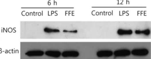
FFE의 iNOS 발현에 미치는 영향. FFE가 산화질소 생성을 증 가시킴을 확인하고 산화질소의 생성을 촉진하는 주효소인 iNOS 생성 량을 웨스턴 블럿으로 확인하였다. 대식세포에 LPS (1 µg/
mL) 또는 40 µg/mL의 FFE를 세포에 처리한 다음 6시간과 12 시간 후에 iNOS 발현을 웨스턴 블럿으로 확인하였다. Fig. 3의 결과와 같이 iNOS의 발현은 비록 LPS 보다는 약하지만 무처 리 대조군과 비교하여 유의한 증가를 나타내었다.
FFE의 cytokine의 발현에 미치는 영향. FFE에 의한 대식세포 의 또 다른 면역 증강 효과를 확인하기 위하여 활성이 증가된 Fig. 1 Cytotoxicity of FFE on RAW 264.7 cells. RAW 264.7 cells were treated with various concentrations (5, 10, 20 and 40µg/mL) of FFE.
After 24 h cytotoxicity was determined by measuring the absorbance at 450 nm after WST-8 regent addition. The values shown here are means ± SD of three independent experiments. **p <0.01 as compared to the untreated cells (control).
대식세포로부터 분비되는 면역 활성 cytokine, TNF-α, IL-1β, IL-6의 분비량을 효소면역분비법으로 분석하였다. cytokine의 양 을 측정하기 위하여 세포에 FFE를 농도 별로 처리 한 후 18시 간 후 배양 상층액에서 cytokine의 양을 측정하였다. Fig. 4에 서와같이 무처리 대조군(Control)은 세 종류의 cytokine이 거의 분비되지 않은 반면, LPS의 처리에 의해 cytokine의 생성은 급 격히 증가되는 것을 확인할 수 있었다. IL-1β와 IL-6의 생성은 FFE의 최고 농도에서 LPS와 유사한 수준까지 증가된 반면, TNF-α는 FFE의 최고 농도인 40 µg/mL에서 LPS보다 많이 분 비되었다.
FFE의 IκB 발현에 미치는 영향. NFκB는 면역 반응에 중요한 세포신호전달 단백질로서 활성이 증가되기 위해서는 먼저 IκBα 가 분해되어야 한다. 그러므로 FFE의 면역증강활성에 대한 기 전연구로서 NFκB의 활성을 IκBα의 량으로 확인 하였다. IκBα 의 존재량을 확인하기 위해서 FFE를 40 µg/mL의 농도로 처리 한 다음, 30분과 1시간 후에 IκBα의 발현량을 웨스턴 블럿으 로 확인한 결과, FFE처리 후 30분에서 IκBα가 LPS를처리했을 때와 유사하게 되는 것을 확인 할 수 있었다(Fig. 5). 반면 1시 간이 경과한 후에는 IκBα의 발현이 회복되었다.
FFE의 MAP kinases 발현 효과. MAP kinases는 NFκB와 더 불어 면역반응을 일으키는데 중요한 역할을 하는 것으로 알려 져 있어서, FFE의 처리에 의해 RAW 264.7 세포에서 MAP kinases의 신호가 변화되는지를 웨스턴 블럿으로 확인 하였다.
IκBα에서와 동일한 조건으로 세포에 FFE를 처리한 후 10, 20, 30분 후에 세포를 회수하여 MAP kinases에 속하는 p38, ERK, Fig. 4 Effects of FFE on TNF-α, IL-1β, and IL-6 production in RAW 264.7 cells. RAW 264.7 cells were treated with 5−40 µg/mL of samples or LPS (1µg/mL) for 24 h. The culture supernatant, which was subject to enzyme immunoassay of cytokines, was harvested. The values shown are means ± SD for three independent experiments. **p <0.01 as compared to the untreated cells (control).
Fig. 5 Effects of FFE on IκB degradation. RAW 264.7 cells were treated with 40 µg/mL of FFE or LPS (1 µg/mL). The total protein lysate of each RAW 264.7 cell was prepared after 30 min and 1 h, and cell signaling proteins were detected by Western blot analysis. The expression levels of IκB were compared with β-actin as a control. Western blot analysis using a specific antibody was performed in triplicate and all of them showed similar results.
Fig. 3 Effects of FFE on the iNOS production of the RAW 264.7 cells.
The cells were treated with LPS (1µg/mL) or FFE (40 µg/mL) for 6 or 12 h. Total protein lysate of each RAW 264.7 cell was prepared and the resolved proteins by western blot were analyzed by anti-iNOS antibody.
Anti-β-actin antibody was used as a control. Western blot analysis was performed in triplicate and all of them showed similar results.
Fig. 2 Effects of FFE on the nitric oxide production of the RAW 264.7 cells. RAW 264.7 cells were treated with various concentrations (5, 10, 20 and 40µg/mL) of FFE. After 24 hour the culture supernatant were subjected the nitric oxide assay. The values shown here are means ± SD of three independent experiments. **p <0.01 as compared to the untreated cells (control).
JNK의 인산화 정도를 측정한 결과, FFE를 처리한 경우 LPS를 처리한 것과 동일하게 ERK와 p38는 10분 후부터 인산화가 증 가되어 30분 까지 지속되었다. 반면 JNK는 LPS와 FFE 모두 20분 이후에 인산화가 증가되었다.
고 찰
버섯은 탄수화물, 단백질, 지질, 무기질, 및 비타민 등의 영양소 를 골고루 함유하고 있어서 항암 작용, 면역증강 효과, 항산화 효과, 혈당 강하, 콜레스테롤 저하, 뇌졸중 방지, 신장병 완화 등의 여러 가지 생리활성이 있는 것으로 알려져 있다(Lie 등, 1997; Pyo 등, 2001; Mau 등, 2002; Yang 등, 2002; Cheung 등, 2003; Lee 등, 2003b; Oh와 Lee, 2005). 말굽버섯도 다양 한 활성이 알려져 있다. 말굽버섯 유래 β-glucan 복합체는 항생 제와 동일한 수준의 세균억제 효과가 보고 된 바 있다(Seniuk 등, 2011). 뿐만 아니라 말굽버섯은 항산화와 혈당 및 콜레스테 롤 저하에 좋은 효과를 보였다(Lee, 2005). 그러나 말굽버섯의 면역증강효과에 대한 연구결과는 보고된 바가 없다.
산화질소는 아미노산인 arginine으로부터 생성되며 라디칼 물 질로서 세포 내에서 면역신호 전달자로서 중요한 역할을 한다 (Palmer 등, 1988). iNOS는 산화질소의 생성을 유도하는 주효 소로서(Kubes, 2000) LPS, IFN-γ, IL-1β, TNF-α 등의 자극에 의해 생성이 증가되며, 대식세포, 내피세포, 심근세포에서 발현 되어 다량의 산화질소 생성을 유도하는 것으로 알려져 있다. 산
Lee 등, 2000; Seo 등, 2000; Chiou 등 2001; Seo 등, 2001), 대식세포로부터 분비되는 산화질소는(Griffith 등, 1993) 암세포 에 대한 독성을 나타내며, cytokine의 분비를 촉진시켜 면역 반 응을 유도할 수 있는 물질이다(Thomas와 Edginton, 1984;
Hibbs 등, 1998). 그러므로 산화질소의 또 다른 대표 활성은 면 역증강 효과라고 할 수 있다. 본 연구에서는 FFE를 대식세포에 처리하면 iNOS의 발현이 증가되고, 결국 산화질소의 생성을 증 가시킴을 밝혔다. 비록 40 µg/mL의 FFE가 1 µg/mL의 LPS 보 다 약한 산화질소 분비 유도 효과를 보였지만 지나친 면역증강 은 염증을 초래할 수 있다는 측면을 고려한다면, FFE의 활성은 긍정적이라고 할 수 있다.
대식세포에서 분비되는 TNF-α는 대표적인 면역계의 활성을 증가시키는 cytokine으로서 대식세포에서 생성 되어 면역반응에 필요한 인자들의 생성을 촉진 한다(Yoon 등, 2007). 특히 대식 세포에 의해 주도되는 내재면역반응에서 TNF-α는 가장 중요한 역할을 담당하게 된다(Lee 등, 2003a). 본 연구에서 FFE는 전 농도에서 TNF-α의 생성을 증가시켰으며, 최고 농도에서는 LPS 와 동일한 수준의 활성을 나타내었다. IL-1β는 TNF-α와 함께 대 부분의 면역반응에 관여하는 cytokine으로서, 특히 T 세포, NK 세포, B 세포 활성에 직접적으로 관여하며 다양한 활성인자의 분비를 유도하여 세포증식과 세포 외 기질 축적을 자극 한다 (Papayianni, 1996; Delgado 등, 2003). IL-6는 대식세포로부터 분비되며 면역반응, 신경세포의 기능, 조혈작용(hematopoiesis) 등 에서 중요한 역할을 수행한다(Hirano 등, 1994; Hibi 등, 1996). 본 연구에서 FFE는 실험에 사용된 전 농도에서 농도 의 존적으로 대식세포로부터 IL-1β와 IL-6의 생성을 증가시켰다.
이 결과로 미루어 볼 때 FFE는 면역증강 활성은 농도 의존적 임을 알 수 있었다.
본 연구에서는 FFE에 의한 산화질소 및 cytokine의 분비증가 효과의 작용 기전을 확인하기 위해 대표적인 세포 내부 면역관 련 신호 분자인 NFκB와 MAP Kinases (ERK, p-38, JNK)의 활성을 웨스턴 블럿을 통해 확인해 보았다. NFκB는 세포의 분 화 조절, 발암 유전자의 생성에도 관련이 있을 뿐만 아니라 (Chen등, 2001), 면역세포에서 활성화 되면 iNOS나 TNF-α 등 의 유전자 발현에 관여한다(Lee 등, 2003a). MAP kinases에 속하는 신호분자로는 p38, ERK, JNK가 있으며, 이 세 분자 모 두 면역활성의 변화에 큰 영향을 주고 있음이 밝혀진 바 있다 (Garrington과 Johnson, 1999; Lee 등, 2003a; Seo 등, 2004).
FFE는 40 µg/mL의 농도에서 IκB의 분해를 통하여 NFκB의 활 성을 증가 시킴을 확인 할 수 있었다. 또한 FFE는 ERK와 p- 38의 신속한 인산화를 유도하였으며 비록 ERK와 p-38 보다는 약하지만 JNK의 인산화도 촉진됨을 보여 주었다. 물론 좀더 구 체적인 근거를 얻기 위한 연구가 추가적으로 진행되어야 하지 만, 이 결과들로 미루어 볼 때 FFE에 의한 면역증강 효과는 NFκB와 MAP Kinases 신호에 의존적임을 유추할 수 있었다.
기존에 발표된 논문에 의하면(Park 등, 2004) 말굽버섯 메탄 올 추출물은 RAW 264.7 세포에 대하여 항염증 효과가 있는 것으로 보고되었다. 이 연구는 강력한 염증 유도 물질을 처리 한 후 말굽버섯 메탄올 추출물을 처리 함으로써 지나치게 분비 되는 면역 관련 인자들의 생성이 억제됨을 확인 하였다. 그러 므로 이 연구 결과와 본 논문의 연구 결과가 서로 모순 되는 것으로 보일 수 있으나, 본 논문에서는 염증 상황이 아닌 정상 적인 상황에서 말굽버섯의 면역증강 효과를 검증한 것이라면 기 Fig. 6 Effects of FFE on phosphorylation of MAP kinases. RAW 264.7
cells were treated with 40µg/mL FFE or LPS (1 µg/mL). The total protein lysate of each RAW 264.7 cell was prepared after 10, 20, 30 min, and cell signaling proteins were detected by Western blot analysis. The expression levels of ERK, p38, and JNK were compared with phosphorylated proteins. Western blot analysis using a specific antibody was performed in triplicate and all of them showed similar results.
존에 발표된 논문은 염증 유도 물질과 말굽버섯의 상호작용에 의한 항염증 효과를 검증한 것이므로 각 연구 결과는 모순된 것이 아니라 서로 다른 의미를 지니고 있다고 볼 수 있다.
본 연구에서는 아직 까지 연구되지 않은 말굽버섯의 면역증 강 효과를 대식세포에서 검증하였다. 연구결과에 따르면 FFE의 처리에 의해 대식세포의 산화질소 및 면역성 cytokines의 생성 이 증가 되었으며, 이 같은 면역증강 활성은 면역활성을 중계 하는 세포 신호전달분자 중 NF-κB와 MAP Kinases의 활성증 가에 의한 것임을 확인 할 수 있었다. 이 연구 결과는 말굽버 섯의 임상적 적용에 중요한 자료로 활용될 것으로 생각된다.
초 록
본 연구에서는 아직 까지 연구되지 않은 말굽버섯의 면역증강 효과를 설치류 대식세포에서 검증하였다. 연구결과에 따르면 말 굽버섯 메탄올 추출물(FFE)의 처리에 의해 대식세포의 산화질 소의 생성이 농도 의존적으로 증가되었다. 산화질소의 생성이 증가된 이유는 산호질소의 생성을 유도하는 효소인 iNOS의 발 현이 FFE에 의해 증가되었기 때문이었다. FFE는 면역반응에 중 요한 cytokine인 TNF-α, IL-1β, IL-6의 생성도 농도 의존적으 로 증가 시켰다. 이 같은 FFE의 면역증강 활성은 면역활성을 중계하는 세포 신호전달분자 중 NF-κB와 MAP Kinases의 활 성증가에 의한 것임을 확인 할 수 있었다. 본 연구 결과는 말 굽버섯의 임상적 적용에 중요한 자료로 활용될 것이며, 만일 말 굽버섯으로부터 분리한 단일성분에서 면역증강활성을 검증 하 게 된다면 새로운 식·의약소재로서의 가치를 인정 받을 수 있 을 것으로 생각된다.
감사의 글 본 연구에 사용한 말굽버섯 시료는 미래창조과학부의 한-몽골 과 학기술협력센터사업의 지원으로 준비되었음.
References
Chen F, Castranova V, and Shi X (2001) New insights into the role of nuclear factor-kappaB in cell growth regulation. Am J Pathol 159, 387–97.
Cheung LM, Cheung PCK, and Ooi VEC (2003) Antioxidant activity and total phenolics of edible mushroom extracts. Food Chem 80, 1–7.
Chiou WF, Chou CJ, and Chen CF (2001) Camptothecin suppresses nitric oxide biosynthesis in RAW 264.7 macrophages. Life Sci 69, 625–35.
Delgado AV, McManus AT, and Chambers JP (2003) Production of tumor necrosis factor-alpha, interleukin 1-beta, interleukin 2, and interleukin 6 by rat leukocyte subpopulations after exposure to substance P.
Neuropeptides 37, 355–61.
Garrington TP and Johnson GL (1999) Organization and regulation of mitogen-activated protein kinase signaling pathways. Curr Cell Biol 11, 211–8.
Hibbs JB, Taintor RR, Vavrin I, and Rachlin EM (1998) Nitric oxide: A cytotoxic activated macrophage effector molecule. Biochembiophys Rescommun 157, 87–92.
Hibi M, Nakajima K, and Hirano T (1996) IL-6 cytokine family and signal transduction: a model of the cytokine system. J Mol Med 74, 1–12.
Hirano T, Matsuda T, and Nakajima K (1994) Signal transduction through gp130 that is shared among the receptors for the interleukin 6 related cytokine subfamily. Stem Cells 12, 262–77.
Kawamata H, Ochiai H, Mantani N, and Terasawa K (2000) Enhanced expression of inducible nitric oxide synthase by Juzen-taiho-to in LPS-
activated RAW 264.7 cells, a murine macrophage cell line. Am J Chin Med 28, 217–26.
Kim N-Y, Jung H-K, Park M-J, Kim S-J, Kim S-H, Choi J-W et al. (2005) Effects of Fomesfomentarius Extract on Blood Glucose, Lipid Profile and Immune Cell in Streptozotocin - Induced Diabetic Rats. J Korean Soc Food Sci Nutr 34, 825–32.
Kubes P (2000) Inducible nitric oxide synthase; a little bit of good in all of us.
Gut 7, 6–9.
Lee AK, Sung SH, Kim YC, and Kim SG (2003a) Inhibition of lipopolysaccharide-inducible nitric oxide synthase, TNF-α and COX-2 expression by sauchinone effects on I-κBα phosphorylation, C/EBP and AP-1 activation. British J Pharmacol, 139, 11–20.
Lee BC, Bae JT, Pyo HB, Choe TB, Kim SW, Hwang HJ et al. (2003b) Biological activities of the polysaccharides produced from submerged culture of the edible Basidimycete Grifola frondosa. Enz Microb Technol 6274, 1–8.
Lee BG, Kim SH, Zee OP, Lee KR, Lee HY, Han JW et al. (2000) Suppression of inducible nitric oxide synthase expression in RAW 264.7 macrophages by two-carboline alkaloids extracted from Meliaazedarach.
Eur J Pharmacol 406, 301–9.
Lee JS (2005) Enzyme activities, blood glucose, and lipid profile in streptozotocin-indeced diabetic rats. Nutrition research 34, 182–95.
Lie F, Oo VEC, and Chang ST (1997) Free radical scavenging activities of mushroom polysaccharide extracts. Life Sci 60, 763–71.
Mau JL, Lin HC, and Song SF (2002) Antioxidant properties of several speciality mushroom. Food Res Int 35, 519–26.
Oh SI and Lee MS (2005) Antioxidative and antimutagenic effects of Ganoderma lucidum Krast Extracts. Korean J Food &Nutr 18, 54–62.
Palmer RM, Ashton DS, and Moncada S (1988) Vascular endothelial cells synthesize nitric oxide from L-arginine. Nature 333, 664–6.
Papayianni A (1996) Cytokines, growth factors, and other inflammatory mediators in glomerulonephritis. Ren Fail 18, 725–40.
Park YH and Lee HD (1999) In Coloured Korea Medical mushrooms, Kyohaksa, Korea.
Park YM, Kim IT, Park HJ, Choi JW, Park KY, Lee JD et al. (2004), Anti- inflammatory and anti-nociceptive effects of the methanol extract of Fomes fomentarius. Biol Pharm Bull 27, 1588−93.
Pyo MY, Hyun SM, and Yang KS (2001), Effects of Ohellinuslinteus Extracts on the humoral immune response in normal and cyclophosphamide- treated mice. J Appl Pharmacol 9, 194–200.
Seniuk OF, Gorovoj LF, Beketova GV, Savichuk HO, Rytik PG, Kucherov II et al. (2011) Anti-infective properties of the melanin-glucan complex obtained from medicinal tinder bracket mushroom, Fomes fomentarius Int J Med Mushrooms 13, 7–18.
Seo JH, Lim JW, Kim H, and Kim KH (2004) Helicobacter pylori in a Korean isolate activates mitogen-activated protein kinases. AP-1 and NF-kappaB and induces chemokine expression in gastric epithelial AGS cells. Lab Invest 84, 49–62.
Seo WG, Pae HO, Oh GS, Chai KY, Yun YG, Kwon TO et al. (2000) Inhibitory effect of ethyl acetate fraction from Cudraniatricuspidata on the expression of nitric oxide synthase gene in RAW 264.7 macrophages stimulated with interferon-and lipopolysaccharide. Gen Pharmacol 35, 21–8.
Seo WG, Pae HO, Oh GS, Kim NY, Kwon TO, Shin MK et al. (2001) The aqueous extract of Rhodiolasachalinensis root enhances the expression of inducible nitric oxide synthase gene in RAW 264.7 macrophages. J Ethnopharmacol 76, 119–23.
Thomas PM and Edginton S (1984) Human monocyte mediated tumor cytotoxicity. JImmunol 132, 1980–6.
Yang JH, Li HC, and Mau JL (2002) Antioxidant properties of several commercial mushroom. Food Chem 77, 229–35.
Yoon HJ, Moon ME, Park HS, Im SY, Lee JH, and Kim YH (2007) Effects of Chitosan oligosaccharide on the C. albicans-induced Inflammatory Effect in Mice and RAW 264.7 Macrophage Cells. J Chitin Chitosan 12, 15–
20.