www.krspine.org
Measurement of Canal Encroachment Using Axial and Sagittal-Reconstructed Computed Tomographic Images
in Thoracolumbar Burst Fractures
Jin-Ho Kim, M.D., Nam-Su Chung, M.D., Oh-Kyung Lim, M.D., Ph.D.
Hyong-Rae Roh, M.D., Chang-Hoon Jeon, M.D., Ph.D.
J Korean Soc Spine Surg 2011 Sep;18(3):111-116.
Originally published online September 30, 2011;
http://dx.doi.org/10.4184/jkss.2011.18.3.111
Korean Society of Spine Surgery
Department of Orthopedic Surgery, Inha University School of Medicine
#7-206, 3rd ST. Sinheung-Dong, Jung-Gu, Incheon, 400-711, Korea Tel: 82-32-890-3044 Fax: 82-32-890-3467
©Copyright 2011 Korean Society of Spine Surgery pISSN 2093-4378 eISSN 2093-4386
The online version of this article, along with updated information and services, is located on the World Wide Web at:
http://www.krspine.org/DOIx.php?id=10.4184/jkss.2011.18.3.111
This is an Open Access article distributed under the terms of the Creative Commons Attribution Non-Commercial License (http://
creativecommons.org/licenses/by-nc/3.0) which permits unrestricted non-commercial use, distribution, and reproduction in any medium, provided the original work is properly cited.
Journal of Korean Society of
Spine Surgery
Received: July 12, 2011 Revised: August 10, 2011 Accepted: August 12, 2011
Published Online: September 30, 2011
Corresponding author: Chang-Hoon Jeon, M.D., Ph.D.
Department of Orthopedic Surgery, Ajou University School of Medicine, San 5 Wonchon-dong, Youngtong-gu, Suwon, Kyounggi-do, Korea
TEL: 82-31-219-5220, FAX: 82-31-219-5229 E-mail: [email protected]
“This is an Open Access article distributed under the terms of the Creative Commons Attribution Non-Commercial License (http://
creativecommons.org/licenses/by-nc/3.0/) which permits unrestricted non-commercial use, distribution, and reproduction in any medium, provided the original work is properly cited.”
Measurement of Canal Encroachment Using Axial and Sagittal-Reconstructed Computed Tomographic Images in Thoracolumbar Burst Fractures
Jin-Ho Kim, M.D., Nam-Su Chung, M.D., Oh-Kyung Lim, M.D., Ph.D.
*Hyong-Rae Roh, M.D., Chang-Hoon Jeon, M.D., Ph.D.
Department of Orthopaedic Surgery, Ajou University School of Medicine, Suwon, Korea Department of Rehabilitation, Gachon University of Medicine and Science, Incheon, Korea*
Study Design: A retrospective study.
Objectives: The aim of this study was to examine the usefulness of axial and sagittal-reconstructed CT images in the evaluation of spinal canal encroachment by thoracolumbar burst fractures.
Summary of Literature Review: The dimensions of spinal canal encroachment by burst fractures have been described using axial CT images in the thoracolumbar region and sagittal-reconstructed images in the lower cervical region. However, the validity and reliability, depending on the measuring method, have not been fully evaluated.
Materials and Methods: A hundred and ninety-nine patients, who had diagnosed as a thoracolumbar burst fracture, were included in this study. Three orthopedic surgeons independently measured the canal encroachment of the burst fragment in the axial CT images and the sagittal-reconstructed images using the ratio of spinal length (method 1) and the ratio of area (method 2). The validity for the evaluation of the deformity and fracture stability was evaluated. In addition, the reliability of each method was assessed.
Results: Sixty-seven stable burst fractures and 132 unstable burst fractures were assessed. The mean kyphotic angle of stable and unstable burst fracture were 11.89 ± 8.49°and 15.90 ± 9.63°(P=0.005). The mean canal encroachment ratios of stable fracture were 17.21 ± 15.82 % (axial-method 1), 16.71 ±16.49 % (axial-method 2), 19.54 ± 17.03 % (sagittal reconstructed-method 1), and 11.75 ± 12.33
% (sagittal reconstructed-method 2). The mean canal encroachment ratios of unstable fracture were 31.54 ± 17.10 % (axial-method 1), 29.67 ± 18.47 % (axial-method 2), 28.53 ± 18.60 % (sagittal reconstructed-method 1), and 21.20 ± 15.11 % (sagittal reconstructed- method 2). There was no relationship between the fracture deformity and the canal encroachment ratio in all 4 methods. All ratios in the 4 method showed significant differences in the evaluation of fracture stability. All methods except method 1 in the sagittal-reconstructed images showed significant differences in the assessment of neurologic compromise.
Conclusions: The measurement of a canal encroachment area using axial and sagittal-reconstructed images was valid in the description of fracture stability.
Key Words: Thoracolumbar, Burst fracture, Canal encroachment, Computed tomography
서 론
흉요추부 방출성 골절에서 전산화 단층촬영은 척추 골절의 손 상 범위와 골절의 형태를 관찰할 수 있어, 수상 기전에 따른 골 절의 분류와 삼주론에 의거한 안정성 평가에 매우 유용하게 이 용되고 있다.1,2) 전산화 단층 촬영에서는 골편의 척추강내 감입 도 측정할 수 있어 신경 조직의 손상을 판단할 수도 있다.3-11) 최 근 Spine trauma study group에서는 방출성 척추 골편의 척추강 내 감입 기술에 대해, 하부 경추에서는 시상면 재건 영상을,12) 흉 요추부에서는 축상 영상으로 측정할 것을 제안하고 있다.13) 골 편의 척추강내 감입에 대해 척추 골절의 발생 부위에 따라서 각
Jin-Ho Kim et al Volume 18 • Number 3 • September 2011
www.krspine.org
112
각 다른 측정 방법을 적용하는 것에 대한 근거나 타당도, 신뢰도 를 고찰한 보고는 드물며, 축상 영상과 시상면 재건 영상을 모두 측정하여 비교한 연구는 없었다.
저자들은 흉요추부 방출성 골절에 대해서 축상 영상과 시상면 재건 영상에서 척추강내 골편 감입 정도를 측정하였고, 후만 변 형, 척추강내 골편 감입 정도와 골절의 안정성과의 상관관계를 분석하였으며, 측정 방법에 따른 신뢰도 차이를 비교하였다.
대상 및 방법
1. 연구대상
본 연구는 후향적 연구로서, 2002년 7월부터 2009년 4월까지 흉요추부 (제 11 흉추부터 제 2 요추)의 급성 방출성 골절로 치 료 받고, 최소 2년 이상 추시된 199명의 환자들을 대상으로 하였 다. 본원의 기관연구윤리 심의위원회는 본 연구를 승인하였다.
수술적 치료를 요하는 불안정성 골절로는 후주의 손상이 동반 되었거나 신경학적 증상을 보이는 경우로 판단하였다. 신경학적 증상과 후주의 손상이 없는 경우는 안정성 골절로 판단하여 흉 요천추 보조기를 이용한 보존적 치료를 시행하였다. 안정성 방 출성 골절은 67예였으며, 불안정성 방출설 골절은 132예였다.
불안정성 골절로 판정된 132 예 중 2예의 경우 동반된 다발성 외 상으로 인해 수술적 치료를 시행하지 못하였다. 전체 대상 환자 의 평균 연령은 45.8 ± 15.2 세 (15-80세)였고, 남자 환자가 111 명, 여자 환자가 88명이었다. 수상 기전은 추락이 91예, 교통 사 고가 87예, 스포츠 손상이 14예, 기타 외상이 7예였다. 수상 부위 는 제 11 흉추 15 예, 제 12 흉추가 67 예, 제 1 요추가 69 예, 제 2 요추 48 예였다.
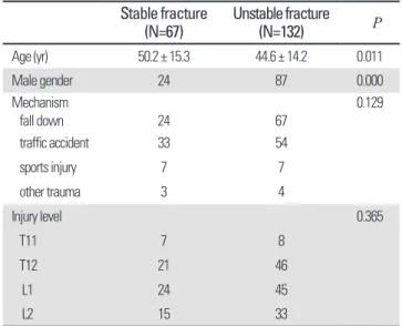
안정성 골절 군과 불안정성 골절 군 간의 환자 군 비교로는 평 균 연령 (P=0.011)과 성별 (P<0.000)의 차이를 보였으나 수상 기 전, 수상 부위의 차이는 보이지 않았다 (Table 1).
2. 방사선학적 측정
내원 당시에 시행한 전후면 및 측면 단순 방사선 사진에서 골 절 추체 상, 하 추체의 상, 하 종판을 이용하여 Cobb 각도를 측 정하여 측만 및 후만 변형을 평가하였다 (Fig. 1).
최대 골편 감입이 관찰되는 전산화 단층 영상에서 축상 영상 과 시상면 재건 영상에서 골편의 척추강내 감입의 정도를 측정 하였다. 골편의 척추강내 감입 정도는 인접 상부 및 하부 추체 척추강의 평균 전후 직경 [(L1+L2)/2]에 대한 감입된 골편의 최 대 직경 (L3)의 비를 이용하는 방법 (방법 1; [(L1+L2)/2-L3]
/ [(L1+L2)/2] x 100)과 인접 상부 추체 척추강의 평균 면적에 [(A1+A2)/2]대한 감입된 골편의 최대 면적 (A3)의 비를 이용하 는 방법 (방법 2; [(A1+A2)/2-A3] / [(A1+A2)/2] x 100)으로 각
각 측정하였다 (Fig. 2).
세 명의 정형외과 의사들이 각각 3 회씩 측정하였으며, PACS [π view starⓇ (Infinitt, Seoul, Korea)]디지털 계측 프로그램을 이 용하여 측정하였다. 각 계측값의 평균을 대표값으로 설정하였 다.
3. 통계학적 분석
전산화 단층 사진의 축상 영상과 시상면 재건 영상에서 방 법 1, 2로 각각 측정한 척추강내 골편 감입정도의 비교 분석은 paired t-test를 사용하였다. 안정성 골절 군과 불안정성 골절 군 의 각종 측정 지표의 비교 분석은 student t-test를 사용하였다.
후만 변형, 안정성 여부와 축상 및 시상면 재건 영상에서 2가
Fig. 1. Measurement of coronal and sagittal deformity using Cobb method.
Table 1. Patient Demographics
Stable fracture
(N=67) Unstable fracture
(N=132)
PAge (yr) 50.2 ± 15.3 44.6 ± 14.2 0.011
Male gender 24 87 0.000
Mechanism
fall down 24 67 0.129
traffic accident 33 54
sports injury 7 7
other trauma 3 4
Injury level 0.365
T11 7 8
T12 21 46
L1 24 45
L2 15 33
*Unless otherwise noted, data are number of participants.
지 방법으로 각각 측정한 척추강내 골편 감입 정도와의 상관 관 계는 Pearson correlation test와 student t-test를 이용하여 분석하 였다.
측정자 3인간의 신뢰도와 측정자 내 3 회의 반복 측정에 대한 신뢰도는 intraclass correlation coefficient (ICC)로 분석하였다.
통계 프로그램은 statistical package for social sciences (SPSS) version 12.0 (SPSS Inc, Chicago, IL)을 이용하였으며, p value는 0.05 이하로 설정하였다.
결 과
1. 후만 변형 측정 및 척추강내 골편 감입과의 상관 관계 분석 안정성 방출성 골절 군과 불안정성 방출성 골절의 후만 변형
은 각각 11.89 ± 8.49°와 15.90 ± 9.63°로 의미있는 차이를 보였다 (p=0.005). (Table 2)
후만 변형과 척추강내 골편 감입의 정도와의 상관 관계 는 축상 영상에서 측정한 방법 1 (R= 0.1120, p=0.116), 방법 2 (R=0.080, p=0.261), 시상면 재건 영상에서 측정한 방법 1 (R=0.117, p=0.104), 방법 2 (R=0.130, p=0.068) 모두 통계학적 으로 유의한 상관 관계를 보이지 않았다.
2. 골절 안정성과 척추강내 골편 감입의 비교
축상 영상에서 측정한 안정성 방출성 골절에서 평균 척추강내 골편 감입 정도는 방법 1로 측정했을 경우에 17.21 ± 15.82 % 였고, 방법 2로 측정한 경우 16.71 ± 16.49 %였으며, 불안정성 방출성 골절에서는 각각 31.54 ± 17.10 %와 29.67 ± 18.47 %
Fig. 2. Canal encroachment ratio was calculated by the upper and lower adjacent levels (A) using the length ratio on the axial images, (B) using the area ratio on the axial Table 2. Radiologic measurement
Stable fracture Unstable fracture
PPlain radiograph Cobb angle (°)
AP 2.25 ± 2.24 3.15 ± 3.11 0.058
lateral 11.89 ± 8.49 15.90 ±9.63 0.005
Computed tomography Canal encroachment (%)
axial (method 1) 17.21 ± 15.82 31.54 ± 17.10 0.000
axial (method 2) 16.71 ± 16.49 29.67 ± 18.47 0.000
sagittal (method 1) 19.54 ± 17.03 28.53 ± 18.60 0.001
sagittal (method 2) 11.75 ± 12.33 21.20 ± 15.11 0.000
Jin-Ho Kim et al Volume 18 • Number 3 • September 2011
www.krspine.org
114
였다.
시상면 재건 영상에서 측정한 안정성 방출성 골절에서 평 균 척추강내 골편 감입 정도는 방법 1로 측정한 경우 19.54 ± 17.03 %였고, 방법 2로 측정한 경우에 11.75 ± 12.33 %였으며, 불안정성 방출성 골절에서는 각각 28.53 ± 18.60 %와 21.20 ± 15.11%였다.
안정성 여부와 척추강내 골편 감입은 축상 영상과 시상 면 재건 영상 각각, 방법 1, 2가 모두 통계학적 차이를 보였다 (p<0.001).
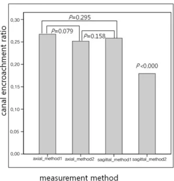
3. 측정 방법에 따른 골편 감입율의 차이
축상 영상의 방법 1, 2 와 시상면 재건 영상의 방법 1로 측정한 골편 감입 비율은 통계학적 차이를 보이지 않았으나, 시상면 재 건 영상의 방법 2는 다른 방법들로 측정한 평균치보다 작게 측 정되었다 (p<0.001). (Fig. 3)
4. 신뢰도 분석
측정자 3인간의 ICC는 0.61-0.81였으며, 측정자 내 3회 측정 에 대한 ICC는 0.69-0.88로 fair to good agreement를 보였다.
측정 방법 별로 가장 일치도가 높았던 측정 항목은 축상 영상 의 방법 1 이었으며 (ICC = 0.79-0.87), 가장 낮았던 항목은 시 상면 재건 영상의 방법 2였다 (ICC = 0.60-0.65).
고 찰
척추 방출성 골절에서 척추에 가해진 수직 압박력이 원환 응 력 (hoop stress)로 작용하면,14) 척추체는 사방으로 파열되는 양 상과 함께 척추체의 후벽 골편은 척추강내로 감입되어 신경 손 상의 원인이 되기도 한다.
과거에는 단순 방사선 검사를 이용하여 압박 변형이나 후만 변형의 의미 있는 크기를 규정하였으며,15-17) 전산화 단층촬영이 진단에 이용되면서 척추 골절의 생역학적인 면에서 연구가 시행 되었다.1) 자기공명영상을 이용한 척추 골절의 연구에서는 후방 인대복합체의 중요성이 결과가 보고되었으며,18-,20) 척추골절의 진단에서 자기공명영상의 유용성이 입증되었다.21) 수상 기전과 신경학적 손상, 후방 인대복합체의 손상을 종합적으로 고려한
“흉요추부 손상 심각 지수 (thoracolumbar injury severity score;
TLISS)”가 제안되어22) 척추 골절의 불안정성을 진단하기 위한 흉요추부 손상 심각 지수의 타당도와 신뢰도에 대한 연구가 진 행되고 있다.23)
방출성 골절에서 척추강내로 갑입된 골편의 감입정도의 평가 는 척주의 손상 정도와 신경 조직 손상과의 관계에 대한 연구가 진행되었다.3-11) 본 연구에서는 전산화 단층 영상에서 축상 영상
과 시상면 재건 영상에서 척추강내로 갑입된 골편의 감입 정도 를 여러 가지 방법으로 분석하여, 이런 측정 방법의 유용성을 입 증하였다. 척추 골절의 불안정성과 척추강내로 갑입된 골편의 감입정도와의 상관관계는 축상 영상과 시상면 재건 영상에서 모 두 차이가 있었다. 시상면 재건 영상에서 방출 면적을 이용하여 척추강내로 갑입된 골편의 감입정도를 평가한 방법 (방법 2)은 다른 방법들에 의한 측정치에 비해 평균 값이 낮게 측정되었으 며, 신뢰도가 낮은 것으로 분석되어 흉요추부 방출성 골절의 기 술에 유용성이 낮음을 확인하였다.
척추강내로 갑입된 골편의 크기와 신경 손상 정도과의 상관 관계에 대해서는 논란이 되고 있다. 축상 영상의 골편 감입률이 신경 손상과 상관 관계가 있다고 보고한 연구들도 있으며,3,5,24) 골편 감입률이 신경 손상과 무관하였다는 연구도 있다. 본 연구 에서는 축상 영상에서의 측정값과 시상면 재건 영상에서 측정한 면적은 신경 손상 정도와 상관 관계를 보였으나, 시상면 재건 영 상에서의 길이의 측정값은 신경 손상 정도에 따른 차이를 보이 지 않았다.
저자들의 연구에서는 축상 영상이나 시상면 재건 영상의 길 이 비율 측정법과 면적 비율 측정법은 골절로 인한 후만 변형과 는 상관 관계를 보이지 않았으며, 척추 골절의 안정성 여부와는 상관 관계가 있었다. 또한, 시상면 재건 영상의 면적을 측정하는 방법은 반복 측정의 신뢰도 (reliability)와 타당도 (validity)가 낮 아 그 유용성이 낮다고 판단되었다.
본 연구도 다른 대부분의 연구들처럼 증례 수집을 통한 후 향적 분석이었으므로 회상 편견 (recall bias) 나 선택 편견
Fig. 3. Paired t-tests revealed that the canal encroachment ratio using the area ratio on the sagittal-reconstructed images was different from those of other methods.
(selection bias)이 작용했을 가능성이 있다. 향후 전향적으로 기 획된 적절한 비교 연구가 필요하며, 척추강내 골편 감입 요소 이 외의 다른 위험 인자를 포함한 다인자 분석이 필요하다고 사료 된다.
결 론
흉요추부 방출성 골절의 전산화 단층 영상에서 축상 영상과 시상면 재건 영상에서 면적과 길이의 비를 이용한 척추강내 골 편 감입률은 골절로 인한 후만 변형과는 모두 상관 관계를 보이 지 않았으나, 골절의 안정성에 대해서는 모두 유의한 상관 관계 가 있었다. 시상면 재건 영상의 면적을 측정하는 방법은 유용성 이 낮게 나타났다.
REFERENCE
1. Denis F. The three column spine and its significance in the classification of acute thoracolumbar spinal injuries. Spine (Phila Pa 1976). 1983;8:817-31.
2. McAfee PC, Yuan HA, Fredrickson BE, Lubicky JP. The value of computed tomography in thoracolumbar fractures.
An analysis of one hundred consecutive cases and a new classification. J Bone Joint Surg Am. 1983;65:461-73.
3. Vaccaro AR, Nachwalter RS, Klein GR, Sewards JM, Albert TJ, Garfin SR. The significance of thoracolumbar spinal canal size in spinal cord injury patients. Spine (Phila Pa 1976). 2001;26:371-6.
4. Dai LY, Wang XY, Jiang LS. Neurologic recovery from thoracolumbar burst fractures: is it predicted by the amount of initial canal encroachment and kyphotic deformity? Surg Neurol. 2007;67:232-7.
5. Meves R and Avanzi O. Correlation between neurological deficit and spinal canal compromise in 198 patients with thoracolumbar and lumbar fractures. Spine (Phila Pa 1976).
2005;30:787-91.
6. Dall BE, Stauffer ES. Neurologic injury and recovery patterns in burst fractures at the T12 or L1 motion segment.
Clin Orthop Relat Res. 1988;171-6.
7. Eberl R, Kaminski A, Muller EJ, Muhr G. Importance of the cross-sectional area of the spinal canal in thoracolumbar and lumbar fractures. Is there any correlation between the degree of stenosis and neurological deficit? Orthopade.
2003;32:859-64.
8. Herndon WA, Galloway D. Neurologic return versus cross-
sectional canal area in incomplete thoracolumbar spinal cord injuries. J Trauma. 1988;28:680-3.
9. Keene JS, Fischer SP, Vanderby R Jr, Drummond DS, Turski PA. Significance of acute posttraumatic bony encroachment of the neural canal. Spine (Phila Pa 1976). 1989;14:799- 802.
10. Mohanty SP, Venkatram N. Does neurological recovery in thoracolumbar and lumbar burst fractures depend on the extent of canal compromise? Spinal Cord. 2002;40:295-9.
11. Zhao X, Fang XQ, Zhao FD, Fan SW. Traumatic canal stenosis should not be an indication for surgical decompression in thoracolumbar burst fracture. Med Hypotheses. 2010;75:550-2.
12. Bono CM, Vaccaro AR, Fehlings M, et al. Measurement techniques for lower cervical spine injuries: consensus statement of the Spine Trauma Study Group. Spine (Phila Pa 1976). 2006;31:603-9.
13. Keynan O, Fisher CG, Vaccaro A, et al. Radiographic measurement parameters in thoracolumbar fractures: a systematic review and consensus statement of the spine trauma study group. Spine (Phila Pa 1976). 2006;31:E156- 65.
14. Tran NT, Watson NA, Tencer AF, Ching RP, Anderson PA. Mechanism of the burst fracture in the thoracolumbar spine. The effect of loading rate. Spine (Phila Pa 1976).
1995;20:1984-8.
15. Willen J, Lindahl S, Nordwall A. Unstable thoracolumbar fractures. A comparative clinical study of conservative treatment and Harrington instrumentation. Spine (Phila Pa 1976). 1985;10:111-22.
16. Krompinger WJ, Fredrickson BE, Mino DE, Yuan HA.
Conservative treatment of fractures of the thoracic and lumbar spine. Orthop Clin North Am. 1986:17;161-70.
17. Gertzbein SD. Scoliosis Research Society. Multicenter spine fracture study. Spine (Phila Pa 1976). 1992;17:582-40.
18. Petersilge CA, Pathria MN, Emery SE, Masaryk TJ.
Thoracolumbar burst fractures: evaluation with MR imaging. Radiology. 1995;194:49-54.
19. Oner FC, van Gils AP, Faber JA, Dhert WJ, Verbout AJ. Some complications of common treatment schemes of thoracolumbar spine fractures can be predicted with magnetic resonance imaging: prospective study of 53 patients with 71 fractures. Spine (Phila Pa 1976).
2002;27:629-36.
20. Haba H, Taneichi H, Kotani Y, et al. Diagnostic accuracy
Jin-Ho Kim et al Volume 18 • Number 3 • September 2011
www.krspine.org
116
of magnetic resonance imaging for detecting posterior ligamentous complex injury associated with thoracic and lumbar fractures. J Neurosurg. 2003;99:20-6.
21. Oner FC, van Gils AP, Dhert WJ, Verbout AJ. MRI findings of thoracolumbar spine fractures: a categorisation based on MRI examinations of 100 fractures. Skeletal Radiol.
1999;28:433-43.
22. Vaccaro AR, Zeiller SC, Hulbert RJ, et al. The thoracolumbar injury severity score: a proposed treatment
algorithm. J Spinal Disord Tech. 2005;18:209-15.
23. Vaccaro AR, Baron EM, Sanfilippo J, et al. Reliability of a novel classification system for thoracolumbar injuries: the Thoracolumbar Injury Severity Score. Spine (Phila Pa 1976).
2006;31(11 Suppl):S62-9.
24. Meves R and Avanzi O. Correlation among canal compromise, neurologic deficit, and injury severity in thoracolumbar burst fractures. Spine (Phila Pa 1976).
2006;31:2137-41.
축상 및 시상면 재건 전산화 단층촬영 영상을 이용한 흉요추부 방출성 골절의 척추강내 골편 감입 측정
김진호 • 정남수 • 임오경* • 노형래 • 전창훈
아주대학교 의과대학 정형외과학교실, 가천대학교 의학전문대학원 재활의학교실*
연구 계획: 후향적 방사선학 연구
목적: 흉요추부 방출성 골절의 전산화 단층 영상에서 축상 영상과 시상면 재건 영상에서 척추강내 골편 감입 측정의 유용성을 평가하고자 하였다.
선행문헌의 요약: 흉요추부 방출성 골절에서 방출성 골편의 척추강내 감입 측정에 대해 축상 영상과 시상면 재건 영상을 이용한 측정 방법의 차이에 대 한 타당도와 신뢰도는 아직 보고되지 않았다.
대상 및 방법: 2002년 7월부터 2009년 4월까지 흉요추부 방출성 척추 골절로 치료 받은 199예의 환자들을 대상으로 하였다. 단순 방사선 영상에서 후 만 변형을 측정하였으며, 전산화 단층촬영의 축상 영상과 시상면 재건 영상에서 감입된 골편의 최대 길이의 비(방법 1)와 면적의 비(방법 2)를 이용하여 척추강내 골편의 감입 정도를 측정하였다. 후만 변형, 축상 영상과 시상면 재건 영상에서 각각 측정한 골편 감입 정도과 골절 안정성 과의 상관관계를 분석하였다.
결과: 안정성 방출성 골절 67예와 불안정성 방출성 골절 132예가 본 연구에 포함되었다. 안정성 방출성 골절 군과 불안정성 방출성 골절의 후만 변형은 각각 11.89 ± 8.49°와 15.90 ± 9.63°로 의미있는 차이를 보였다 (p=0.005). 안정성 방출성 골절군의 평균 골편 감입 정도는 축상 영상에서 방법 1로 측정시 17.21 ± 15.82 %, 방법 2는 16.71 ±16.49 %, 시상면 재건 영상에서 방법 1로 측정한 경우 19.54 ± 17.03 %, 방법 2로 측정한 경우 11.75 ± 12.33 %로 나타났다. 불안정성 방출성 골절군의 평균 골편 감입 정도는 축상 영상에서 방법 1로 측정시 31.54 ± 17.10 %, 방법 2는29.67 ± 18.47 %, 시상면 재건 영상에서 방법 1로 측정한 경우 28.53 ± 18.60 %, 방법 2로 측정한 경우 21.20 ± 15.11 %로 나타났다. 후만 변형과 척추강내 골편 감입 의 상관관계는 축상 영상과 시상면 재건 영상 각각, 방법 1, 2가 모두 통계학적으로 유의한 상관관계를 보이지 않았다 (p>0.05). 안정성 여부와 척추강내 골편 감입은 축상 영상과 시상면 재건 영상 각각, 방법 1, 2가 모두 통계학적 차이를 보였다 (p<0.001). 신뢰도가 높았던 측정 방법은 축상 영상의 방법 1 이었으며 (ICC = 0.79-0.87), 신뢰도가 가장 낮았던 방법은 시상면 재건 영상의 방법 2였다 (ICC = 0.60-0.65).
결론: 흉요추부 방출성 골절에서 전산화 단층 촬영의 축상 영상과 시상면 재건 영상을 이용하여 감입된 골편의 길이 및 면적의 비를 측정한 결과, 모든 측정에서 골절의 안정성 여부에 따른 차이를 나타냈다.
색인단어: 흉요추부 골절, 전산화 단층촬영, 축상 영상, 시상면 재건 영상 약칭제목: 흉요추부 방출성 골절의 전산화 단층촬영