J o u r n a l o f R h e u m a t i c D i s e a s e s V o l . 2 0 , N o . 1 , F e b r u a r y 2 0 1 3
http://dx.doi.org/10.4078/jrd.2013.20.1.52 □ Case Report □
52
<Received:December 19, 2011, Revised (1st: February 24, 2012, 2nd: April 2, 2012), Accepted:April 4, 2012>
Corresponding to:Young Ho Lee, Division of Rheumatology, Department of Internal Medicine, Korea University Anam Hospital, Korea University College of Medicine, 126-1, Anam-dong 5-ga, Seongbuk-gu, Seoul 136-705, Korea. E-mail:
[email protected] pISSN: 2093-940X, eISSN: 2233-4718
Copyright ⓒ 2013 by The Korean College of Rheumatology
This is a Free Access article, which permits unrestricted non-commerical use, distribution, and reproduction in any medium, provided the original work is properly cited.
A Case of Behcet's Disease Complicated with a Pulmonary Artery Aneurysm and Deep Vein Thrombosis, Separately
Jae Min Lee, Jemma Ahn, Young Jae Hwang, Seung Han Kim, Jong Su Lee, Sung Jae Choi, Young Ho Lee, Jong Dae Ji, Gwan Gyu Song
Division of Rheumatology, Department of Internal Medicine, Korea University Medical College, Seoul, Korea
Behcet'sdisease is a chronic inflammatory disease charac- terized by oral ulcers, genital ulcers, uveitis, and skin lesions. Furthermore, Behcet's disease can manifest as vas- cular lesions, such as, those of vasculitis, venous thrombo- sis, or thrombophlebitis or as an arterial aneurysm. Here, the authors report the case of a pulmonary artery aneur- ysm and deep vein thrombosis in a 41-year-old woman with a previous diagnosis of Behcet's disease. The patient presented with hemoptysis and a cough, and was found to
have a bleeding pulmonary artery aneurysm at the right lower lung. Pulmonary arteriography was performed and the aneurysm was embolized with coils. As a result, he- moptysis did not subsequently recur. However, five years later, deep vein thrombosis occurred in the left leg. Left leg pain improved after the regional infusion of thromb- olytics.
Key Words. Behcet's disease, Pulmonary artery aneurysm, Endovascular embolization, Deep vein thrombosis
Introduction
Behcet’s disease is a chronic, systemic, inflammatory disease of unknown origin. Its major clinical manifestations are oral ulcers, genital ulcers, uveitis, and skin lesions, and its other manifestations are multiple arthritis, gastrointestinal lesions, central nervous system involvement, and vascular lesions.
Small or large vessel vasculitis in Behcet’s disease leads to vessel occlusion or aneurysm formation (1), but arterial in- volvement occurs infrequently and presents as aortitis or a pe- ripheral arterial aneurysm with arterial thrombosis. The pul- monary artery is the second most common site of arterial in- volvement in Behcet’s disease (2), and pulmonary artery aneurysms can cause massive hemoptysis by rupture in bronchi. Venous thrombosis is also present in one-fourth of Behcet’s disease patients, and deep venous thrombosis of the lower limb is the most frequent venous manifestation. Few cases of Behcet's disease in a female patient combined with
a pulmonary artery aneurysm and deep venous thrombosis have been reported (3). In this article, we describe a case of Behcet's disease that was complicated by a pulmonary artery aneurysm and deep venous thrombosis, separately occurred with 5 years interval.
Case Report
A 41 year-old woman was admitted with a 2-month history of hemoptysis and sustained cough. Initially, amount of he- moptysis was small manifesting as blood-tinged sputum. Four hours before admission, she presented 30 mL of fresh hemopt- ysis accompanying with intractable cough. The patient had been diagnosed with Behcet's disease about 4 years previou- sly. Her diagnosis was based on oral ulcers, genital ulcers and pathergy test. She was being treated with colchicine 1.2 mg per day and Dapsone 25 mg per day. The patient was a non-smoker and did not consume alcohol regularly. On phys-
Pulmonary Artery Aneurysm and Deep Vein Thrombosis in Behcet's Disease 53
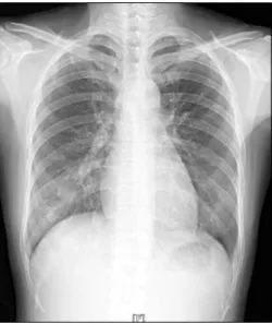
Figure 1. Chest X-ray showing ill-defined increased opacity in the right lower lung.
Figure 2. Contrast computerized tomography (CT) scan of the thorax showing a round enhancing lesion, measuring 2.2×2.0 cm in size, in the right lower lung (arrow).
Figure 3. Pulmonary arteriograph showing Nester coils in the pulmonary artery aneurysm.
ical examination, vital signs were stable, but respiratory sounds were decreased in the right lower lung field. A periph- eral blood examination revealed; hemoglobin 10.2 g/dL, hem- atocrit 32.6%, platelet count 545,000/μL, erythrocyte sed- imentation rate (ESR) 47 mm/hr, and C-reactive protein (CRP) 19.7 mg/dL. Biochemical analyses of blood showed no pathological findings. Prothrombin and partial thromboplastin times were normal. But HLA-B51 test was not executed.
Chest radiography showed an ill-defined increased opacity in the right lower lung (Figure 1), and a computerized tomog- raphy (CT) scan of the thorax with intravenous contrast re- vealed a round enhancing lesion, measuring 2.2×2.0 cm, in right lower lung (Figure 2). The lesion was connected to the
pulmonary artery, and a hypo-dense lesion was seen in the proximal pulmonary artery. Pulmonary artery angiography was performed for diagnosis and treatment. A pulmonary ar- tery aneurysm and broncho-pulmonary shunt was demon- strated at lateral segment of right lower lung. The aneurysm was embolized with two 6 mm×14 cm coils (NESTER, Cook, USA) and eight 4 mm×14 cm coils (NESTER, Cook, USA) (Figure 3). After coil embolization, methylprednisolone was administered during 3 days at 1 g per day delivered by intra- venous boluses. After steroid pulse therapy, intravenous meth- ylprednisolone was changed to oral prednisolone 55 mg (1 mg/kg) per day. Post-procedural recovery was uneventful, and the patient was discharged eight days after embolization. The patient was treated with a combination of cyclophosphamide and prednisolone after discharge in an outpatient clinic.
Cyclophosphamide was administered as monthly intravenous boluses of 1 g and oral prednisolone was tapered from 55 mg to 5 mg over 10 months. Cyclophosphamide was continued for 6 months and then changed to azathioprine 100 mg per day. Because of the possibility of a hemorrhagic complication, anticoagulant medication was not given.
Five years after discharge, the patient was transferred to emergency room with left leg pain presumed to be due to deep vein thrombosis in the left lower limb. Laboratory tests showed; hemoglobin 12.9 g/dL, platelets 399,000/μL, WBC 8,400/μL, prothrombin time 86%, partial thromboplastin time 38.4 seconds, ESR 36 mm/hr, CRP 15.4 mg/dL and D-dimer increased to 0.83 μg/mL. Protein C/S activity and antigen levels was within normal range. Lupus anticoagulant was neg- ative, but antiphospholipid antibody IgG/IgM was not tested.
54 Jae Min Lee et al.
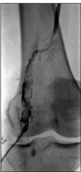
Figure 4. Lower extremity venograph showing a thrombus in the left popliteal vein and the catheter used for the peripheral venous approach.
CT angiovenography revealed a thrombus in the left super- ficial femoral vein and popliteal vein (Figure 4). IVC filter (OPTEASE, Cordis, USA) insertion was performed for endo- vascular management. After filter insertion, loading dose of urokinase 100,000 IU and heparin 5,000 IU was infused through the distal portion of the left popliteal vein. After in- fusing regional thrombolytics, the remnant thrombus was man- ually removed. Two weeks later, IVC filter was removed. Leg pain and swelling improved after flow-directed regional thrombolytic therapy, and there were no evidence of a secon- dary pulmonary embolism or bleeding. After discharge, she was treated with azathioprine 100 mg per day and colchicine 1.2 mg per day for 3 months, and no evidence of hemoptysis recurrence was encountered after pulmonary artery emboliza- tion, warfarin was administered for 3 months, and then changed to aspirin. At 6 months after discharge, the patient remained asymptomatic with no recurrence of the leg pain.
Discussion
Behcet’s disease is a chronic, systemic, inflammatory disease. Its major manifestations are recurrent oral and genital aphthous ulcerations. Furthermore, Behcet’s disease may in- volve neurologic, cardiovascular, pulmonary, and muscu- loskeletal systems, and cardiovascular manifestations have been described in 7∼49% of patients. Veins are frequently affected, and their involvements result in both superficial thrombophlebitis and deep venous thrombosis in 30∼40% of patients. Arterial complications occur in 1∼7% of patients and pulmonary artery involvement is observed in 1% (4-6).
Men are much more likely to be affected by arterial disease.
Pulmonary artery aneurysm has a poor prognosis and is one of the leading causes of death in Behcet’s disease patients.
Hemoptysis, when massive and untreated, has a mortality rate of >50% (7). Medical treatments based on steroids and on immunosuppressive drugs, such as cyclophosphamide or aza- thioprine, have been tried, but although some have reported successful treatment results and aneurysmal regression for im- munosuppressive treatments (8,9), embolization with medical therapy more widely accepted (10). Surgical treatment may be considered for refractory vessel disease.
Venous thrombosis of the lower extremities and superior or inferior vena cava occlusion frequently occur in Behcet’s dis- ease patients. Unfractionated heparin and anticoagulation his- torically represent the mainstay treatment for deep vein throm- bosis, but anticoagulation therapy cannot be used in patients with a high risk of hemorrhagic complications. Thus, anti- coagulation therapy can be administered via flow-directed re- gional thrombolytic therapy, which is based on the direct re- gional infusion of concentrated thrombolytic agent from an ip- silateral peripheral vein into the deep venous system (11).
Because secondary pulmonary embolism due to small frag- ments can occur, IVC filtration must be performed before di- rect regional thrombolytic therapy.
Immunosuppressive agents are recommended for the man- agement of acute deep vein thrombosis in Behcet’s disease, but anticoagulation is not recommended by the EULAR guide- lines (12). The ‘vasculo-Behcet’ concept has been adopted when vascular complications dominate clinical features (13).
There is some debate about use of non-soluble coil or soluble gelfoam. In this case, pulmonary artery aneurysm was treated successfully by coil embolization and deep vein thrombosis was treated by regional thrombolytics infusion. Because of co- existence bleeding risk and thrombotic complication, we must concern to determine treatment in vasculo-Behcet's disease.
The early detection of vascular lesions and appropriate treat- ment are essential for the optimal care of these patients.
References
1. Cil BE, Geyik S, Akmangit I, Cekirge S, Besbas N, Balkanci F. Embolization of a giant pulmonary artery aneurysm from Behcet disease with use of cyanoacrylate and the "bubble technique". J Vasc Interv Radiol 2005;16:
1545-9.
2. Erkan F, Kiyan E, Tunaci A. Pulmonary complications of Behçet's disease. Clin Chest Med 2002;23:493-503.
3. Kim YG, Ko HK, Ko OB, Kim TS, Kim HW, Lee CK, et al. Bronchial artery embolization for massive hemopt- ysis in a patient with Behcet's disease. J Korean Rheum
Pulmonary Artery Aneurysm and Deep Vein Thrombosis in Behcet's Disease 55
Assoc 2005;12:311-4.
4. Kural-Seyahi E, Fresko I, Seyahi N, Ozyazgan Y, Mat C, Hamuryudan V, et al. The long-term mortality and mor- bidity of Behçet syndrome: a 2-decade outcome survey of 387 patients followed at a dedicated center. Medicine (Baltimore) 2003;82:60-76.
5. Hamuryudan V, Er T, Seyahi E, Akman C, Tüzün H, Fresko I, et al. Pulmonary artery aneurysms in Behçet syndrome. Am J Med 2004;117:867-70.
6. Atzeni F, Sarzi-Puttini P, Doria A, Boiardi L, Pipitone N, Salvarani C. Behçet's disease and cardiovascular involvement. Lupus 2005;14:723-6.
7. Jean-Baptiste E. Clinical assessment and management of massive hemoptysis. Crit Care Med 2000;28:1642-7.
8. Tunaci M, Ozkorkmaz B, Tunaci A, Gül A, Engin G, Acunaş B. CT findings of pulmonary artery aneurysms during treatment for Behçet's disease. AJR Am J Roentgenol 1999;172:729-33.
9. Lê Thi Huong D, Wechsler B, Papo T, Piette JC, Bletry
O, Vitoux JM, et al. Arterial lesions in Behçet's disease.
A study in 25 patients. J Rheumatol 1995;22:2103-13.
10. Swanson KL, Johnson CM, Prakash UB, McKusick MA, Andrews JC, Stanson AW. Bronchial artery embolization:
experience with 54 patients. Chest 2002;121:789-95.
11. Sharafuddin MJ, Sun S, Hoballah JJ, Youness FM, Sharp WJ, Roh BS. Endovascular management of venous throm- botic and occlusive diseases of the lower extremities. J Vasc Interv Radiol 2003;14:405-23.
12. Hatemi G, Silman A, Bang D, Bodaghi B, Chamberlain AM, Gul A, et al. Management of Behçet disease: a sys- tematic literature review for the European League Against Rheumatism evidence-based recommendations for the management of Behçet disease. Ann Rheum Dis 2009;68:
1528-34.
13. Calamia KT, Schirmer M, Melikoglu M. Major vessel in- volvement in Behçet's disease: an update. Curr Opin Rheumatol 2011;23:24-31.