177 Introduction
Venous thromboembolism (VTE) occurs to be one of the most serious complications after undergoing total joint arthro- plasty.1) Generally, pulmonary thromboembolism is generated as the secondary by-product from deep vein thrombosis (DVT) while its emergence may possibly trigger the chronic throm- boembolic pulmonary hypertension as well as post-thrombotic syndrome.2) Patent foramen ovale (PFO), a residue of fetal cir- culation, is found approximately about 25–30% among adults;
and, it is also known that thrombus from the venous circula- tion rarely causes arterial thromboembolism through right-to- left shunt.3) In this case, the authors experienced a patient with PFO and several thrombotic disease such as pulmonary throm- boembolism, DVT, and right atrial thrombus along with cryp- togenic ischemic stroke after total knee arthroplasty (TKA).
pISSN 1975-4612/ eISSN 2005-9655 Copyright © 2015 Korean Society of Echocardiography www.kse-jcu.org http://dx.doi.org/10.4250/jcu.2015.23.3.177
CASE REPORT J Cardiovasc Ultrasound 2015;23(3):177-180
Case
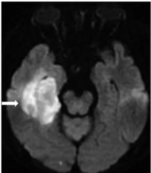
A 64-year-old female presented to the emergency room for the shortness of breath. She underwent a left total knee replacement 3 weeks ago at other hospital. She had been taking antiplatelet drugs (aspirin 100 mg/day, clopidogrel 75 mg/day) for the treatment of the right posterior temporal lobe infarction de- tected in brain magnetic resonance imaging for the disorienta- tion on the third day after the operation (Fig. 1). Carotid and brain computed tomography (CT) angiography showed no ev- idence of atherosclerosis. Afterwards, she experienced dyspnea in the second week after the operation and was performed the transthoracic echocardiography (TTE), which showed right atrial thrombi and was treated with the low molecular weight heparin (LMWH). The symptoms, nonetheless, remained so that she had been transferred to our hospital. In the medical his- tory she was being treated with medication for hypertension and diabetes. Preoperative electrocardiogram (ECG) and TTE
Post-Operative Multiple Thrombosis
Associated with Patent Foramen Ovale:
Embolic Stroke, Right Atrial Thrombi, Pulmonary Embolism and Deep Vein
Thrombosis
Sun-Young Cho, MD1, Ho-Joong Youn, MD2, Mi-Youn Park, MD1, Byung-Ju Shim, MD1, Seung-Jae Lee, MD1, Jeong-Ho Kim, MD1, Jung-Ku Park, MD1, Chang-Yul Oh, MD1, So-Hyun Ahn, MD1, and Woo-Hyun Cho, MD1
1Division of Cardiology, Department of Internal Medicine, Pohang St. Mary’s Hospital, Pohang, Korea
2Division of Cardiology, Department of Internal Medicine, Seoul St. Mary’s Hospital, School of Medicine, The Catholic University of Korea, Seoul, Korea
Patients undergoing total joint arthroplasty frequently develop post-operative complication, such as deep vein thrombosis and pulmonary thromboembolism. However, it is not common coexisting deep vein thrombosis, pulmonary thromboembolisms, right atrial thrombus and acute cerebral infarction raised by thrombus through patent foramen ovale. We reported the patient who had multiple thrombi which were accompanied with a cryptogenic ischemic stroke and associated with patent foramen ovale after operation.
KEY WORDS: Deep vein thrombosis · Pulmonary thromboembolism · Patent foramen ovale.
• Received: April 23, 2015 • Revised: June 12, 2015 • Accepted: July 22, 2015
• Address for Correspondence: Ho-Joong Youn, Division of Cardiology, Department of Internal Medicine, Seoul St. Mary’s Hospital, School of Medicine, The Catholic University of Korea, 222 Banpo-daero, Seocho-gu, Seoul 06591, Korea Tel: +82-2-2258-1134, Fax: +82-2-2258-1506, E-mail: [email protected]
• This is an Open Access article distributed under the terms of the Creative Commons Attribution Non-Commercial License (http://creativecommons.org/licenses/by-nc/3.0) which permits unrestricted non-commercial use, distribution, and reproduction in any medium, provided the original work is properly cited.
Journal of Cardiovascular Ultrasound 23 | September 2015
178
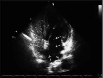
were normal. At the hospitalization, her blood pressure was 127/79 mm Hg while her heart rate was 89 bpm. On physical, pitting edema in her left lower leg was noted and neurologic examination was unremarkable. The chest X-rays did not show any signs of cardiomegaly as well as pulmonary edema. Both d- dimer and N-terminal pro-brain natriuretic peptide were in- creased respectively at 2686 ng/mL, and 3310 pg/mL and on the other hand, cardiac enzymes, such as creatinine kinase-MB and troponin-I were within a normal range. TTE demonstrat- ed normal left ventricular systolic function (ejection fraction of 63%), but in the right atrium two highly mobile masses (1.67
× 2.18 cm, 0.69 × 0.80 cm) suggesting possible thrombi were observed. The echocardiogram also revealed right atrium and right ventricle enlargement, severe pulmonary hypertension (pulmonary artery systolic pressure 102 mm Hg), and D- shaped left ventricle (Fig. 2). Her chest CT showed that pul- monary thromboembolism was found in both main pulmonary arteries while DVT was observed in left femoral vein (Fig. 3).
24-hour Holter ECG did not reveal arrhythmia except rare atrial premature complex and ventricle premature complex. The patient was administered to warfarin (target INR 2–3) after us-
Fig. 1. An embolic infarction in right posterior temporal lobe is seen in diffusion brain magnetic resonance imaging (arrow).
Fig. 2. Transthoracic echocardiographic finding. A: D-shaped left ventricle is seen in the short axis view. B: Two thrombi (arrows) are present in the right atrium; and, the size of right atrium and right ventricle is enlarged (in four chamber view). C: Tricuspid regurgitation and severe pulmonary hypertension (pulmonary artery systolic pressure 102 mm Hg) are shown in the Doppler echocardiogram. D: Inferior vena cava is dilated (in subcostal view).
A B
C D
Post-Operative Thrombosis Associated with PFO | Sun-Young Cho, et al.
179 ing LMWH. In follow-up TTE 3 weeks later, the two previous
thrombi in right atrium (RA) disappeared. Along with the im- provement in RA, there was also a positive change on the extent of pulmonary hypertension. The follow up CT taken 4 weeks after the hospitalization did not reveal pulmonary thromboem- bolism and DVT (Fig. 3). Since the cause of the cerebral infarc- tion was unclear, additional evaluation were needed. In order to examine the intracardiac shunt, TTE was performed on the 5th week. The agitated saline test was also conducted along the way. The results confirmed the transference of microbubble from right atrium to left atrium without showing atrial septal defect; and therefore, we diagnosed the patient with PFO (Fig. 4). Currently, the patient is taking warfarin continuously without any signs of abnormalities in outpatient clinic. We rec- ommended further treatment for PFO including device closure but patient refused because of poor general condition.
Discussion
As VTE is one of the fatal complications after undergoing
TKA, DVT, and pulmonary embolism (PE) are known to occur approximately around 13% and 3%, respectively.4) The occur-
A
C
B
D
Fig. 3. Chest computed tomography (CT) angiogram. A and B: Thrombi (arrows) exist in both pulmonary arteries according to chest CT. C and D:
Thrombi was not found at the follow up chest CT.
Fig. 4. The passage of microbubbles (arrows) from right atrium to left atrium is seen on the agitated saline test.
Journal of Cardiovascular Ultrasound 23 | September 2015
180
rence period of VTE after TKA is reported to be 3 to 9 days af- ter the operation5) and, there are mostly cases that accompany DVT and PE and occasionally thrombi observed on several or- gans. Particularly, it was reported that the patients with PFO even might develop arterial thromboembolism with crypto- genic stroke.3)6)
In the present case, the authors suspected that cryptogenic stroke 3 days after the operation might possibly have been caused by the VTE changed into arterial thromboembolism through PFO.
PFO, one of the congenital heart diseases that can be observed even in adulthood, typically have no symptom. Nonetheless, it can affect as the risk factor for several conditions including ischemic stroke, platypnea-orthodeoxia syndrome, and decom- pression sickness.7) PFO as the cause of cryptogenic stroke is not uncommon; hence, the occurrence of ischemic stroke with either PE or DVT can be considered as the possible example of para- doxical embolism through right-to-left shunt.8)
Among the treatments on the prevention of frequent strokes within a patient who has PFO, there are medical options and in- vasive methods.9)
The medical options include the use of antiplatelet agents as well as the prescription of warfarin in the patients with hyper- coagulable state or venous thrombosis. In terms of invasive methods, it was used to have an operation in the past while the application of device closure is being used recently.10) In this case, cryptogenic stroke took place within 3 days after TKA while right atrial thrombus, DVT, and PE were present in a way that the passage through right to left shunt was speculated and the result of subsequent echocardiography led to the diagnosis of a positive reaction from the agitated saline test for PFO. The patient was treated with LMWH and then administered war- farin; and the patient discharged with the recovered conditions.
In conclusion, the occurrence of pulmonary thromboembo- lism or DVT in a patient with PFO can trigger the develop- ment of cryptogenic stroke through right to left shunt.
Therefore, further evaluation of cause are essential in patients with the occurrence of cryptogenic stroke who are associated with a high risk of VTE after total joint arthroplasty. Also, anti- coagulation treatment is necessary if a patient has a right to left
shunt, such as PFO.
References
1. Geerts WH, Bergqvist D, Pineo GF, Heit JA, Samama CM, Lassen MR, Colwell CW; American College of Chest Physicians. Prevention of venous thromboembolism: American College of Chest Physicians Evidence- Based Clinical Practice Guidelines (8th Edition). Chest 2008;133(6 Suppl):381S-453S.
2. Goldhaber SZ, Bounameaux H. Pulmonary embolism and deep vein thrombosis. Lancet 2012;379:1835-46.
3. Chun KJ. Patent foramen ovale and cryptogenic stroke. Korean Circ J 2008;38:631-7.
4. Watanabe H, Sekiya H, Kariya Y, Hoshino Y, Sugimoto H, Hayas- aka S. The incidence of venous thromboembolism before and after total knee arthroplasty using 16-row multidetector computed tomography. J Arthroplas- ty 2011;26:1488-93.
5. Warwick D, Friedman RJ, Agnelli G, Gil-Garay E, Johnson K, FitzGerald G, Turibio FM. Insufficient duration of venous thromboembo- lism prophylaxis after total hip or knee replacement when compared with the time course of thromboembolic events: findings from the Global Orthopaedic Registry. J Bone Joint Surg Br 2007;89:799-807.
6. Park MS, Park JP, Yun SH, Lee JU, Kim JK, Lee NE, Song JE, Lee SE, John SH, Lim JH, Rhew JY. A case of cryptogenic stroke associated with patent foramen ovale coexisting with pulmonary embolisms, deep vein thromboses, and renal artery infarctions. Korean Circ J 2012;42:853-6.
7. Drighil A, El Mosalami H, Elbadaoui N, Chraibi S, Bennis A. Patent foramen ovale: a new disease? Int J Cardiol 2007;122:1-9.
8. Homma S, Sacco RL. Patent foramen ovale and stroke. Circulation 2005;
112:1063-72.
9. Sacco RL, Adams R, Albers G, Alberts MJ, Benavente O, Furie K, Goldstein LB, Gorelick P, Halperin J, Harbaugh R, Johnston SC, Katzan I, Kelly-Hayes M, Kenton EJ, Marks M, Schwamm LH, Tomsick T; American Heart Association; American Stroke Associa- tion Council on Stroke; Council on Cardiovascular Radiology and Intervention; American Academy of Neurology. Guidelines for pre- vention of stroke in patients with ischemic stroke or transient ischemic attack:
a statement for healthcare professionals from the American Heart Associa- tion/American Stroke Association Council on Stroke: co-sponsored by the Council on Cardiovascular Radiology and Intervention: the American Acad- emy of Neurology affirms the value of this guideline. Stroke 2006;37:577- 617.
10. Windecker S, Wahl A, Nedeltchev K, Arnold M, Schwerzmann M, Seiler C, Mattle HP, Meier B. Comparison of medical treatment with percutaneous closure of patent foramen ovale in patients with cryptogenic stroke. J Am Coll Cardiol 2004;44:750-8.