• 검색 결과가 없습니다.

A Case of Antiphospholipid Syndrome Refractory to Secondary Anticoagulating Prophylaxis after Deep Vein Thrombosis-Pulmonary Embolism

N/A
N/A
Protected

Academic year: 2021

Share "A Case of Antiphospholipid Syndrome Refractory to Secondary Anticoagulating Prophylaxis after Deep Vein Thrombosis-Pulmonary Embolism"

Copied!
5
0
0

로드 중.... (전체 텍스트 보기)

전체 글

(1)

A Case of Antiphospholipid Syndrome Refractory to Secondary Anticoagulating Prophylaxis after Deep Vein Thrombosis- Pulmonary Embolism

Kang Mo Gu, M.D., Jong Wook Shin, M.D., Ph.D. and In Won Park, M.D., Ph.D.

Division of Allergy and Pulmonary Medicine, Department of Internal Medicine, Chung-Ang Universitiy Hospital, Chung-Ang University College of Medicine, Seoul, Korea

Antiphospholipid syndrome (APS) is an acquired systemic autoimmune disorder characterized by a combination of clinical criteria, including vascular thrombosis or pregnancy morbidity and elevated antiphospholipid antibody titers. It is one of the causes of deep vein thrombosis and pulmonary embolism that can be critical due to the mortality risk. Overall recurrence of thromboembolism is very low with adequate anticoagulation prophylaxis. The most effective treatment to prevent recurrent thrombosis is long-term anticoagulation. We report on a 17-year-old male with APS, who manifested blue toe syndrome, deep vein thrombosis, pulmonary thromboembolism, and cerebral infarction despite adequate long- term anticoagulation therapy.

Keywords: Antiphospholipid Syndrome; Venous Thrombosis; Pulmonary Embolism; Cerebrovascular Disorders; Blue Toe Syndrome

bidity, and elevated titers of antiphospholipid (aPL) antibod- ies, which are the lupus anticoagulant (LA), anticardiolipin (aCL) antibodies, and/or anti-β2 glycoprotein-I antibodies

1

. One of the common clinical manifestations of APS is venous thrombosis. At least 20% of cases of deep vein thrombosis, with and without pulmonary embolism, may be associated with aPL

2

.

After the first thromboembolic event, warfarin is recom- mended for secondary prevention in APS patients
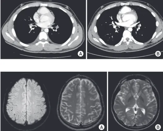
3

. We report a patient who was confirmed to have APS, of which manifested on blue toe syndrome, deep vein thrombosis, and pulmonary thromboembolism. For this patient, appropriate long-term anticoagulation with warfarin was administered but cerebrovascular accident developed during anticoagulation period.

Case Report

A 17-year-old male patient was admitted with dyspnea on exertion 10 days ago.

Fifteen days ago, this patient visited outpatient clinic due to Copyright © 2014

The Korean Academy of Tuberculosis and Respiratory Diseases.

All rights reserved.

Introduction

The antiphospholipid syndrome (APS) is an acquired sys- temic autoimmune disorder characterized by a combination of clinical criteria of vascular thrombosis or pregnancy mor-

Address for correspondence: In Won Park, M.D., Ph.D.

Division of Allergy and Pulmonary Medicine, Department of Internal Medicine, Chung-Ang Universitiy Hospital, Chung-Ang University College of Medicine, 102 Heukseok-ro, Dongjak-gu, Seoul 156-755, Korea

Phone: 82-2-6299-1401, Fax: 82-2-825-7571 E-mail: [email protected]

Received: Oct. 3, 2014 Revised: Dec. 10, 2014 Accepted: Dec. 10, 2014

cc

It is identical to the Creative Commons Attribution Non-Commercial

License (http://creativecommons.org/licenses/by-nc/3.0/).

(2)

both lower leg pitting edema. This was caused by deep vein thrombosis. Ten days ago, he felt exertional dyspnea. Chest pain was also combined with the character of non-specific, non-angina symptom.

One year ago, blue toe syndrome was detected, of which manifestations were left first toe tingling pain and erythema- tous macular eruption for 2 months.

The patient had non-specific drug history and his father had hypertension. He was high school student and had no history of alcohol and smoking.

1. Physical examination

His vital signs were as followings: systolic and diastolic blood pressure, 120/70 mm Hg; respiratory rate, 20/min; heart rate, 80/min; and body temperature, 36.3

o

C. Body weight was 67.0 kg, height was 170 cm. He presented acute-ill appearance and alert mental status. In auscultation, breathing and cardiac sounds was normal without adventitious sounds. His face was not cyanotic. Painless non-pitting edema was noted on his right lower extremity.

2. Laboratory findings

Complete blood counts were follows: white blood cells, 5,600/μL (neutrophils 49.7%); hemoglobin, 13.0 g/dL; and platelets, 153,000/μL. Coagulation studies were as follows:

prothrombin time (PT), 13.8 seconds (normal, 10.4−12.5 sec- onds); PT international normalized ratio (INR), 1.21 (normal, 0.9−1.21); activated partial thromboplastin time, 46.9 seconds (normal, 26−41 seconds); D-dimer, 2.8 μg/mL (normal, 0−0.5 μg/mL); and anti-thrombin III, 98.7% (normal, 75%−125%).

Factor V mutation was not detected. Factor VIII activity was mild decreased to 46% (normal, 80%−140%). Protein C and S activity were 96% (normal, 70%−130%) and 34% (normal, 73.7%−146.3%). Other blood chemical data (liver function test and electrolytes) were within normal range. Arterial blood gas analysis in room air showed the followings: pH, 7.38; pCO

2

, 38.6 mm Hg; PaO

2

, 110 mm Hg; HCO

3

, 23.0 mmol/L; and SpO

2

, 99.5%.

aPL antibody of IgM was negative as the level of 4.0 MPL (normal, 0−10 MPL). But aPL antibody of IgG was positive measured as 57 GPL (normal, 0−10 GPL). aCL antibody of

Figure 1. (A) Doppler ultrasonography (US). Uncompressed distal superficial femoral and popliteal veins with internal iso- to hyperechoic material. (B) No blood signal or flow was detected on Doppler US, suggesting a deep vein thrombosis.

Figure 2. A lung perfusion scan was per- formed, and large-size perfusion defects were found in the anterior segment of the right upper lobe and superior and inferior segments of the left upper lobe. ANT: an- terior; POST: posterior; LT: left; RT: right;

LAT: lateral; RPO: right posterior oblique;

LPO: left posterior oblique; RAO: right an-

terior oblique; LAO: left anterior oblique.

(3)

IgM was negative (measured result, 5 MPL; normal, 0−7 MPL).

Notably, aCL antibody of IgG was positive (59 GPL; normal, 0−10 GPL). LA was positive in plasma. All the other autoim- mune antibodies were negative (anti-nuclear antibody 1:40, anti-dsDNA, anti-Sm, anti-RNP, anti SS-A/Ro, anti SS-B/La, anti-Scl-70, anti-Jo-1, and anti SS-Ro).

Leg Doppler ultrasonography defined the deep vein throm- bosis in his right femoral vein (Figure 1A, B). Lung perfusion scan was also evident in the compatible lesions (Figure 2).

3. Treatment

Initially, 1 mg/kg of low-molecular weight heparin (LMWH;

enoxaparin) was administered subcutaneously twice a day for bridging warfarin therapy for 5 days. Warfarin was adminis- tered with a dose of 5 mg once a day thereafter.

4. Clinical course

In chest computerized tomography taken 11 months later after discharge, remnant embolus was detected in left lower lobar pulmonary artery (Figure 3A). In the 5-year follow-up computed tomography, pulmonary thromboembolism was not detected (Figure 3B). However, the patient continued to take oral warfarin. Four years later, he felt numbness on right arm and ipsilateral face. In brain-neck angiography and brain diffusion magnetic resonance imaging, two patchy lesions

were noted on left frontal cortex and thalamus subacute cere- bral infarction (Figure 4). In carotid arterial untrasonography, no abnormal thrombosis was found. At that time, his PT INR was 1.71 which was therapeutic maintenance range. In spite of adequate anticoagulation, new vascular thrombosis (cere- bral infarction) developed. Clinically, it was transient ischemic attack and clinical symptoms were recovered completely thereafter with supportive care with continuing anticoagula- tion and steroid therapy.

Discussion

With this case, we intend to focus on the long-term follow- up experience that was not easy to manage the multiple sequential thrombosis in the multi-organ involvements of dysfunctional coagulation systems in APS.

The APS is characterized by recurrent arterial or venous thrombosis at any level of the vascular trees and the presence of circulating aPL

2

. About one-third of patients present with venous thromboembolism at the time of diagnosis of APS

3

. Among patients with aPL, the absolute risk of developing new thrombosis is low (1% per year) in otherwise healthy patients without prior thrombotic events

4

.

The treatment of acute venous thromboembolism in pa- tients with APS is based on the similar principles for the usual patients with venous thromboembolis for whom the antico- Figure 3. (A) A lesion was detected in the left lower lobar pulmonary artery on a chest computed tomography (CT) scan taken 11 months after discharge, which may have been the remaining thrombus.

(B) No evidence of deep vein thrombosis or pulmonary thromboembolism was detected on a CT scan 5 years after anti- coagulation therapy was initiated.

Figure 4. Brain magnetic resonance im-

age. Hyper-intense foci are seen in the

left centrum semiovale on T2-weighted

image (A) and diffusion-weighted images

(B).

(4)

agulation with unfractionated heparin or LMWH, followed by warfarin is the standard of care

3

.

Patients with venous thromboembolic episodes and aPL have a high risk for recurrent venous thromboembolic epi- sodes. After a first thromboembolic event, warfarin is recom- mended for secondary prevention in APS patients

5

. APS is a substantial high-risk factor for thrombosis recurrence;

prospective studies have reported an incidence of recurrent thrombosis of 3%−24% per year

6

. Experts recommend long- term anticoagulation for prophylaxis, although the optimal du- ration of anticoagulation remains unclear. Patients receiving oral anticoagulants had a 100% probability of survival without recurrence at eight years, whereas patients in whom antico- agulant drugs were stopped had a 50% probability of a recur- rent venous thromboembolic episode at two years, and a 78%

probability of recurrence at eight years

7

. LA was found to be a strong risk factor for thrombosis with odds ratios ranging from 5 to 16. A weaker association was found for aCL antibody and did not reach statistical significance

8

.

Some retrospective studies of APS patients suggested that a high-intensity regimen with an INR or 3.0 or higher was more effective than less intensive treatments

9

. The warfarin in the antiphospholipid syndrome trial, a randomized prospective study with a 3.6-year follow-up, reported no significant differ- ence between high and moderate intensity oral anticoagula- tion for preventing recurrent thrombosis

6

. Crowther et al.

7

found that a high intensity warfarin regimen is not better than a moderate intensity warfarin regimen (INR, 2.0−3.0) in pre- venting recurrent thrombosis.

The most frequent arterial manifestation occurring in APS patients involves cerebral infarction and APS is a risk factor in cerebrovascular ischemia in young people. There are no data from prospective controlled studies in APS patients with arterial event, reporting the superiority of high intensity over moderate intensity anticoagulation regimens.

If recurrence of thrombosis occurs under anticoagula- tion with adequate INR, warfarin failure must be considered and therapy option including switching to LMWH, adding antiplatelet drugs to the therapy schedule or increasing the intensity of anticoagulation (INR target, 3.0−4.0). New oral an- tithrombotic agents (oral Factor Xa and thrombin inhibitors) are being tested as an alternative to warfarin but no significant data was detected.

Taken together, the mainstream of clinical management of thrombosis in APS is anticoagulation with warfarin. Moder- ate-intensity warfarin regimen is recommended for majority APS patients with thrombosis. However, no definite data can support for optimal duration and proper therapy for recurrent thrombosis with anticoagulation and arterial thrombosis. It is essential to carry out well-designed randomized clinical trials for these topics.

In this case, the APS was not catastrophic, but the multiple events of venous and arterial thrombotic events were similarly

significant morbidity for such long time in his life. Although recurrent vascular events attack his life, the final outcome was still excellent with the prolonged anticoagulation. Proper time to cease the anticoagulation must be concerned for prevent- ing the inadvertent hemorrhagic complication with the appro- priate clinical decision and laboratory data.

In summary, doctors should have at least the two important prospectives for this patient in the points of vascular events and autoimmune phenomenon; the recurrent main or periph- eral vascular thromboembolic diseases in the patients with APS who has hypercoagulable autoantibodies such as LA and aPL.

Conflicts of Interest

No potential conflict of interest relevant to this article was reported.

References

1. Miyakis S, Lockshin MD, Atsumi T, Branch DW, Brey RL, Cer- vera R, et al. International consensus statement on an update of the classification criteria for definite antiphospholipid syn- drome (APS). J Thromb Haemost 2006;4:295-306.

2. Cervera R, Piette JC, Font J, Khamashta MA, Shoenfeld Y, Camps MT, et al. Antiphospholipid syndrome: clinical and immunologic manifestations and patterns of disease expres- sion in a cohort of 1,000 patients. Arthritis Rheum 2002;46:

1019-27.

3. Kearon C, Kahn SR, Agnelli G, Goldhaber S, Raskob GE, Comerota AJ, et al. Antithrombotic therapy for venous throm- boembolic disease: American College of Chest Physicians Evidence-Based Clinical Practice Guidelines (8th Edition).

Chest 2008; 133(6 Suppl):454S-545S.

4. Lim W, Crowther MA, Eikelboom JW. Management of an- tiphospholipid antibody syndrome: a systematic review.

JAMA 2006;295:1050-7.

5. Puente D, Pombo G, Forastiero R. Current management of antiphospholipid syndrome-related thrombosis. Expert Rev Cardiovasc Ther 2009;7:1551-8.

6. Finazzi G, Marchioli R, Brancaccio V, Schinco P, Wisloff F, Musial J, et al. A randomized clinical trial of high-intensity warfarin vs. conventional antithrombotic therapy for the pre- vention of recurrent thrombosis in patients with the antiphos- pholipid syndrome (WAPS). J Thromb Haemost 2005;3:848- 53.

7. Crowther MA, Ginsberg JS, Julian J, Denburg J, Hirsh J, Douke- tis J, et al. A comparison of two intensities of warfarin for the prevention of recurrent thrombosis in patients with the an- tiphospholipid antibody syndrome. N Engl J Med 2003;349:

1133-8.

(5)

8. Cervera R. 8th International Congress on Autoimmunity: new perspectives for refractory catastrophic antiphospholipid syndrome. Expert Rev Clin Immunol 2012;8:617-9.

9. Khamashta MA, Cuadrado MJ, Mujic F, Taub NA, Hunt BJ,

Hughes GR. The management of thrombosis in the antiphos-

pholipid-antibody syndrome. N Engl J Med 1995;332:993-7.

수치

Figure 1. (A) Doppler ultrasonography (US). Uncompressed distal superficial femoral and popliteal veins with internal iso- to hyperechoic  material
Figure 4. Brain magnetic resonance im- im-age. Hyper-intense foci are seen in the  left centrum semiovale on T2-weighted  image (A) and diffusion-weighted images  (B)

참조

관련 문서

Long-term outcome after an early invasive versus selective invasive treatment strategy in patients with non.ST-elevation acute coronary syndrome and elevated

Near Nong Khai, on a terrace of the Mae Nam Khong, adequate water from shallow dug wells, 30 to 50 feet deep in alluvial sand and gravel, supply municipal

Percutaneous polymethylmethacrylate vertebroplasty in the treatment of osteoporotic vertebral body compression fractures: technical aspects. Pulmonary embolism caused

Undiagnosed malignancy in patients with deep vein thrombosis: incidence, risk indicators, and diagnosis.. Monreal M, Fernandez-Llamazares J, Perandreu

It is compiled through extensive improvisation for the masses, oral sung gradually formed and developed, and has a long historical tradition of deep and

Table 2. Schatzker and Lambert assessment --- 18.. A), B) Preoperative anteroposterior and lateral radiograph of the left knee of a sixty-two-year-old man who

Serum uric acid levels and risk for vascular disease in patients with metabolic syndrome... Prevalence if the metabolic syndrome in a Turkish

A Study on the Power Metering Data Missing Correction Model Based on Deep Learning..