DOI:10.5125/jkaoms.2010.36.3.214-6
214
Abstract (J Korean Assoc Oral Maxillofac Surg 2010;36:214-6)
Ⅰ.
서 론
골연골종(osteochondroma, solitary osteocartilaginous exos- tosis)은 연골로 둘러싸인 골성 융기(osseous projection)를 특징으로 하는 양성 신생물로 양성 골종양의 35.8%, 전체 골종양의 8.5%를 차지하며 대부분 장골에서 발생한다1. 악 안면골은 막내 골화를 통해 형성되므로, 골연골종이 악안 면골에 발생하는 경우는 상대적으로 드물다. 골연골종은 주로 연골성 기원(cartilaginous derivation)을 갖는 부위에서 생긴다고 알려져 있으며 이에 따라 하악골에서는 90% 이 상이 과두(외측 익돌근이 부착되는 전내면)와 근돌기(측두 근이 부착되는 내면)에 발생한다2.
골연골종이 과두와 근돌기를 제외한 하악골의 다른 부위 에서 발생된 예는 극히 드물다. 저자 등은 하악골 하연에서 발생한 골연골종을 경험하였기에 이를 보고하고자 한다.
Ⅱ.
증례보고
17세 남자 환자가 좌측 하악골 하연에서 뼈가 돌출된다 는 주소로 본과에 내원하였다.(Fig. 1) 환자는 약 1년 반 전 에 병소를 인지하였으며 내원하기 전까지 병소의 크기가 점점 커지는 것 외에는 특이할 만한 증상이 없었다. 환자는 특기할 과거력이 없었고 안면 외상에 대한 경험도 없었다.
검진 결과 좌측 하악골 하연에 발생한 약 1.5 cm 크기의 유 경 골융기(pedunculated osseous protuberance)가 관찰되었 다.(Fig. 2. A) 압통은 없었고 상부 피부와의 유착이나 피부 의 색 변화도 없었다.
종괴 절제술은 전신마취하에 구강 내 접근법으로 시행되 었다. 우리는 종괴의 목 부분을 절단하고 줄(rasp)을 이용하 여 돌출부를 다듬었다.(Fig. 3) 환자는 수술 후 특별한 문제 없이 퇴원하였다. 수술 직후 촬영한 파노라마 방사선사진 을 보면 골융기가 완전히 제거되지 않은 것을 알 수 있 다.(Fig. 2. B) 그러나 술후 6개월 경과 관찰 시 촬영한 사진 에서는 돌출부가 모두 없어져 하악골 하연의 연속성이 완 전하게 회복된 것을 알 수 있다.(Fig. 2. C) 술후 1년 경과 관 찰 시까지 재발 소견은 없었다.
조직병리학적 검사 결과 연골성 덮개(cartilaginous cap)와 해면골로 이루어진 골연골종으로 진단하였다.(Fig. 4) 남 웅
120-752 서울특별시 서대문구 성산로250
연세대학교 치과대학 구강악안면외과학교실, 구강종양연구소 Woong Nam
Department of Oral and Maxillofacial Surgery,
Oral Cancer Research Institute, College of Dentistry, Yonsei University 250 Sungsan-no, Seodaemun-gu, Seoul, 120-752, Korea,
TEL: +82-2-2228-2971 FAX: +82-2-2227-8022 E-mail: [email protected]
하악골 하연에 발생한 골연골종: 증례보고
길태준1∙김재영1∙김소미1∙김학진1∙남 웅1,2 연세대학교 치과대학 1구강악안면외과학교실, 2구강종양연구소
Osteochondroma of the mandibular inferior border: an atypical case
Tae-Jun Kil1, Jae-Young Kim1, Somi Kim1, Hak-Jin Kim1, Woong Nam1,2
1Department of Oral and Maxillofacial Surgery and 2Oral Cancer Research Institute, College of Dentistry, Yonsei University, Seoul, Korea
An osteochondroma is an osseous protuberance with cartilaginous growth potential, usually arising in skeletal bone and relatively uncommon in the craniofacial bone. Osteochondroma of the craniofacial region usually occurs at the condyle or the tip of the coronoid process, and rarely arises in the mandibular body, symphysis, ramus, and similar areas. Excision of the lesion including the periosteum is curative, and recurrence or malignant change (usually to a chondrosarcoma) after treatment is rare. We present an atypical case of osteochondroma in the left mandibular inferior border with review of literature.
Key words: Osteochondroma, Mandible, Inferior border
[원고접수일 2010.3.24 / 1차수정일 2010.4.18 / 2차수정일 2010.5.3 / 게재확정일 2010.5.20]
하악골 하연에 발생한 골연골종: 증례보고
215
Fig. 1.Osseous protuberance on the left mandibular inferior border.(arrow)
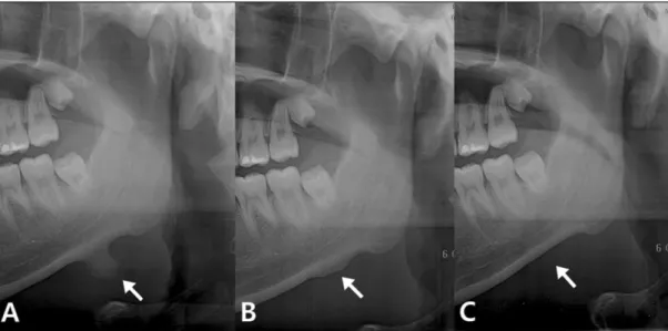
Fig. 2. A. Radiographic examination revealed a radiopaque bony mass from the mandibular inferior border.(arrow) There was neither surface erosion nor intra-lesional discrepancy in density.
B. Panoramic view taken immediately after surgery.(arrow)
C. Panoramic view 6 months after excision. The continuity of the mandibular inferior border was recovered by bony remodeling. There was no recurrence.(arrow)
Fig. 3.The excised tumor was 10 mm in length and 13 mm in width, with a whitish smooth surface.
Fig. 4. Microscopic appearance illustrating a bony structure covered with a cartilaginous cap. The cartilage is covered by fibrovascular connective tissue. Below that, the cartilage is being replaced by mature lamellar bone.(endochondral bone formation) (H&E staining, original magnification ×10)
대구외지 2010;36:214-6
216
Ⅲ.
고 찰
악안면 영역에서 발생되는 골연골종은 대부분 과두와 근 돌기에서 발생하며 환자들은 안면 비대칭이나 개구 제한, 통증, 관절음 등을 호소한다. 장골에서 발생한 골연골종은 종종 무증상으로 수술하지 않는데 반해 과두와 근돌기의 골연골종은 이러한 기능적∙심미적 결함으로 인해 일반적 으로 수술로 제거해야 한다.
과두나 근돌기를 제외하면 상하악골에서 발생한 골연골 종은 매우 드물며 무통성의 골융기로 인해 환자들이 인지 하게 되는 경우가 많다3-6. 저자 등이 살펴본바, 현재까지 보 고된 증례는 하악골체에서 5예, 정중부에서 2예 등이며 평 균 크기는 16×16 mm2이다. 모두 종괴 절제술로 치료했으 며, 재발이나 악성 변이는 없었다.
조직학적으로 보면 골연골종은 피질골과 해면골로 이루 어져 있으며, 유리질(hyaline) 연골성 덮개를 갖는 것이 특 징이다. 병소가 오래 된 경우, 연골 내 골화 또는 마손(wear and tear abrasion)에 의해 연골성 덮개가 없어지거나 아주 얇아져 성숙골(mature bone)만 남게 된다6,7. 이로 인해 연골 성 덮개가 없는 골연골종이 골종(osteoma)으로 진단되는 경우도 있다.
골연골종의 발병기전은 아직 불명확하다. Geshickter 등 은 건(tendon)이 부착되는 곳에 있는 세포는 연골성 성장 잠재력(cartilaginous growth potential)을 가지고 있으며, 긴 장과 압박(stress and strain)이 세포의 과활성 변화(hyper- plastic change)와 연골성 전환(cartilaginous transformation) 을 유발한다고 하였다8. 하악골 중 과두와 근돌기에서 골연 골종이 호발하는 것은 이 때문일 것이다. 또한 우리가 살펴 본바 하악골의 비호발 부위에서 발생한 골연골종도 대개 근육이나 건에 의한 견인력이 작용하는 곳에서 발생하였 다. 또 다른 가설로는 상당한 연골-골아세포성 잠재력 (chondro-osteoblastic potential)을 갖는 골막이 골연골종을
발생시킨다는 것과4,9 배아 연골성 잔유물(embryonic carti- laginous remnant)이 지속적으로 성장하여 병소가 된다는 견해가 있다10.
골연골종의 연골세포는 정상골단(epiphysis)의 세포가 갖 는 방향성과 유사한 모양으로 정렬되어 있으며2종양의 성 장은 골융기의 첨부에 있는 연골의 성장과 그에 따른 골화 로 이루어진다. 본 증례에서 수술 직후 촬영한 파노라마 방 사선사진을 보면 인접한 하악골 표면의 골융기가 완전히 제거되지 않은 것을 알 수 있다. 그러나 저자 등은 성장 잠 재력을 갖는 연골 부분(cartilaginous portion)이 제거되었기 때문에 재발없이 골개조(bony remodeling)가 완료된 것으 로 생각한다.
References
1. Dahlin DC, Unni KK, eds. Bone tumors: general aspects and data on 8,542 cases. 4th ed. Springfield, IL: Charles C Thomas; 1986.
2. Barnes L, ed. Surgical pathology of the head and neck.
NewYork: Dekker; 1985.
3. Anupam M, Shukla GK, Mishra SC, Bhatia N, Srivastava AN, Mishra N. Unusual solitary osteochondroma of the mandibular ramus. J Laryngol Otol 2002;116:65-6.
4. Brady FA, Sapp JP, Christensen RE. Extracondylar osteochon- dromas of the jaws. Oral Surg Oral Med Oral Pathol 1978;46:
658-68.
5. Erc¸oc¸en AR, Yilmaz S, Tas¸F. Osteochondroma arising from the mandibular symphysis. Ann Plast Surg 2002;48:336-7.
6. Tanaka E, Iida S, Tsuji H, Kogo M, Morita M. Solitary osteo- chondroma of the mandibular symphysis. Int J Oral Maxillofac Surg 2004;33:625-6.
7. Gnepp DR, ed. Diagnostic surgical pathology of the head and neck. Philadelphia: Saunders; 2001.
8. Geschickter CF, Copeland MM. Tumors of bone. NewYork: The American Journal of Cancer; 1931.
9. Lichtenstein L. Bone tumors. 5th ed. St. Louis: Mosby; 1977.
10. Kermer C, Rasse M, Undt G, Lang S. Cartilaginous exostoses of the mandible. Int J Oral Maxillofac Surg 1996;25:373-5.