CASE REPORT
식도 암육종 환자에서 이시적으로 발생한 식도 편평상피세포암 1예
차라리1,3, 정운태1,3, 오혜원1,3, 김희진1,3, 하창윤1,3, 김홍준1,3, 김태효1,3, 고경혁2,3
경상대학교 의학전문대학원 내과학교실1, 병리학교실2, 건강과학연구원3
A Case of Metachronous Development of Esophageal Squamous Cell Carcinoma in the Patient with Esophageal Carcinosarcoma
Ra Ri Cha1,3, Woon Tae Jung1,3, Hye Won Oh1,3, Hee Jin Kim1,3, Chang Yoon Ha1,3, Hong Jun Kim1,3, Tae Hyo Kim1,3 and Gyung Hyuck Ko2,3 Departments of Internal Medicine1 and Pathology2, Institute of Health Sciences3, Gyeongsang National University School of Medicine, Jinju, Korea
Esophageal carcinosarcoma is a rare malignant esophageal neoplasm consisting of both carcinomatous and sarcomatous elements, with an incidence of 0.5%. There have been only a few case reports of carcinosarcoma and squamous cell carcinoma coexisting in the esophagus. However, all of these are cases of synchronous or metachronous development of carcinosarcoma after chemoradiotherapy in patients of esophageal squamous cell carcinoma. A 53-year-old man underwent esophagogas- troduodenoscopy because of chest pain for several months. Endoscopic examination revealed a huge pedunculated esophageal polypoid mass. Endoscopic submucosal dissection (ESD) was performed and histopathologic examination confirmed spindle cell carcinoma (carcinosarcoma). He refused additional esophagectomy. After 21 months, third follow-up endoscopy showed poorly-demarcated flat, faint discolored lesions at different location from the previous ESD site and endoscopic biopsies confirmed squamous cell carcinoma. To the best of our knowledge, this is the first case of metachronous development of esophageal squamous cell carcinoma in a patient with esophageal carcinosarcoma. (Korean J Gastroenterol 2014;64:364-369) Key Words: Esophageal squamous cell carcinoma; Carcinosarcoma; Spindle cell carcinoma
Received May 19, 2014. Revised June 9, 2014. Accepted June 23, 2014.
CC This is an open access article distributed under the terms of the Creative Commons Attribution Non-Commercial License (http://creativecommons.org/licenses/
by-nc/3.0) which permits unrestricted non-commercial use, distribution, and reproduction in any medium, provided the original work is properly cited.
교신저자: 정운태, 660-751, 진주시 진주대로 816번길 15, 경상대학교 의학전문대학원 소화기내과
Correspondence to: Woon Tae Jung, Department of Internal Medicine, Gyeongsang National University School of Medicine, 15 Jinju-daero 816beon-gil, Jinju 660-751, Korea. Tel: +82-55-750-8617, Fax: +82-55-758-9122, E-mail: wtjung@gnu.ac.kr
Financial support: None. Conflict of interest: None.
INTRODUCTION
Carcinosarcoma of the esophagus is a rare malignant neo- plasm consisting of both carcinomatous and sarcomatous elements. Its reported incidence is approximately 0.5% of all esophageal neoplasms.1 There have been only a few case re- ports on development of both esophageal carcinosarcoma and squamous cell carcinomas in a patient, all of which were cases of synchronous or metachronous development of car- cinosarcoma after chemoradiotherapy in patients of esoph- ageal squamous cell carcinoma. There were no reports on de-
velopment of esophageal squamous cell carcinoma follow- ing carcinosarcoma. In most cases involving development of esophageal carcinosarcoma following squamous cell carci- noma, it has been suggested that the sarcomatous element of esophageal carcinosarcoma generally results from differ- entiation of carcinoma cells into mesenchymal tumor cells.2 In contrast, in the histogenesis of squamous cell carcinoma from carcinosarcoma, it is unclear whether both of them have a common clonal origin or not. Here, we present a case of metachronous development of esophageal squamous cell carcinoma in a patient who had undergone endoscopic re-
Cha RR, et al. Esophageal Carcinosarcoma and Squamous Cell Carcinoma 365
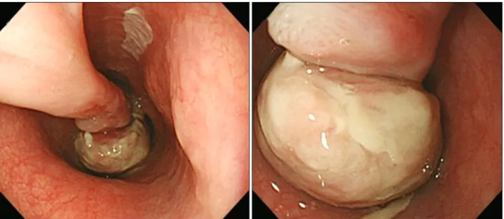
Fig. 1. Endoscopic examination reveals a huge pedunculated esophageal polyp. The polyp stalk originates at 25 cm from the upper incisors and the polyp head extends down to 33 cm.
section of esophageal carcinosarcoma, 21 months ago.
CASE REPORT
A 53-year-old man presented to our hospital complaining of epigastric pain for several months. He was diagnosed with hepatitis B associated liver cirrhosis 10 years ago and under- went endoscopic variceal ligation twice three years ago. He had smoked one-third pack of cigarettes a day and drank heavily for 30 years but had stopped binge drinking three years ago.
An endoscopic examination revealed a huge pedunculated esophageal polyp, polyp stalk originating at 25 cm from the upper incisors. The polyp head extended down to 33 cm from the upper incisors and was almost filling the lumen (Fig. 1).
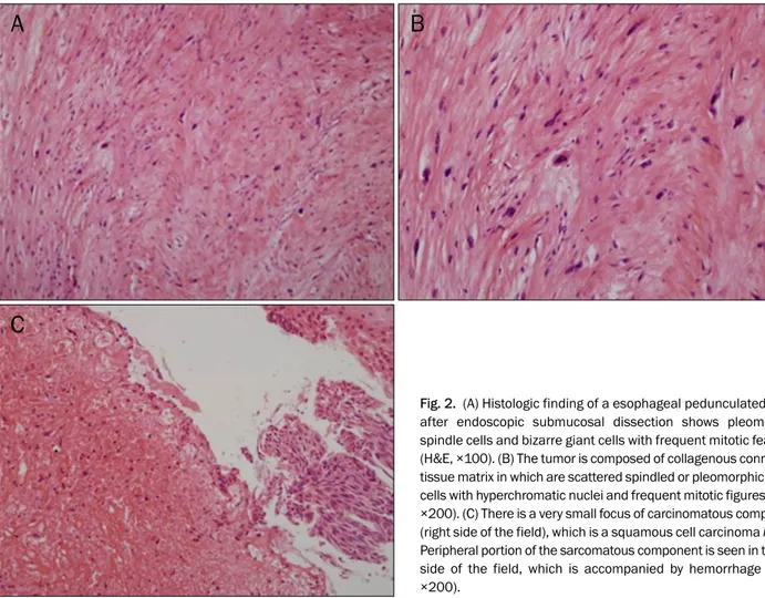
The endoscopic biopsies showed some atypical spindle cells, therefore, spindle cell carcinoma or other undifferentiated carcinoma was suspected. A CT scan of the chest and PET-CT were negative for metastasis. Endoscopic submucosal dis- section (ESD) was performed. The size of the resected speci- men was 4.2×3.0×2.5 cm and the histopathologic examina- tion confirmed spindle cell carcinoma (carcinosarcoma) with more than seven mitoses per high power field (HPF) (Fig. 2).
Immunohistochemically, the spindle-shaped sarcomatous cells showed negative reaction to cytokeratin, S-100 protein, smooth muscle actin, c-Kit, DOG-1, and epithelial membrane antigen (EMA), but a positive reaction to vimentin. No transi-
tional zone was seen between sarcomatous and carcinoma- tous elements (Fig. 3). The patient was finally diagnosed with true esophageal carcinosarcoma. The tumor had involve- ment of deep resection margin, with no evidence of vascular or lymphatic invasion. We recommended additional esoph- agectomy because of the possibility of remnant tumor. He re- fused the esophagectomy, thus, follow-up endoscopy and CT were performed at 2 months, 5 months, and 1 year after ESD.
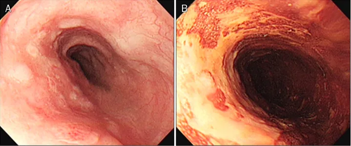
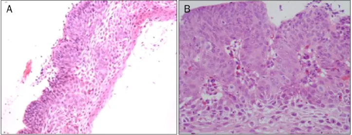
At 21 months after ESD, the patient complained of dyspha- gia over one month. An endoscopic examination showed a whitish fibrotic scar by previous ESD at 25 cm from the upper incisors and poorly-demarcated flat, faint discolored lesions, which were unstained by Lugol solution, at 30-34 cm from up- per incisors (Fig. 4). The endoscopic biopsy of faint discolored lesions indicated squamous cell carcinoma with well differ- entiated type (Fig. 5). He wanted and transferred to another hospital. A chest CT showed small paraesophageal lympha- denopathy at subcarina and a PET-CT showed a hypermeta- bolic nodule of the left retropharynx. He underwent ESD at the esophagus and piecemeal endoscopic mucosal re- section (EMR) at the left hypopharynx in another hospital.
Histopathologic examination of the esophagus showed squ- amous cell carcinoma, moderately differentiated with sub- mucosal invasion at a depth of 150 m. Histopathologic ex- amination of the left hypopharyngeal mass also showed in- vasive squamous cell carcinoma with moderately differ- entiated type. Immunohistochemically, the squamous cell
Fig. 2. (A) Histologic finding of a esophageal pedunculated polyp after endoscopic submucosal dissection shows pleomorphic spindle cells and bizarre giant cells with frequent mitotic features (H&E, ×100). (B) The tumor is composed of collagenous connective tissue matrix in which are scattered spindled or pleomorphic tumor cells with hyperchromatic nuclei and frequent mitotic figures (H&E,
×200). (C) There is a very small focus of carcinomatous component (right side of the field), which is a squamous cell carcinoma in situ.
Peripheral portion of the sarcomatous component is seen in the left side of the field, which is accompanied by hemorrhage (H&E,
×200).
carcinoma in the esophagus had shown a diffuse and strong positive reaction to p53 protein. There was no further man- agement of the paraesophageal lymph node. He transferred back to our hospital and underwent two cycles of chemo- radiation therapy with 5-fluorouracil and cisplatin. He died from sepsis after one month from last chemotherapy and radiotherapy.
DISCUSSION
The current case was the metachronous development of squamous cell carcinoma following carcinosarcoma of the esophagus in a patient on the basis of histologic evidence.
There have been several case reports on synchronous carci- nosarcoma and squamous cell carcinoma in the esoph- agus,3-5 and a case report of esophageal carcinosarcoma arising from squamous cell carcinoma after chemoradia- tion.6 In a study with Taiwanese patients, Kuo et al.7 reported that approximately 33% (4/12) of carcinosarcoma had pre-
vious head and neck squamous cell carcinoma that occurred metachronously. However, there has been no previous report on metachronous development of esophageal squamous cell carcinoma in a patient with carcinosarcoma.
Like squamous cell carcinoma of the esophagus, esoph- ageal carcinosarcoma occurs most often in middle-aged men with a history of smoking or drinking or both.8 The cur- rent patient has been a heavy drinker and smoker, and did not have previous history of any other malignancy before diag- nosis of esophageal carcinosarcoma.
Carcinosarcoma of the esophagus, also termed sarcoma- toid carcinoma, pseudosarcoma, spindle cell carcinoma, or polypoid carcinoma, is an unusual malignant tumor of the esophagus, consisting of both carcinomatous and sarcoma- tous components. The various terms in use reflect the un- certain pathogenesis of this tumor.9
Chino et al.10 reported that the carcinomatous component was differentiated squamous cell carcinoma and the sarcom- atous component was spindle cell carcinoma. Histological
Cha RR, et al. Esophageal Carcinosarcoma and Squamous Cell Carcinoma 367
Fig. 3. (A) The tumor cells are negative for cytokeratin (×200). (B) The tumor cells are negative for S-100 protein (×200). (C) The tumor cells are negative for smooth muscle actin. In contrast, normal smooth muscle cells around blood vessels are positive for smooth muscle actin (×200).
(D) Many tumor cells are positive for vimetin (×200). (E) The tumor cells are negative for epithelial membrane antigen (×200).
Fig. 4. Twenty-one months later, endoscopic examination demonstrated poorly-demarcated flat, faint discolored lesions at 30-34 cm from the upper incisors (A), which are unstained by Lugol solution (B).
analyses showed that the majority of the protruding tumors consisted of the sarcomatous component, while the ulcer- ating tumor consisted mainly of squamous cell carcinoma. In our case, the tumor was a huge pedunculated protruding mass consisting of an almost sarcomatous component.
According to the Japanese Society for Esophageal Disease, three main hypotheses have been proposed for the patho- genesis of carcinosarcomas. The first is the metaplastic con- cept, which proposes that the individual components of this malignancy may be derived from a single, common ancestor
Fig. 5. A forcep biopsy specimen from flat discolored lesions in the esophagus shows well-differentiated squamous cell carcinoma (H&E; A,
×100, B, ×400).
cell (so-called carcinosarcoma), in other words, the sarcoma- tous components are considered to result from metaplasia of carcinoma cells.9 This theory is supported by the frequent finding of a transition zone between the two cell populations and identical genetic alterations are observed in both components.11 The second is the collision concept, which hy- pothesizes that two individual stem cells may undergo malig- nant transformation (true carcinosarcoma) independently and simultaneously, and are actually separate tumors that have merged.9 Expression of cytokeratin and vimentin were mutually exclusive in the carcinomatous or sarcomatous ele- ments, which may support the collision concept. The third theory suggests that the spindle cell component is a reaction to the carcinoma.9 In most cases, the metaplastic concept is generally accepted. In the current case, there was no tran- sition zone between carcinomatous and sarcomatous com- ponents and the tumor cells showed an immunochemically negative reaction to cytokeratin and a positive reaction to vimentin. Therefore, the patient was finally diagnosed with true carcinosarcoma.
The clinical presentation of esophageal carcinosarcoma is similar to that of squamous cell carcinoma with dysphagia as the most prominent and frequent symptom.12 They are typi- cally large (mean, 6-7 cm) and polypoid at presentation.13 Because of accelerated intraluminal growth, esophageal car- cinosarcoma often presents relatively early.9 Despite the huge size of the tumor, it does not invade as deeply as an esophageal squamous cell carcinoma. In spite of their pro-
pensity to cause symptoms earlier because of their large size and the fact that more than 80% of tumors are limited to the submucosa or muscularis propria at presentation,8,13 these tumors might behave aggressively. Sasajima et al.14 reported the doubling time of their case to be 2.2 months, whereas that of ordinary esophageal squamous cell carcinoma was 5 months.
The treatment of esophageal carcinosarcoma did not dif- fer from that of other esophageal malignant lesions. Esopha- gectomy has traditionally been considered as the first option for esophageal carcinosarcoma patients.13 With the ad- vances in micro-invasive techniques, endoscopic procedures, including endoscopic polypectomy, endoscopic mucosal re- section or ESD, may represent an alternative to esophagec- tomy for superficial esophageal carcinosarcoma.15 Chemo- therapy and concomitant radiation therapy must also be con- sidered for residual microscopic disease and local control.
Esophageal carcinosarcomas do not necessarily have a better prognosis and some authors showed that there is no significant difference in the 5-year survival rates.1,3 In a study with 20 cases of esophageal carcinosarcoma, Iyomasa et al.1 reported that recurrence due to hematogenous metastasis was more frequent in esophageal carcinosarcoma than esophageal squamous cell carcinoma. They emphasized that radical resection with lymph node dissection was neces- sary for treatment of carcinosarcoma regardless of the depth of invasion.
In conclusion, this is a first case report of metachronous
Cha RR, et al. Esophageal Carcinosarcoma and Squamous Cell Carcinoma 369
development of esophageal squamous cell carcinoma fol- lowing carcinosarcoma in a patient. The clinicopathological characteristics and histogenesis of both tumors are not clearly known; therefore, further molecular studies and more cases are needed.
REFERENCES
1. Iyomasa S, Kato H, Tachimori Y, Watanabe H, Yamaguchi H, Itabashi M. Carcinosarcoma of the esophagus: a twenty-case study. Jpn J Clin Oncol 1990;20:99-106.
2. Wang ZY, Itabashi M, Hirota T, Watanabe H, Kato H.
Immunohistochemical study of the histogenesis of esophageal carcinosarcoma. Jpn J Clin Oncol 1992;22:377-386.
3. Uchiyama S, Imai S, Hoshino A, et al. Rapid-growing carcino- sarcoma of the esophagus arising from intraepithelial squ- amous cell carcinoma: report of a case. Surg Today 2000;
30:173-176.
4. Zhao S, Xue Q, Ye B, Lu H, He J, Zhao H. Synchronous primary carci- nosarcoma and adenosquamous carcinoma of the esophagus.
Ann Thorac Surg 2011;91:926-928.
5. Tomizawa Y, Taniguchi M, Mori M. An unusual case of intra- luminally growing esophageal tumor. Diagnosis: carcinosarcoma of the esophagus. Gastroenterology 2011;141:e10-e11.
6. Nakagawa S, Yabusaki H, Tanaka O. Rapid-growth carcinosarco- ma of the esophagus arising from 0-IIc squamous cell carcino-
ma after definitive chemoradiotherapy: a case report. Esopha- gus 2009;6:123-126.
7. Kuo CJ, Lin TN, Lin CJ, et al. Clinical manifestation of esophageal carcinosarcoma: a Taiwan experience. Dis Esophagus 2010;23:
122-127.
8. Kimura H, Konishi K, Kawamura T, et al. Esophageal sarcomas:
report of three cases. Dig Surg 1999;16:244-247.
9. Madan AK, Long AE, Weldon CB, Jaffe BM. Esophageal carcino- sarcoma. J Gastrointest Surg 2001;5:414-417.
10. Chino O, Kijima H, Shimada H, et al. Clinicopathological studies of esophageal carcinosarcoma: analyses of its morphological characteristics using endoscopic, histological, and immunohis- tochemical procedures. Endoscopy 2000;32:706-711.
11. Iascone C, Barreca M. Carcinosarcoma and pseudosarcoma of the esophagus: two names, one disease--comprehensive review of the literature. World J Surg 1999;23:153-157.
12. Ziauddin MF, Rodriguez HE, Quiros ED, Connolly MM, Podbielski FJ. Carcinosarcoma of the esophagus--pattern of recurrence.
Dig Surg 2001;18:216-218.
13. Ji F, Xu YM, Xu CF. Endoscopic polypectomy: a promising ther- apeutic choice for esophageal carcinosarcoma. World J Gastro- enterol 2009;15:3448-3450.
14. Sasajima K, Taniguchi Y, Morino K, et al. Rapid growth of a pseu- dosarcoma of the esophagus. J Clin Gastroenterol 1988;10:
533-536.
15. Pesenti C, Bories E, Danisi C, Monges G, Giovannini M. Endosco- pic treatment of esophageal carcinosarcoma: report of a case.
Endoscopy 2004;36:95.